Comparative Study
. 2016 Jul;68(1):123-32.
doi: 10.1161/HYPERTENSIONAHA.116.07237. Epub 2016 May 23.

Activation of Human T Cells in Hypertension: Studies of Humanized Mice and Hypertensive Humans

Affiliations
Comparative Study

Activation of Human T Cells in Hypertension: Studies of Humanized Mice and Hypertensive Humans

Hana A Itani et al. Hypertension. 2016 Jul.

Abstract

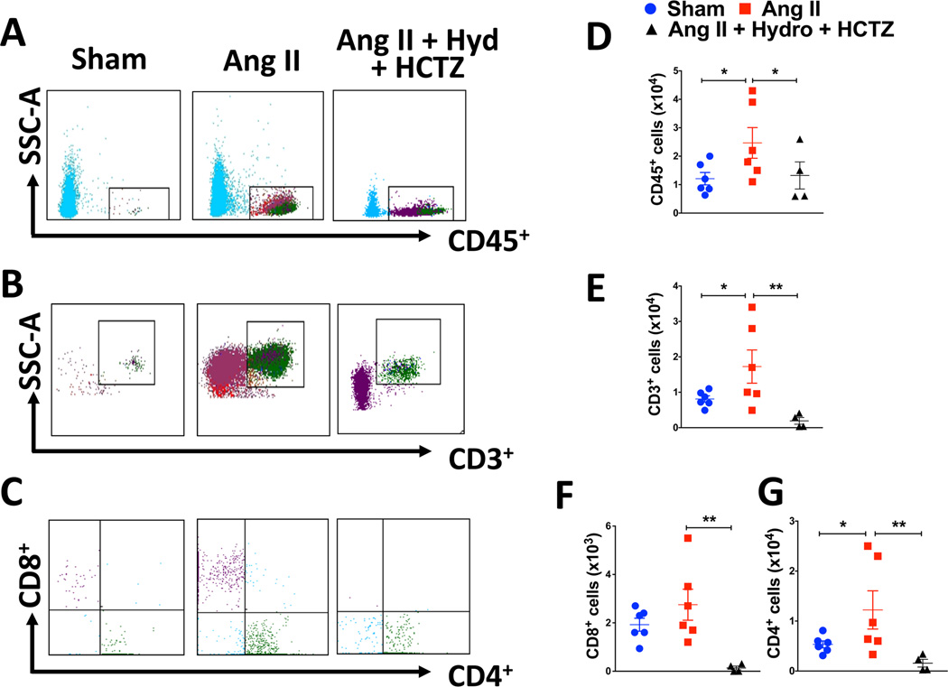
Emerging evidence supports an important role for T cells in the genesis of hypertension. Because this work has predominantly been performed in experimental animals, we sought to determine whether human T cells are activated in hypertension. We used a humanized mouse model in which the murine immune system is replaced by the human immune system. Angiotensin II increased systolic pressure to 162 versus 116 mm Hg for sham-treated animals. Flow cytometry of thoracic lymph nodes, thoracic aorta, and kidney revealed increased infiltration of human leukocytes (CD45(+)) and T lymphocytes (CD3(+) and CD4(+)) in response to angiotensin II infusion. Interestingly, there was also an increase in the memory T cells (CD3(+)/CD45RO(+)) in the aortas and lymph nodes. Prevention of hypertension using hydralazine and hydrochlorothiazide prevented the accumulation of T cells in these tissues. Studies of isolated human T cells and monocytes indicated that angiotensin II had no direct effect on cytokine production by T cells or the ability of dendritic cells to drive T-cell proliferation. We also observed an increase in circulating interleukin-17A producing CD4(+) T cells and both CD4(+) and CD8(+) T cells that produce interferon-γ in hypertensive compared with normotensive humans. Thus, human T cells become activated and invade critical end-organ tissues in response to hypertension in a humanized mouse model. This response likely reflects the hypertensive milieu encountered in vivo and is not a direct effect of the hormone angiotensin II.

Keywords: antigens, CD45; dendritic cells; inflammation; lymph nodes; myeloid cells.


Conflict of interest statement

Conflict (s) of interest/disclosure (s) statement: The authors have no conflict of interests.

Figures

Figure 1
Figure 1. Effects of hypertension on renal leukocyte and T cell infiltration
Humanized mice received sham or ang II infusion (490 ng/kg/min) for two weeks with or without co-administration of hydralazine (Hyd) and hydrochlorothiazide (HCTZ). Single cell suspensions were prepared from kidneys of humanized mice and analyzed for total human leukocytes and T cells. Representative dot plots are of human live, singlets gated for (A) total leukocytes (CD45+), (B) total T cells (CD3+), (C) CD8+/CD4+ T cells. The solid black lines in the histogram indicate the gates used for analysis. Summary data are shown for renal accumulation of total leukocytes (D), total T cells (E), CD8+ T cells (F), and CD4+ T cells (G) in response to either a sham or ang II infusion. Data were analyzed using an unpaired T test or the non-parametric Mann Whitney test when variances between groups were not equal, n= 4–6 per group.
Figure 2
Figure 2. Effects of hypertension on leukocyte and T cell infiltration into the descending, thoracic aorta
Mice underwent sham or Ang II infusion with or without co-administration of hydralazine (Hyd) and hydrochlorothiazide (HCTZ) as in figure 1. Representative dot plots are shown of live, human, singlet cells gated for (A) total leukocytes (CD45+), (B) total T cells (CD3+), (C) CD8+/CD4+ T cells. Summary data are shown for aortic accumulation of total leukocytes (D), total T cells (E), CD8+ T cells (F) and CD4+ T cells (G) in response to either a sham or ang II infusion. Data were analyzed using an unpaired T test or the non-parametric Mann Whitney test when variances between groups were not equal, n= 4–6 per group.
Figure 3
Figure 3. Effect of hypertension on leukocyte and T cell infiltration into thoracic lymph nodes
Mice underwent sham or Ang II infusion with or without co-administration of hydralazine (Hyd) and hydrochlorothiazide (HCTZ) as in figure 1 and thoracic lymph nodes were harvested from the periaortic region. Representative images are of human live, singlets gated for (A) total leukocytes (CD45+), (B) total T cells (CD3+), (C) CD8+ / CD4+ T cells. The solid black lines in the histogram indicate the gates that used for all of our analysis. Summary data are shown for the presence of total leukocytes (D), total T cells (E), CD8+ T cells (F), and CD4+ T cells (G). Data were analyzed using an unpaired T test or the non-parametric Mann Whitney test when variances between groups were not equal, n= 4–6 per group.
Figure 4
Figure 4. Effect of hypertension on myeloid differentiation in the kidney, aorta and lymph nodes
Summary data from mice following sham or angiotensin II (ang II) infusion (490 ng/kg/min) of non-CD3+ CD45+ cells in the kidney (A), descending, thoracic aorta (B), and thoracic lymph nodes (C). The flow cytometry gating strategy used to identify myeloid cells is shown (D). The kidney, aorta and lymph nodes of the humanized mice all showed differentiation of the monocytes to dendritic cells (E). Myeloid differentiation was not significantly shifted in any of the tissues when we compared monocyte (CD14+CD83) vs. activated monocyte (CD14+CD83+) vs. dendritic cell (CD14CD83+) populations.
Figure 5
Figure 5. Direct effect of ang II on T cell phenotype and dendritic cell activation
Human T cells were cultured on anti-CD3 plates containing anti-DC28 (2 µg/ml) without and with ang II (0.1 µMol/L) for 7 days and intracellular staining was performed to detect the cytokines IFN-γ and IL-17A and the transcription factor FoxP3 (A–D). Human monocytes were differentiated into dendritic cells by exposure to GM-CSF and IL-4 for 7 days without and with ang II (0.1 µMol/L). The differentiated DCs were then co-cultured with CFSE-labeled T cells from the same donor at a ratio of 1:10. Proliferation was measured by CFSE dilution (E). Isoketal-protein adducts in DCs were determined by intracellular staining with isoketal adduct antibody D11 (F).
Figure 6
Figure 6
Characterization of circulating T cells in humans with and without hypertension (n = 20 for each). Blood was obtained from patients with hypertension (HTN) and normotensive controls (CTR) and T cells were stained for CD3, CD4, CD8 and the memory marker CD45RO (panels A and B). Intracellular staining was performed to identify cells producing IL-17A (panels C and D) and for IFN-γ and TNFα (panel E and F). Data were analyzed using an unpaired T test with Bonferroni correction for multiple comparisons.

References

    1. Guzik TJ, Hoch NE, Brown KA, McCann LA, Rahman A, Dikalov S, Goronzy J, Weyand C, Harrison DG. Role of the t cell in the genesis of angiotensin ii induced hypertension and vascular dysfunction. J Exp Med. 2007:2449–2460. - PMC - PubMed
    1. Marvar PJ, Thabet SR, Guzik TJ, Lob HE, McCann LA, Weyand C, Gordon FJ, Harrison DG. Central and peripheral mechanisms of t-lymphocyte activation and vascular inflammation produced by angiotensin ii-induced hypertension. Circ Res. 2010:263–270. - PMC - PubMed
    1. Marvar PJ, Vinh A, Thabet S, Lob HE, Geem D, Ressler KJ, Harrison DG. T lymphocytes and vascular inflammation contribute to stress-dependent hypertension. Biol Psychiatry. 2012:774–782. - PMC - PubMed
    1. Crowley SD, Song YS, Lin EE, Griffiths R, Kim HS, Ruiz P. Lymphocyte responses exacerbate angiotensin ii-dependent hypertension. Am J Physiol Regul Integr Comp Physiol. 2010:R1089–R1097. - PMC - PubMed
    1. Mattson DL, James L, Berdan EA, Meister CJ. Immune suppression attenuates hypertension and renal disease in the dahl salt-sensitive rat. Hypertension. 2006:149–156. - PubMed

Publication types

MeSH terms

Substances