. 2016 Jul 21;84(8):2209-2219.
doi: 10.1128/IAI.00147-16. Print 2016 Aug.

HtrA Is Important for Stress Resistance and Virulence in Haemophilus parasuis

Affiliations

HtrA Is Important for Stress Resistance and Virulence in Haemophilus parasuis

Luhua Zhang et al. Infect Immun. .

Abstract

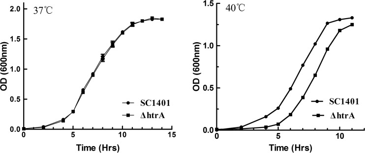
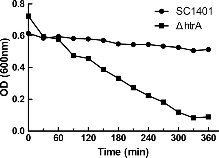
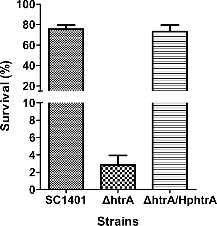
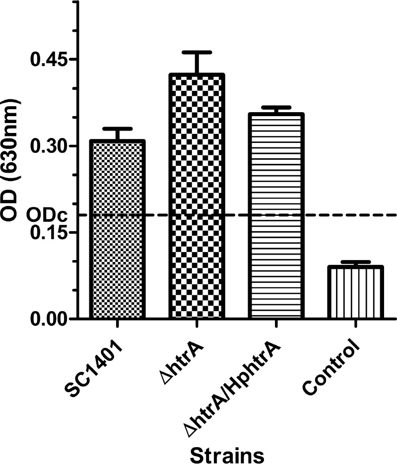
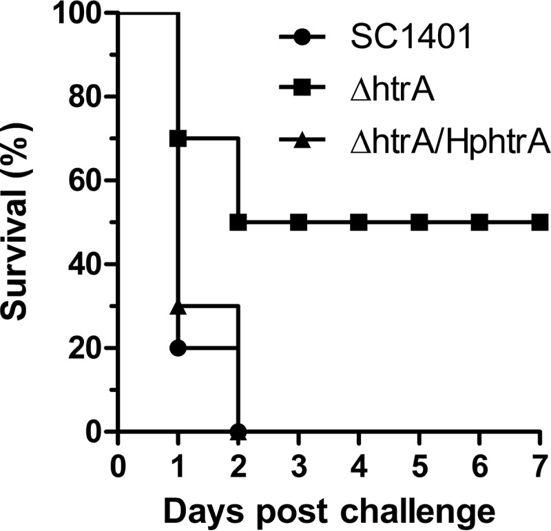
Haemophilus parasuis is an opportunistic pathogen that causes Glässer's disease in swine, with polyserositis, meningitis, and arthritis. The high-temperature requirement A (HtrA)-like protease, which is involved in protein quality control, has been reported to be a virulence factor in many pathogens. In this study, we showed that HtrA of H. parasuis (HpHtrA) exhibited both chaperone and protease activities. Finally, nickel import ATP-binding protein (NikE), periplasmic dipeptide transport protein (DppA), and outer membrane protein A (OmpA) were identified as proteolytic substrates for HpHtrA. The protease activity reached its maximum at 40°C in a time-dependent manner. Disruption of the htrA gene from strain SC1401 affected tolerance to temperature stress and resistance to complement-mediated killing. Furthermore, increased autoagglutination and biofilm formation were detected in the htrA mutant. In addition, the htrA mutant was significantly attenuated in virulence in the murine model of infection. Together, these data demonstrate that HpHtrA plays an important role in the virulence of H. parasuis.


Figures

FIG 1
FIG 1
Sequence analysis of HpHtrA. (A) Schematic of domain architecture showing the various functional domains of HpHtrA. SP, N-terminal signal peptide. (B) Sequence alignment of H. parasuis HtrA (HpHtrA) against H. influenzae HtrA (HiHtrA), E. coli DegP (EcDegP), DegQ (EcDegQ), and DegS (EcDegS), and Legionella fallonii DegQ (LfDegQ). Secondary structures of HpHtrA predicted by computer analysis are indicated by coils for α-helices and arrows for β-strands. Residues forming the catalytic triads are marked with red triangles. Identical residues are highlighted in red, and homologous residues are highlighted in yellow.
FIG 2
FIG 2
Purification of rHpHtrA and Western blotting of the ΔhtrA mutant and its complemented strain. (A) Purification of rHpHtrA. Lane 1, SDS-PAGE analysis verified the existence of purified rHpHtrA in the fraction collected; lane 2, Western blotting verified the two protein bands detected by the anti-6×His tag antibody. (B) Western blotting of the ΔhtrA mutant and its complemented strain with polyclonal anti-rHpHtrA antibodies. Lane 1, wild-type strain SC1401; lane 2, ΔhtrA mutant strain; lane 3, complemented ΔhtrA/HphtrA strain. Western blotting of HpHtrA showed two bands of 48 kDa and 44 kDa, as indicated by the two red arrows, in the wild-type and complemented strains but not in the ΔhtrA mutant strain.
FIG 3
FIG 3
Comparison of the chaperone-like activities of rHpHtrA, rHpHtrAS219A, rHpHtrAH113R, and rHpHtrAD143V in protection of DTT-denatured lysozyme against aggregation. The light-scattering values were recorded at 360 nm. The experiments were performed independently three times in triplicates. The means ± standard deviations from one representative experiment are shown.
FIG 4
FIG 4
Proteolytic activities of rHpHtrA, rHpHtrAS219A, rHpHtrAH113R, and rHpHtrAD143V against β-casein. (A) Proteolytic activities were monitored by incubating β-casein with rHpHtrA at 40°C over a time course of 6 h. M, protein markers; lanes 1 to 7, time points 0, 1, 2, 3, 4, 5, and 6 h. (B) Proteolytic activities were monitored by incubating β-casein with rHpHtrA at different temperatures for different times. Lanes 1 to 5, temperatures of 20, 30, 40, 50, and 60°C. (C) Proteolytic activities were monitored by incubating β-casein with rHpHtrAD143V (lanes 1 to 3), rHpHtrAH113R (lanes 4 to 6), and rHpHtrAS219A (lanes 7 to 9) at 40°C over a time course of 5 h. Lanes 1, 4, and 7, 0 h; lanes 2, 5, and 8, 4 h; lanes 3, 6, and 9, 5 h.
FIG 5
FIG 5
NikE, DppA, and OmpA are substrates for HpHtrA. (A) SDS-PAGE analysis of degradation of H. parasuis membrane proteins by HpHtrA. The extracted membrane proteins were denatured with 20 mM DTT for 12 h at 40°C and then incubated with or without rHpHtrA for 6 h. M, protein markers; lane 1, denatured membrane proteins alone were incubated at 40°C for 6 h before SDS-PAGE analysis; lane 2, denatured membrane proteins were incubated at 40°C for 6 h and then added to rHpHtrA immediately before SDS-PAGE analysis; lane 3, denatured membrane proteins were coincubated with rHpHtrA at 40°C for 6 h before SDS-PAGE analysis. Bands b and c (red arrows) were isolated for analysis by liquid chromatography-mass spectrometry (LC-MS). (B) SDS-PAGE analysis of degradation of rOmpA by rHpHtrA. Lanes 1 and 3, denatured rOmpA was incubated alone at 40°C for 6 h (lane 1) or 12 h (lane 3) and then added to rHpHtrA immediately before SDS-PAGE analysis; lanes 2 and 4, denatured rOmpA was coincubated with rHpHtrA at 40°C for 6 h (lane 2) or 12 h (lane 4) before SDS-PAGE analysis. The red arrow indicates rOmpA protein. (C) SDS-PAGE analysis of degradation of r0079 by rHpHtrA. Lanes 1 and 3, denatured r0079 was incubated alone at 40°C for 12 h (lane 1) or 24 h (lane 3) and then added to rHpHtrA immediately before SDS-PAGE analysis; lanes 2 and 4, denatured r0079 was coincubated with rHpHtrA at 40°C for 12 h (lane 2) or 24 h (lane 4) before SDS-PAGE analysis. The red arrow indicates r0079 protein. (D) SDS-PAGE analysis of degradation of rDppA (lanes 1 to 4) and rNikE (lanes 5 to 8) by rHpHtrA. Lanes 1, 5, 3, and 7, denatured rDppA or rNikE was incubated alone at 40°C for 6 h (lanes 1 and 5) or 12 h (lanes 3 and 7) and then added to rHpHtrA immediately before SDS-PAGE analysis; lanes 2, 6, 4, and 8, denatured rDppA or rNikE was coincubated with rHpHtrA at 40°C for 6 h (lanes 2 and 6) or 12 h (lanes 4 and 8) and then used for SDS-PAGE analysis. The red arrows indicate the substrate proteins.
FIG 6
FIG 6
Growth curves of H. parasuis wild-type strain SC1401 versus the ΔhtrA mutant at 37°C and 40°C. Both the wild-type and mutant strains were cultured in tryptic soy broth supplemented with 0.01% NAD and 5% bovine serum. The experiments were performed three times independently in triplicates. The means ± standard deviations from one representative experiment are shown.
FIG 7
FIG 7
Autoagglutination rates of SC1401 versus the ΔhtrA strain. The cells were harvested and diluted in fresh TSB to an OD600 of ∼0.7 and then allowed to remain static at 25°C. The OD600 of the suspensions was measured every 30 min for 6 h. The experiments were performed three times independently in triplicates. The means ± standard deviations from one representative experiment are shown.
FIG 8
FIG 8
The ΔhtrA strain is more susceptible than SC1401 or the ΔhtrA/HphtrA strain to complement-mediated killing in 50% porcine serum at 37°C. Percent survival was calculated as the ratio of the number of bacteria that survived in normal serum to the number that survived in heat-treated serum. The experiments were performed three times independently in triplicates. Error bars represent the standard errors from three independent experiments. The results for the ΔhtrA strain and SC1401 were significantly different (P < 0.05).
FIG 9
FIG 9
HpHtrA suppresses biofilm formation. The amounts of biofilm formed by H. parasuis SC1401 and the ΔhtrA and ΔhtrA/HphtrA strains were compared. The dotted line shows the cutoff value (ODc = 0.1804) for the determination of a biofilm producer. Based on the OD values, the three strains were all identified as biofilm producers. The experiments were performed three times independently in triplicates. Error bars represent the standard errors from three independent experiments. The results for SC1401 and the ΔhtrA strain were significantly different (P < 0.05).
FIG 10
FIG 10
HphtrA is important in a mouse model of intraperitoneal infection. Survival curves of mice inoculated with SC1401 or the ΔhtrA or ΔhtrA/HphtrA strain are shown. The survival rates of mice infected by SC1401 and the ΔhtrA strain were significantly different (P < 0.05) using the log-rank test.

Similar articles

Cited by

References

    1. Zhang B, Tang C, Liao M, Yue H. 2014. Update on the pathogenesis of Haemophilus parasuis infection and virulence factors. Vet Microbiol 168:1–7. doi:10.1016/j.vetmic.2013.07.027. - DOI - PubMed
    1. Kielstein P, Rapp-Gabrielson V. 1992. Designation of 15 serovars of Haemophilus parasuis on the basis of immunodiffusion using heat-stable antigen extracts. J Clin Microbiol 30:862. - PMC - PubMed
    1. Costa-Hurtado M, Aragon V. 2013. Advances in the quest for virulence factors of Haemophilus parasuis. Vet J 198:571–576. doi:10.1016/j.tvjl.2013.08.027. - DOI - PubMed
    1. Zhang L, Wen Y, Li Y, Wei X, Yan X, Wen X, Wu R, Huang X, Huang Y, Yan Q, Liu M, Cao S. 2014. Comparative proteomic analysis of the membrane proteins of two Haemophilus parasuis strains to identify proteins that may help in habitat adaptation and pathogenesis. Proteome Sci 12:38. doi:10.1186/1477-5956-12-38. - DOI - PMC - PubMed
    1. Pallen MJ, Wren BW. 1997. The HtrA family of serine proteases. Mol Microbiol 26:209–221. doi:10.1046/j.1365-2958.1997.5601928.x. - DOI - PubMed

Publication types

MeSH terms

LinkOut - more resources