. 2016 Jul;171(3):2256-76.
doi: 10.1104/pp.16.00711. Epub 2016 May 23.

A Laser Dissection-RNAseq Analysis Highlights the Activation of Cytokinin Pathways by Nod Factors in the Medicago truncatula Root Epidermis

Affiliations

A Laser Dissection-RNAseq Analysis Highlights the Activation of Cytokinin Pathways by Nod Factors in the Medicago truncatula Root Epidermis

Marie-Françoise Jardinaud et al. Plant Physiol. 2016 Jul.

Abstract

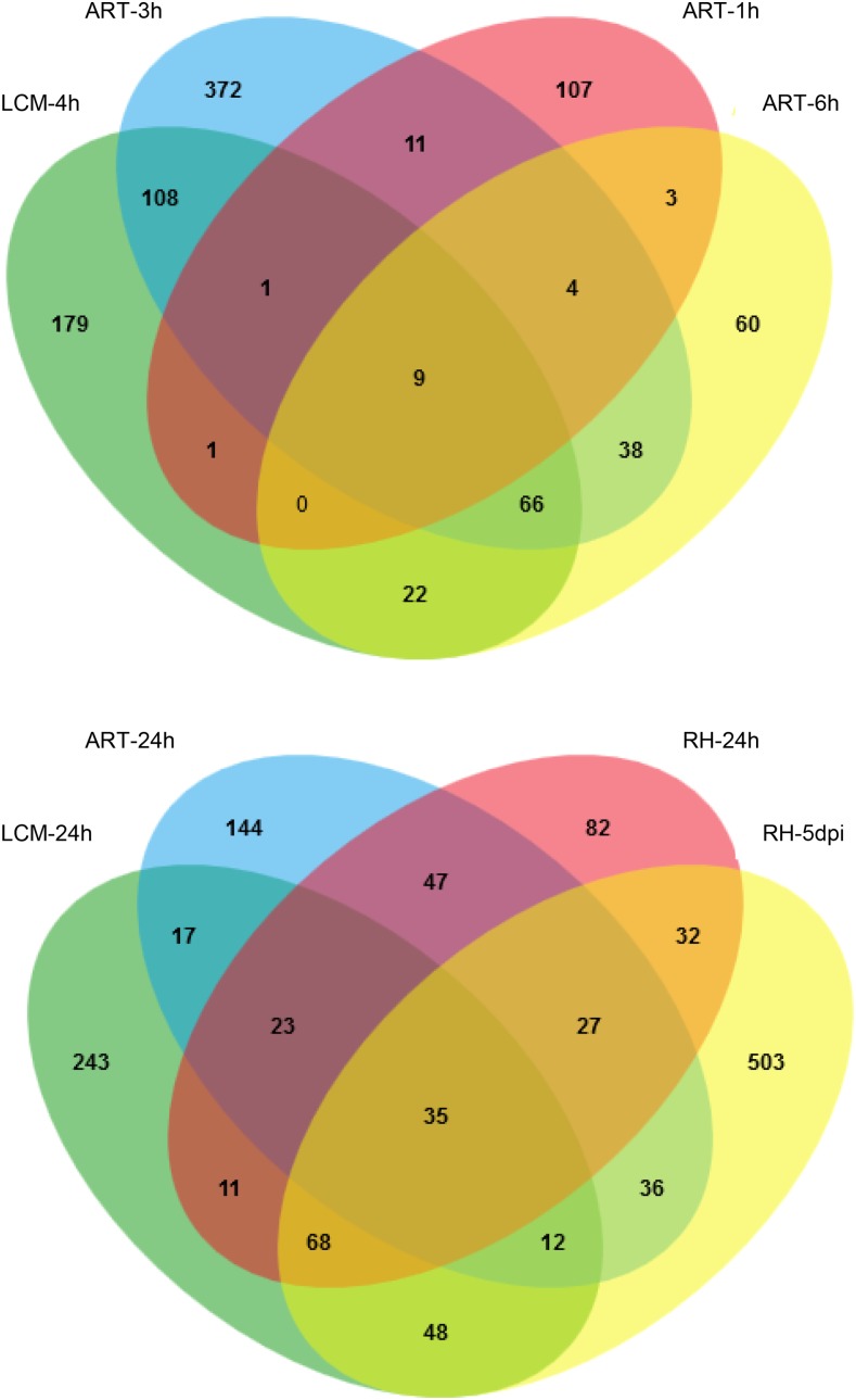
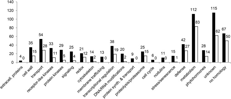
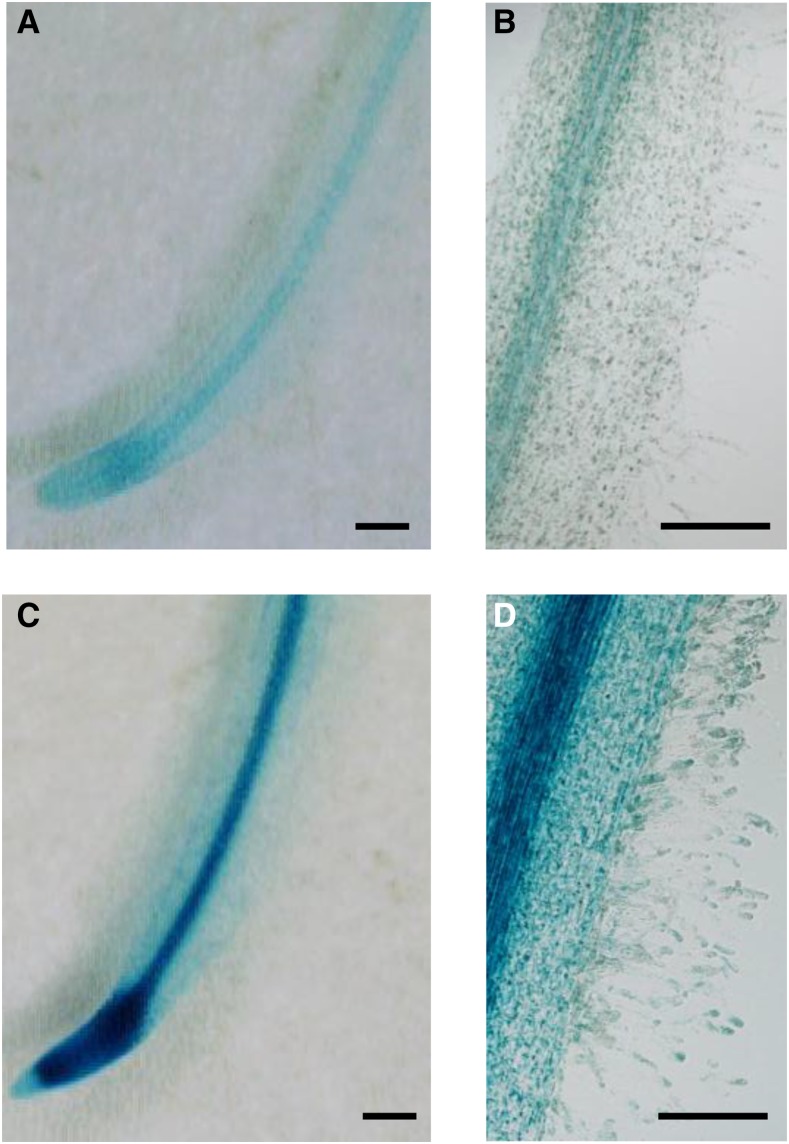
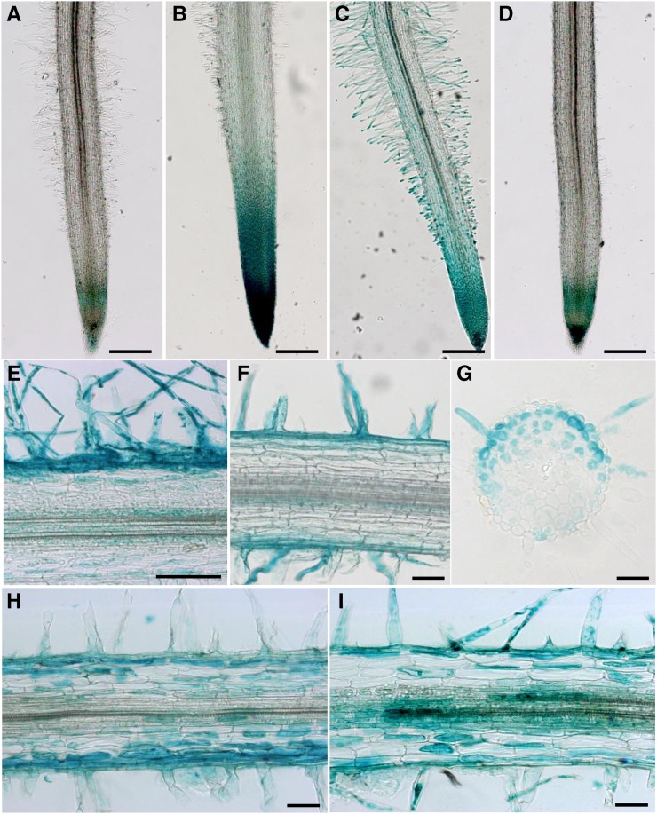
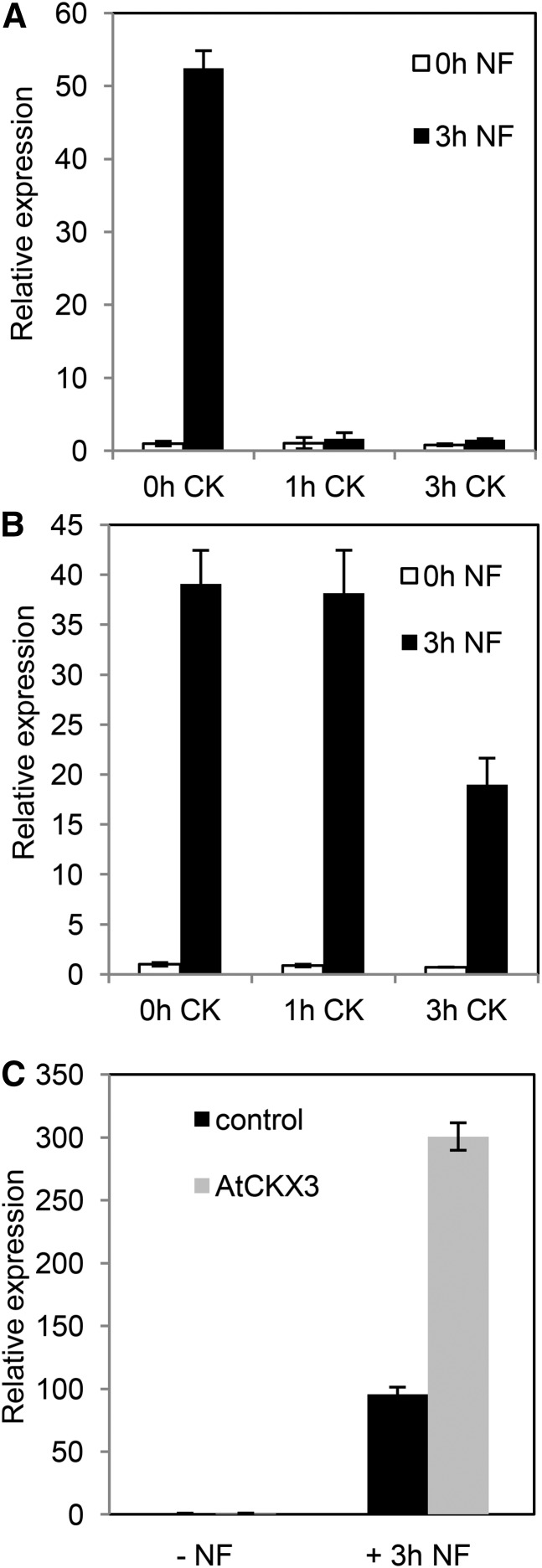
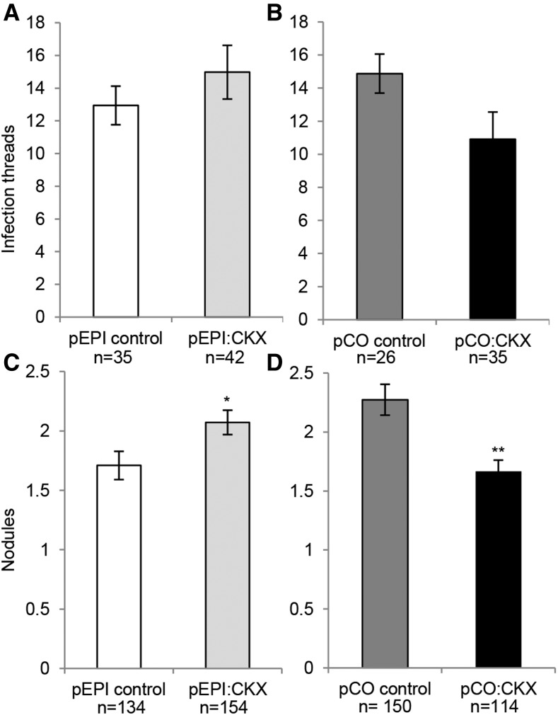
Nod factors (NFs) are lipochitooligosaccharidic signal molecules produced by rhizobia, which play a key role in the rhizobium-legume symbiotic interaction. In this study, we analyzed the gene expression reprogramming induced by purified NF (4 and 24 h of treatment) in the root epidermis of the model legume Medicago truncatula Tissue-specific transcriptome analysis was achieved by laser-capture microdissection coupled to high-depth RNA sequencing. The expression of 17,191 genes was detected in the epidermis, among which 1,070 were found to be regulated by NF addition, including previously characterized NF-induced marker genes. Many genes exhibited strong levels of transcriptional activation, sometimes only transiently at 4 h, indicating highly dynamic regulation. Expression reprogramming affected a variety of cellular processes, including perception, signaling, regulation of gene expression, as well as cell wall, cytoskeleton, transport, metabolism, and defense, with numerous NF-induced genes never identified before. Strikingly, early epidermal activation of cytokinin (CK) pathways was indicated, based on the induction of CK metabolic and signaling genes, including the CRE1 receptor essential to promote nodulation. These transcriptional activations were independently validated using promoter:β-glucuronidase fusions with the MtCRE1 CK receptor gene and a CK response reporter (TWO COMPONENT SIGNALING SENSOR NEW). A CK pretreatment reduced the NF induction of the EARLY NODULIN11 (ENOD11) symbiotic marker, while a CK-degrading enzyme (CYTOKININ OXIDASE/DEHYDROGENASE3) ectopically expressed in the root epidermis led to increased NF induction of ENOD11 and nodulation. Therefore, CK may play both positive and negative roles in M. truncatula nodulation.


Figures

Figure 1.
Figure 1.
Multiple comparisons (Venn diagrams) of NF up-regulated M. truncatula genes detected in recent transcriptomics experiments. Top, 1- to 6-h NF treatments; bottom, 24-h NF treatment and S. meliloti-inoculated RHs 5 d post inoculation (RH-5dpi) in a hyperinfected skl mutant background. ART-1h, 10−8 m NF-treated (1 h) whole roots (Rose et al., 2012); ART-3h, 10−9 m NF-treated (3 h) root segments in the presence of 1 µm aminoethoxyvinylglycine (AVG), an inhibitor of ethylene biosynthesis (van Zeijl et al., 2015); ART-6h and ART-24h, 10−8 m NF-treated root segments (6 and 24 h; Czaja et al., 2012); LCM-4h/-24h, laser-dissected epidermal cells (this study) with 4 or 24 h of 10−8 m NF treatment, respectively; RH-24h, 10−6 m NF-treated RHs (24 h; Breakspear et al., 2014). MtV4.0 identifiers were used in all cases. Thresholds chosen to identify up-regulated genes were defined in the corresponding publications. Venn diagrams were generated with the tool provided by Bardou et al. (2014).
Figure 2.
Figure 2.
Functional classification of M. truncatula NF-regulated genes in the root epidermis. Categories were manually defined, based upon best BLASTP hits using predicted encoded proteins against SWISSPROT and The Arabidopsis Information Resource databases. The number of genes is indicated for each category, with black and white bars corresponding to up- and down-regulated genes, respectively.
Figure 3.
Figure 3.
NFs induce pMtCRE1:GUS in M. truncatula RHs. A and B, GUS staining of untreated roots transformed with a pMtCRE1:GUS fusion. C and D, GUS staining of roots transformed with a pMtCRE1:GUS fusion, treated with NFs (10−9 m) for 4 h. Similar results were obtained with 10−8 m NFs. Bars = 300 µm.
Figure 4.
Figure 4.
CKs, NFs, and S. meliloti induce TCSn:GUS in M. truncatula root tissues. A to D, GUS staining of whole roots transformed with TCSn:GUS fusion via Agrobacterium rhizogenes. Shown are mock-treated roots (A), roots treated with CKs (10−7 m BAP for 4 h; B), and wild type (C) and nfp (D) roots treated with NFs (10−8 m for 4 h). Bars = 500 μm. E to I, GUS staining of longitudinal 25-μm sections (E, F, H, and I) or a transverse 10-μm section (G) of roots transformed via A. rhizogenes with TCSn:GUS (E, G, H, and I) or pENOD11:GUS (F) and treated with NFs (10−8 m for 4 h; E and F) or inoculated with S. meliloti at 8 h (G and H) and 72 h (H) post inoculation, respectively. Bars = 50 μm.
Figure 5.
Figure 5.
The NF induction of ENOD11 is rapidly repressed by CKs via CRE1 and can be increased by expressing AtCKX3 in the root epidermis. A, Real-time reverse transcription-PCR analysis of ENOD11 relative expression in response to a 10−7 m BAP treatment (1 or 3 h) followed by a 10−8 m NF treatment (3 h) in wild-type M. truncatula roots. B, Real-time reverse transcription-PCR analysis of ENOD11 relative expression in response to a 10−7 m BAP treatment (1 or 3 h) followed by a 10−8 m NF treatment (3 h) in a cre1 mutant. C, Real-time reverse transcription-PCR analysis of ENOD11 relative expression in pEPI:AtCKX3 roots treated or not with 10−8 m NFs for 3 h. One biological replicate is shown out of two independent experiments (for the other biological replicate, see Supplemental Fig. S9). Error bars indicate sd of two technical replicates.
Figure 6.
Figure 6.
Expression of the AtCKX3 gene leads to an increased nodulation in the root epidermis and to a decreased nodulation in the root cortex. Infection threads and nodules were counted on individual roots at 6 and 14 d post inoculation with S. meliloti, respectively, using roots transformed by A. rhizogenes with pEPI:CKX3 specifically expressing AtCKX3 in the root epidermis, or the corresponding control vector (A and C), or with pCO:CKX3 specifically expressing AtCKX3 in the root cortex, or the corresponding control vector (B and D). P = 0.1592 and 0.1658 in A and B respectively, and 0.00786 and 0.00010 in C and D, respectively (one-sided Welch’s test). Values shown are means of three biological replicates ± se.

References

    1. Anders S, Huber W (2010) Differential expression analysis for sequence count data. Genome Biol 11: R106. - PMC - PubMed
    1. Andriankaja A, Boisson-Dernier A, Frances L, Sauviac L, Jauneau A, Barker DG, de Carvalho-Niebel F (2007) AP2-ERF transcription factors mediate Nod factor dependent Mt ENOD11 activation in root hairs via a novel cis-regulatory motif. Plant Cell 19: 2866–2885 - PMC - PubMed
    1. Andrio E, Marino D, Marmeys A, de Segonzac MD, Damiani I, Genre A, Huguet S, Frendo P, Puppo A, Pauly N (2013) Hydrogen peroxide-regulated genes in the Medicago truncatula-Sinorhizobium meliloti symbiosis. New Phytol 198: 179–189 - PubMed
    1. Ané JM, Kiss GB, Riely BK, Penmetsa RV, Oldroyd GE, Ayax C, Lévy J, Debellé F, Baek JM, Kalo P, et al. (2004) Medicago truncatula DMI1 required for bacterial and fungal symbioses in legumes. Science 303: 1364–1367 - PubMed
    1. Antolín-Llovera M, Ried MK, Parniske M (2014) Cleavage of the SYMBIOSIS RECEPTOR-LIKE KINASE ectodomain promotes complex formation with Nod factor receptor 5. Curr Biol 24: 422–427 - PubMed

Publication types

MeSH terms

LinkOut - more resources