

Detection and Predictive Value of Fractional Anisotropy Changes of the Corticospinal Tract in the Acute Phase of a Stroke
- PMID: 27217504
- PMCID: PMC4959886
- DOI: 10.1161/STROKEAHA.115.012088
Detection and Predictive Value of Fractional Anisotropy Changes of the Corticospinal Tract in the Acute Phase of a Stroke
Abstract
Background and purpose: A decrease in fractional anisotropy (FA) of the ipsilesional corticospinal tract (CST) distal to stroke lesions in the subacute (eg, 30 days) and chronic phase has been correlated with poor motor outcomes, but it is unclear whether FA values obtained within the acute stroke phase (here defined as 80 hours after onset) can predict later outcome.
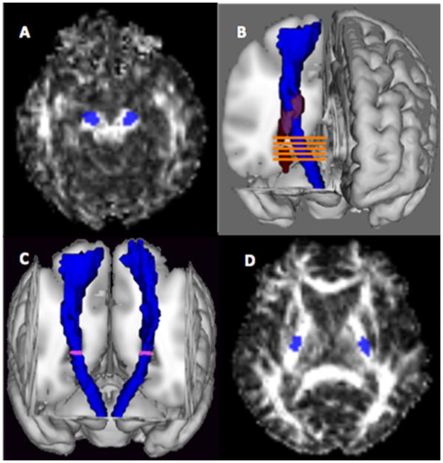
Methods: Fifty-eight patients underwent an assessment of motor impairment in the acute phase and at 3 months using the upper extremity Fugl-Meyer assessment. FA values, obtained within 80 hours after stroke onset, were determined in 2 regions of interest: cerebral peduncle and a stretch of the CST caudal to each stroke lesion (nearest-5-slices).
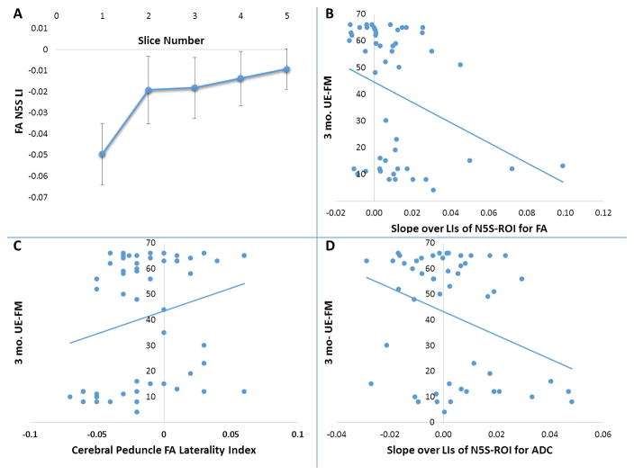
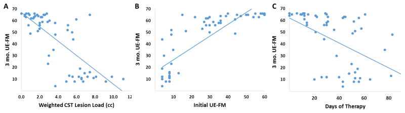
Results: The FA laterality index for the cerebral peduncle-regions of interest was a poor predictor of 3-month outcome (R(2)=0.044; P=0.137), whereas the slope over the FA laterality index of the nearest-5-slices showed a relatively weak but significant prediction (R(2)=0.11; P=0.022) with the affected side having lower FA values. Initial upper extremity Fugl-Meyer (R(2)=0.69; P<0.001) and the weighted CST lesion load (R(2)=0.71; P<0.001) were strong predictors of 3-month outcome. In multivariate analyses, controlling for initial upper extremity Fugl-Meyer, weighted CST lesion load, and days-of-therapy, neither the FA laterality index of the cerebral peduncle nor the slope over the FA laterality index of the nearest-5-slices significantly contributed to the prediction of 86% of the variance in the upper extremity Fugl-Meyer at 3 months.
Conclusions: FA reductions of the CST can be detected near the ischemic lesion in the acute stroke phase, but offer minimal predictive value to motor outcomes at 3 months.
Keywords: corticospinal tract; diffusion tensor imaging; lesion load; lesion mapping; magnetic resonance imaging; outcomes assessment; stroke.
© 2016 American Heart Association, Inc.
Figures
Similar articles
-
Diffusional Kurtosis Imaging and Motor Outcome in Acute Ischemic Stroke.AJNR Am J Neuroradiol. 2017 Jul;38(7):1328-1334. doi: 10.3174/ajnr.A5180. Epub 2017 May 4. AJNR Am J Neuroradiol. 2017. PMID: 28473339 Free PMC article.
-
Early Fiber Number Ratio Is a Surrogate of Corticospinal Tract Integrity and Predicts Motor Recovery After Stroke.Stroke. 2016 Apr;47(4):1053-9. doi: 10.1161/STROKEAHA.115.011576. Epub 2016 Mar 15. Stroke. 2016. PMID: 26979863
-
Motor tract integrity predicts walking recovery: A diffusion MRI study in subacute stroke.Neurology. 2020 Feb 11;94(6):e583-e593. doi: 10.1212/WNL.0000000000008755. Epub 2020 Jan 2. Neurology. 2020. PMID: 31896618
-
An overview of fractional anisotropy as a reliable quantitative measurement for the corticospinal tract (CST) integrity in correlation with a Fugl-Meyer assessment in stroke rehabilitation.J Phys Ther Sci. 2021 Jan;33(1):75-83. doi: 10.1589/jpts.33.75. Epub 2021 Jan 5. J Phys Ther Sci. 2021. PMID: 33519079 Free PMC article. Review.
-
The role of diffusion tensor imaging and fractional anisotropy in the evaluation of patients with idiopathic normal pressure hydrocephalus: a literature review.Neurosurg Focus. 2016 Sep;41(3):E12. doi: 10.3171/2016.6.FOCUS16192. Neurosurg Focus. 2016. PMID: 27581308 Review.
Cited by
-
Neuroimaging Techniques as Potential Tools for Assessment of Angiogenesis and Neuroplasticity Processes after Stroke and Their Clinical Implications for Rehabilitation and Stroke Recovery Prognosis.J Clin Med. 2022 Apr 28;11(9):2473. doi: 10.3390/jcm11092473. J Clin Med. 2022. PMID: 35566599 Free PMC article. Review.
-
Performance Comparison of Different Neuroimaging Methods for Predicting Upper Limb Motor Outcomes in Patients after Stroke.Neural Plast. 2022 Jun 6;2022:4203698. doi: 10.1155/2022/4203698. eCollection 2022. Neural Plast. 2022. PMID: 35707519 Free PMC article.
-
Cerebrolysin as an Early Add-on to Reperfusion Therapy: Risk of Hemorrhagic Transformation after Ischemic Stroke (CEREHETIS), a prospective, randomized, multicenter pilot study.BMC Neurol. 2023 Mar 27;23(1):121. doi: 10.1186/s12883-023-03159-w. BMC Neurol. 2023. PMID: 36973684 Free PMC article. Clinical Trial.
-
Diffusional Kurtosis Imaging and Motor Outcome in Acute Ischemic Stroke.AJNR Am J Neuroradiol. 2017 Jul;38(7):1328-1334. doi: 10.3174/ajnr.A5180. Epub 2017 May 4. AJNR Am J Neuroradiol. 2017. PMID: 28473339 Free PMC article.
-
Diffusion Tensor Imaging as a Prognostic Tool for Recovery in Acute and Hyperacute Stroke.Neurol Int. 2022 Oct 21;14(4):841-874. doi: 10.3390/neurolint14040069. Neurol Int. 2022. PMID: 36278693 Free PMC article. Review.
References
-
- Byblow WD, Stinear CM, Barber PA, Petoe MA, Ackerley SJ. Proportional recovery after stroke depends on corticomotor integrity. Annals of neurology. 2015;78:848–859. - PubMed
-
- Iizuka H, Sakatani K, Young W. Corticofugal axonal degeneration in rats after middle cerebral artery occlusion. Stroke; a journal of cerebral circulation. 1989;20:1396–1402. - PubMed
Publication types
MeSH terms
Grants and funding
LinkOut - more resources
Full Text Sources
Other Literature Sources
Medical
