

SERUM VALUES OF ALKALINE PHOSPHATASE AND LACTATE DEHYDROGENASE IN OSTEOSARCOMA
- PMID: 27217815
- PMCID: PMC4863862
- DOI: 10.1590/1413-785220162403157033
SERUM VALUES OF ALKALINE PHOSPHATASE AND LACTATE DEHYDROGENASE IN OSTEOSARCOMA
Abstract
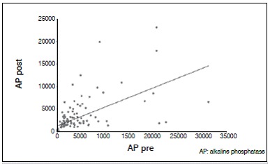
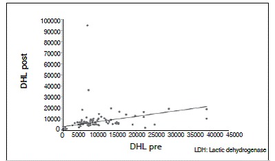
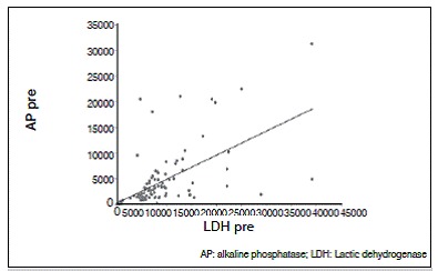
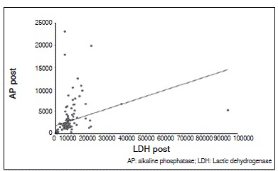
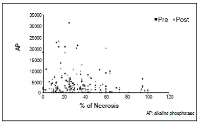
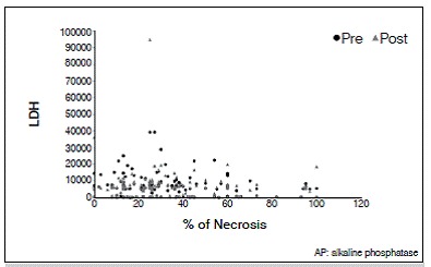
Objective: To study the relationship between the pre and post chemotherapy (CT) serum levels of alkaline phosphatase (AP) and lactate dehydrogenase (LDH), and the percentage of tumor necrosis (TN) found in specimens after the pre surgical CT in patients with osteosarcoma.
Methods: Series of cases with retrospective evaluation of patients diagnosed with osteosarcoma. Participants were divided into two groups according to serum values of both enzymes. The values of AP and LDH were obtained before and after preoperative CT. The percentage of tumor necrosis (TN) of surgical specimens of each patient was also included.
Results: One hundred and thirty seven medical records were included from 1990 to 2013. Both the AP as LDH decreased in the patients studied, being the higher in pre CT than post CT. The average LHD decrease was 795.12U/L and AP decrease was 437.40 U/L. The average TN was 34.10 %. There was no statistically significant correlation between the serums values and the percentage of tumoral necrosis.
Conclusion: The serum levels values of AP and LDH are not good predictors for the chemotherapy-induced necrosis in patients with osteosarcoma. Level of Evidence IV, Case Series.
Keywords: Alkaline phosphatase; Drug therapy; L-Lactate Dehydrogenase; Osteosarcoma; Prognosis.; Tumor necrosis factors.
Conflict of interest statement
All the authors declare that there is no potential conflict of interest referring to this article.
Figures
Similar articles
-
SERUM VALUES OF ALKALINE PHOSPHATASE AND LACTATE DEHYDROGENASE IN EWING'S SARCOMA.Acta Ortop Bras. 2016 Jul-Aug;24(4):196-199. doi: 10.1590/1413-785220162404161312. Acta Ortop Bras. 2016. PMID: 28243173 Free PMC article.
-
Pre-treatment serum lactate dehydrogenase and alkaline phosphatase as predictors of metastases in extremity osteosarcoma.J Bone Oncol. 2015 Oct 30;4(3):80-4. doi: 10.1016/j.jbo.2015.09.002. eCollection 2015 Sep. J Bone Oncol. 2015. PMID: 26587373 Free PMC article.
-
Three hematological indexes that may serve as prognostic indicators in patients with primary, high-grade, appendicular osteosarcoma.Oncotarget. 2017 Jun 27;8(26):43130-43139. doi: 10.18632/oncotarget.17811. Oncotarget. 2017. PMID: 28562345 Free PMC article.
-
[Expressions of ERCC2 and ERCC4 genes in osteosarcoma and peripheral blood lymphocytes and their clinical significance].Beijing Da Xue Xue Bao Yi Xue Ban. 2007 Oct 18;39(5):467-71. Beijing Da Xue Xue Bao Yi Xue Ban. 2007. PMID: 17940561 Chinese.
-
Diagnostic value of combined detection of AKP, TSGF, and LDH for pediatric osteosarcoma: a case-control study.Am J Transl Res. 2024 Aug 15;16(8):3667-3677. doi: 10.62347/IGEA4076. eCollection 2024. Am J Transl Res. 2024. PMID: 39262698 Free PMC article.
Cited by
-
Identification of Prognostic and Predictive Osteosarcoma Biomarkers.Med Sci (Basel). 2019 Feb 11;7(2):28. doi: 10.3390/medsci7020028. Med Sci (Basel). 2019. PMID: 30754703 Free PMC article. Review.
-
Circulating biomarkers in osteosarcoma: new translational tools for diagnosis and treatment.Oncotarget. 2017 Aug 3;8(59):100831-100851. doi: 10.18632/oncotarget.19852. eCollection 2017 Nov 21. Oncotarget. 2017. PMID: 29246026 Free PMC article. Review.
-
Gasdermin D expression and clinicopathologic outcome in primary osteosarcoma patients.Int J Clin Exp Pathol. 2020 Dec 1;13(12):3149-3157. eCollection 2020. Int J Clin Exp Pathol. 2020. PMID: 33425115 Free PMC article.
-
Liquid Biopsy: A New Translational Diagnostic and Monitoring Tool for Musculoskeletal Tumors.Int J Mol Sci. 2021 Oct 26;22(21):11526. doi: 10.3390/ijms222111526. Int J Mol Sci. 2021. PMID: 34768955 Free PMC article. Review.
-
Exosomes in Bone Cancer: Unveiling their Vital Role in Diagnosis, Prognosis, and Therapeutic Advancements.J Cancer. 2024 Jun 3;15(13):4128-4142. doi: 10.7150/jca.95709. eCollection 2024. J Cancer. 2024. PMID: 38947401 Free PMC article. Review.
References
-
- Raymond AK, Ayala AG, Knuutila S. Fletcher CD, Unni KK, Mertens F. Pathology and genetics of tumours of soft tissue and bone. Lyon: IARC Press; 2002. Conventional osteosarcoma; pp. 264–285.
-
- Hornicek FJ. Schwartz HS. Orthopaedic knowledge update: musculoskeletal tumors. 2. Rosemond, IL: American Academy of Orthopaedic Surgeons; 2007. Osteosarcoma of bone; pp. 163–174.
-
- Dorfman HD, Czerniah B. Bone tumors. St. Louis Missouri: Mosby; 1998. Osteosarcoma; pp. 128–252.
-
- Dorfman HD, Czerniah B. St. Louis: Mosby; 1998. Ewing's sarcoma and related entities; pp. 607–663.
-
- Ushigome S, Machinami R, Sorensen PH. Fletcher CD, Unni KK, Mertens F. Pathology and genetics of tumours of soft tissue and bone. Lyon: IARC Press; 2002. Ewing sarcoma / primitive neuroectodermal tumour (PNET) pp. 297–300.
LinkOut - more resources
Full Text Sources
Other Literature Sources