Review
. 2016 Apr 27;6(7):948-68.
doi: 10.7150/thno.15217. eCollection 2016.

Recent Progress in Light-Triggered Nanotheranostics for Cancer Treatment

Affiliations
Review

Recent Progress in Light-Triggered Nanotheranostics for Cancer Treatment

Pengcheng Zhang et al. Theranostics. .

Abstract

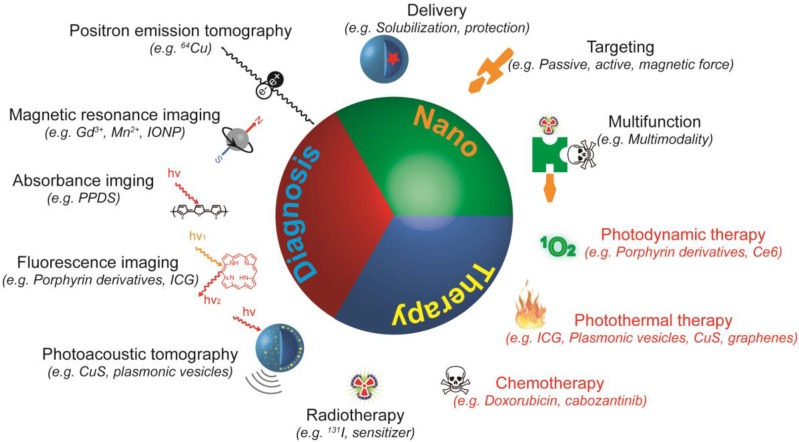
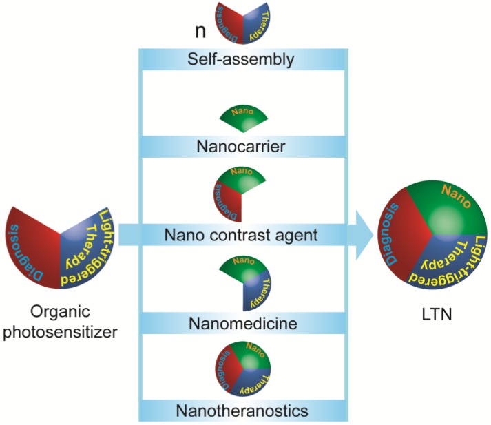
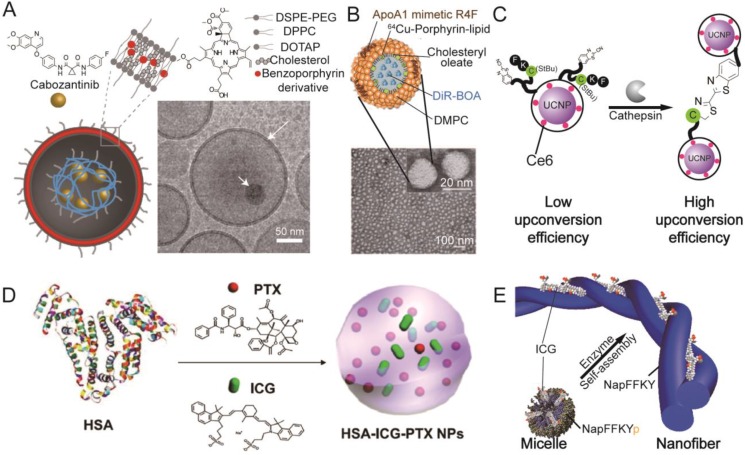
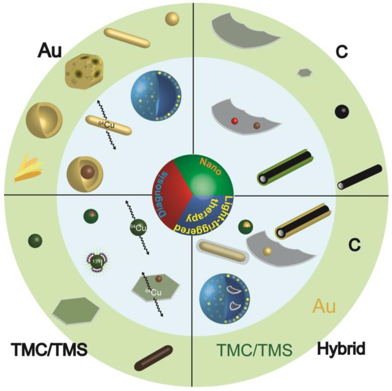
Treatments of high specificity are desirable for cancer therapy. Light-triggered nanotheranostics (LTN) mediated cancer therapy could be one such treatment, as they make it possible to visualize and treat the tumor specifically in a light-controlled manner with a single injection. Because of their great potential in cancer therapy, many novel and powerful LTNs have been developed, and are mainly prepared from photosensitizers (PSs) ranging from small organic dyes such as porphyrin- and cyanine-based dyes, semiconducting polymers, to inorganic nanomaterials such as gold nanoparticles, transition metal chalcogenides, carbon nanotubes and graphene. Using LTNs and localized irradiation in combination, complete tumor ablation could be achieved in tumor-bearing animal models without causing significant toxicity. Given their great advances and promising future, we herein review LTNs that have been tested in vivo with a highlight on progress that has been made in the past a couple of years. The current challenges faced by these LTNs are also briefly discussed.

Keywords: Nanoparticle; imaging; photodynamic; photothermal.; theranostics.


Conflict of interest statement

Competing Interests: The authors have declared that no competing interest exists.

Figures

Figure 1
Figure 1
Schematic illustration of nanotheranostics. A nanotheranostic agent is a single entity with three characteristics: nano-sized particle, therapeutic agent, and diagnostic agent. As nano-sized particles, nanotheranostics help to solubilize and protect cargos, improve accumulation of both contrast and therapeutic agents at the disease site, and enable integration of multiple functions into one single particle. As therapeutic agents, nanotheranostics can eradicate cancer cells via singlet oxygen (1O2) or ROS generation, hyperthermia, radiation, or the release of chemotherapeutics. As diagnostic agents, nanotheranostics provide contrast for positron emission tomography (PET), magnetic resonance imaging (MRI), absorbance imaging, fluorescence imaging, or photoacoustic tomography. Nanotheranostics that exert a therapeutic effect upon light irradiation (red) were termed as light-triggered nanotheranostics or LTNs, which are the focus of this review.
Figure 2
Figure 2
Strategies for constructing light-triggered LTN from small molecular organic photosensitizers (PSs). Small molecular PSs which provide contrast for imaging and a therapeutic effect upon irradiation could be built into LTNs via five strategies: (1) the small molecular PS could be modified and induced to self-assemble into discrete LTNs; (2) nanocarriers, (3) nano-sized contrast agents, (4) nanomedicine, or even (5) LTNs could be used as transporter for small molecular PSs.
Figure 3
Figure 3
Representative organic photosensitizer-based LTNs. (A) Schematic illustration and cryo-TEM imaging of the photoactivable multi-inhibitor nanoliposome. The nanoliposome bilayer was composed of DSPE-PEG, DPPC, DOTAP, cholesterol, and a benzoporphyrin derivative that could be activated by light. The cabozantinib encapsulated in the hollow cavity could be released upon light irradiation. Adapted with permission from . (B) Schematic illustration and TEM imaging of a HDL mimetic nanotheranostic which is highly stable in circulation. Adapted with permission from . (C) Schematic illustration of a tumor microenvironment sensitive LTN which would aggregate in the tumor, resulting in improved fluorescence emission for Ce6 activation. Adapted with permission from . (D) The preparation of an “Abraxane-like” nanotheranostic from HAS, PTX and ICG. Adapted with permission from . (E) Schematic illustration of a tumor microenvironment sensitive ICG-loaded LTN that could undergo a morphological transition from micelle to nanofiber. Adapted with permission from .
Figure 4
Figure 4
Inorganic LTNs. Schematic illustration of the most explored inorganic LTNs built from gold (Au), transition metal chalcogenides or selenides (TMC/TMS), carbon (C), or hybrids thereof. The structures presented in the light green circle are LTNs built from single materials, and are basic building blocks for more complicated LTNs (light blue circle). LTNs built from gold include nanosphere, nanorod, nanocage, nanoshell, and gold beltflower morphologies (from right to the left), which could be made into plasmonic vesicles, labeled with 64Cu or IONPs for enhanced therapeutic and imaging capabilities. LTNs built from TMC/TMS could adopt sphere, sheet, or rod morphologies (from left to right), and could form core/shell structures with IONPs, be labeled with 64Cu or 131I, or doped with IONPs. LTNs made from carbon include graphene, graphene quantum dots, carbon dots, and carbon nanotubes, which could be doped with quantum dots or IONPs, or coated with mesoporous silica for improved tumor imaging or delivery. In addition, hybrid LTNs were created to improve the imaging or therapeutic outcomes, which include Cu9S5 coated gold spheres, gold-coated carbon nanotubes, graphene-coated gold nanorods, gold nanoparticle-doped graphene, and graphene-loaded plasmonic vesicles.
Figure 5
Figure 5
Representative inorganic photosensitizer-based LTNs. (A) Schematic illustration and TEM image of gold-silica quantum rattles. The 2 nm sized gold quantum dots were stabilized in mesoporous silica-based nanoshells. Adapted with permission from . (B) Preparation of plasmonic vesicles from ultrasmall gold nanorods that could dissociate to give PEG-coated gold nanorods after the degradation of PLGA. Adapted with permission from . (C) PVP stabilized 64Cu-labeled CuS nanodots with ultrahigh efficient renal clearance as evidenced by time-resolved PET. Adapted with permission from . (D) The use of SWCNTs in the treatment of 4T1 lung metastases. The intratumorally-injected SWCNTs could accumulate in SLNs which together with the primary tumor must be irradiated with a laser to prevent the lung metastasis of the 4T1 tumor and prolong the survival of the animals. Adapted with permission from .
Figure 6
Figure 6
Representative hybrid LTNs. (A) The schematic illustration and TEM image of a Au-Cu9S5 core-shell LTN. This core-shelled LTN showed an improved photothermal conversion efficiency compared with the mixture of Au nanoparticles and Cu9S5 nanoparticles at the same concentrations. Adapted with permission from (B) Schematic illustration of Au nanorod plasmonic vesicles loaded with rGO and Dox (rGO-AuNRVe-DOX). rGO-AuNRVe-DOX showed improved photothermal conversion efficiency, and the release of Dox from the vesicles could be accelerated by lowering the pH, with laser irradiation, or a combination of the two. Adapted with permission from . (C) Schematic illustration of IR825-loaded and Gd3+-Ce6-conjugated micelles with two therapeutic modalities (PDT and PTT). Treating the tumor sequentially with PTT and PDT was the most effective method for inducing the apoptosis of cancer cells, as evidenced by TUNEL staining. Adapted with permission from .

Similar articles

Cited by

References

    1. Torre LA, Bray F, Siegel RL. et al. Global cancer statistics, 2012. CA Cancer J Clin. 2015;65:87–108. - PubMed
    1. Bedard PL, Hansen AR, Ratain MJ. et al. Tumour heterogeneity in the clinic. Nature. 2013;501:355–64. - PMC - PubMed
    1. Hanahan D, Weinberg RA. Hallmarks of cancer: the next generation. Cell. 2011;144:646–74. - PubMed
    1. Mura S, Couvreur P. Nanotheranostics for personalized medicine. Adv Drug Deliv Rev. 2012;64:1394–416. - PubMed
    1. Cheng Z, Al Zaki A, Hui JZ. et al. Multifunctional nanoparticles: cost versus benefit of adding targeting and imaging capabilities. Science. 2012;338:903–10. - PMC - PubMed

Publication types