Missed Traumatic Thoracic Spondyloptosis With no Neurological Deficit: A Case Report and Literature Review
- PMID: 27218044
- PMCID: PMC4869431
- DOI: 10.5812/traumamon.19841
Missed Traumatic Thoracic Spondyloptosis With no Neurological Deficit: A Case Report and Literature Review
Abstract
Introduction: Traumatic thoracic spondyloptosis is caused by high energy trauma and is usually associated with severe neurological deficit. Cases presenting without any neurological deficit can be difficult to diagnose and manage.
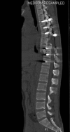
Case presentation: We reported a four-week spondyloptosis of the ninth thoracic vertebra over the tenth thoracic vertebra, in a 20-year-old male without any neurological deficit. The patient had associated chest injuries. The spine injury was managed surgically with in-situ posterior instrumentation and fusion. The patient tolerated the operation well and postoperatively there was no neurological deterioration or surgical complication.
Conclusions: Patients presenting with spondyloptosis with no neurological deficit can be managed with in-situ fusion via pedicle screws, especially when presenting late and with minimal kyphosis.
Keywords: Spondyloptosis; Thoracic; Traumatic.
Figures


Similar articles
-
C3-C4 spondyloptosis without neurological deficit-a case report.Spine J. 2010 Jul;10(7):e16-20. doi: 10.1016/j.spinee.2010.05.002. Spine J. 2010. PMID: 20620981
-
Traumatic spondyloptosis of the thoracolumbar spine.J Neurosurg Spine. 2008 Aug;9(2):145-51. doi: 10.3171/SPI/2008/9/8/145. J Neurosurg Spine. 2008. PMID: 18764746
-
Management of traumatic double-level spondyloptosis of the thoracic spine with posterior spondylectomy: case report.J Neurosurg Spine. 2015 Dec;23(6):715-20. doi: 10.3171/2015.3.SPINE14183. Epub 2015 Aug 21. J Neurosurg Spine. 2015. PMID: 26296192
-
Traumatic lumbosacral spondyloptosis treated five months after injury occurrence: a case report.Spine (Phila Pa 1976). 2012 Oct 15;37(22):E1410-4. doi: 10.1097/BRS.0b013e318268c08a. Spine (Phila Pa 1976). 2012. PMID: 22805340 Review.
-
Traumatic mid-thoracic spondyloptosis without neurological deficit: a case report and review of literature.ANZ J Surg. 2018 Oct;88(10):1083-1085. doi: 10.1111/ans.13747. Epub 2016 Nov 18. ANZ J Surg. 2018. PMID: 27860147 Review. No abstract available.
Cited by
-
Traumatic thoracic spine spondyloptosis treated with spondylectomy and fusion.Surg Neurol Int. 2018 Aug 10;9:158. doi: 10.4103/sni.sni_204_18. eCollection 2018. Surg Neurol Int. 2018. PMID: 30159202 Free PMC article.
-
Thecal sac ligation in the setting of thoracic spondyloptosis with complete cord transection.Surg Neurol Int. 2023 Sep 1;14:304. doi: 10.25259/SNI_360_2023. eCollection 2023. Surg Neurol Int. 2023. PMID: 37810299 Free PMC article.
-
Traumatic Fracture of the Thoracic Spine With Severe Posterolateral Dislocation: A Case Report.Cureus. 2022 Apr 4;14(4):e23830. doi: 10.7759/cureus.23830. eCollection 2022 Apr. Cureus. 2022. PMID: 35530925 Free PMC article.
References
-
- Bohlman HH, Freehafer A, Dejak J. The results of treatment of acute injuries of the upper thoracic spine with paralysis. J Bone Joint Surg Am. 1985;67(3):360–9. - PubMed
-
- Denis F. The three column spine and its significance in the classification of acute thoracolumbar spinal injuries. Spine (Phila Pa 1976). 1983;8(8):817–31. - PubMed
-
- Hanley Jr EN, Eskay ML. Thoracic spine fractures. Orthopedics. 1989;12(5):689–96. - PubMed
-
- Holdsworth F. Fractures, dislocations, and fracture-dislocations of the spine. J Bone Joint Surg Am. 1970;52(8):1534–51. - PubMed
Publication types
LinkOut - more resources
Full Text Sources
Other Literature Sources
