. 2016 Sep;240(1):25-37.
doi: 10.1002/path.4749. Epub 2016 Jul 14.

Idiopathic pulmonary fibrosis fibroblasts become resistant to Fas ligand-dependent apoptosis via the alteration of decoy receptor 3

Affiliations

Idiopathic pulmonary fibrosis fibroblasts become resistant to Fas ligand-dependent apoptosis via the alteration of decoy receptor 3

Jintaek Im et al. J Pathol. 2016 Sep.

Abstract

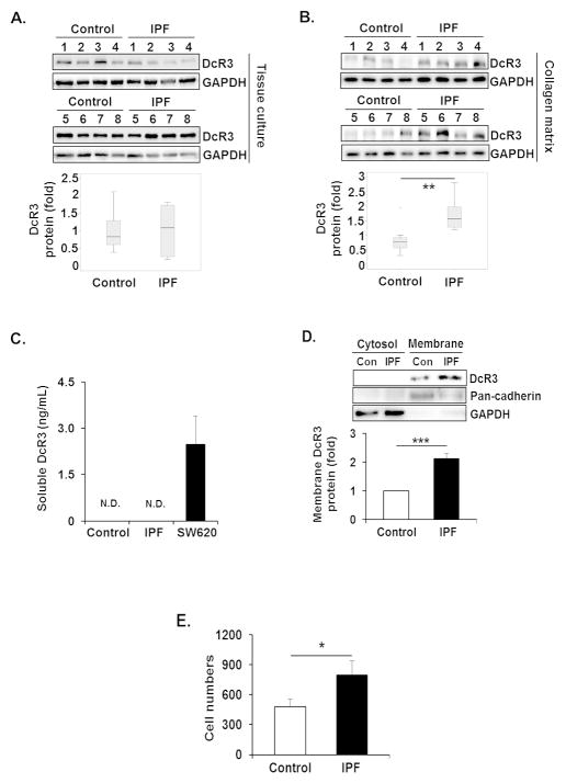
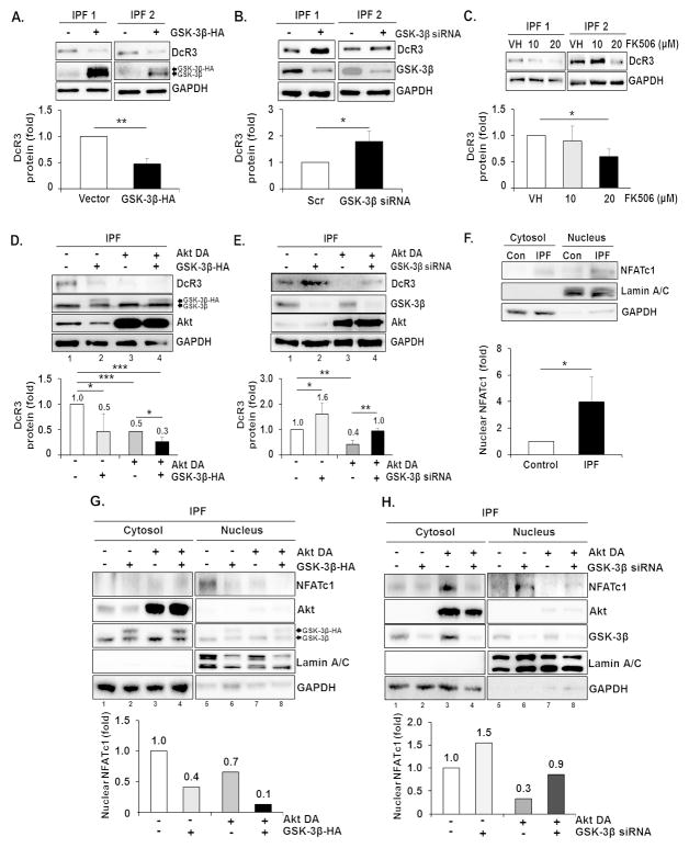
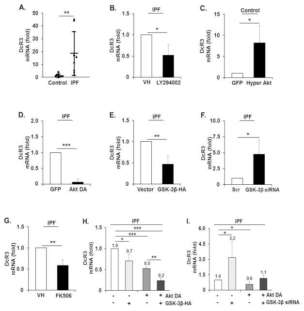
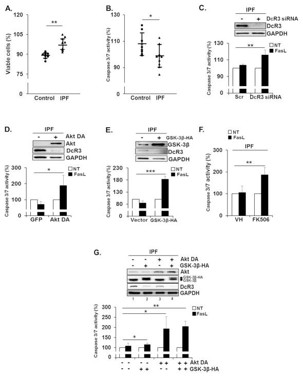
Idiopathic pulmonary fibrosis (IPF) is an irreversible lethal lung disease with an unknown etiology. IPF patients' lung fibroblasts express inappropriately high Akt activity, protecting them in response to an apoptosis-inducing type I collagen matrix. FasL, a ligand for Fas, is known to be increased in the lung tissues of patients with IPF, implicated with the progression of IPF. Expression of Decoy Receptor3 (DcR3), which binds to FasL, thereby subsequently suppressing the FasL-Fas-dependent apoptotic pathway, is frequently altered in various human disease. However, the role of DcR3 in IPF fibroblasts in regulating their viability has not been examined. We found that enhanced DcR3 expression exists in the majority of IPF fibroblasts on collagen matrices, resulting in the protection of IPF fibroblasts from FasL-induced apoptosis. Abnormally high Akt activity suppresses GSK-3β function, thereby accumulating the nuclear factor of activated T-cells cytoplasmic 1 (NFATc1) in the nucleus, increasing DcR3 expression in IPF fibroblasts. This alteration protects IPF cells from FasL-induced apoptosis on collagen. However, the inhibition of Akt or NFATc1 decreases DcR3 mRNA and protein levels, which sensitizes IPF fibroblasts to FasL-mediated apoptosis. Furthermore, enhanced DcR3 and NFATc1 expression is mainly present in myofibroblasts in the fibroblastic foci of lung tissues derived from IPF patients. Our results showed that when IPF cells interact with collagen matrix, aberrantly activated Akt increases DcR3 expression via GSK-3β-NFATc1 and protects IPF cells from the FasL-dependent apoptotic pathway. These findings suggest that the inhibition of DcR3 function may be an effective approach for sensitizing IPF fibroblasts in response to FasL, limiting the progression of lung fibrosis. Copyright © 2016 Pathological Society of Great Britain and Ireland. Published by John Wiley & Sons, Ltd.

Keywords: Akt; DcR3; FasL; GSK-3β; IPF; NFATc1; apoptosis; collagen.


Conflict of interest statement

Authors declare no conflict of interest.

Figures

Figure 1
Figure 1
DcR3 protein expression is aberrantly high in IPF fibroblasts on collagen matrix. (A) Randomly selected 6 × 10⁵ control and IPF fibroblasts (n=8, each) were grown in tissue culture plates (without polymerized collagen) for 48 h in serum free DMEM. Upper, DcR3 protein expression in control and IPF fibroblasts were examined by Western analysis. Lower, box-and-whisker plots of DcR3 normalized to GAPDH in control and IPF fibroblasts. All values of DcR3 in control and IPF fibroblasts were divided by mean value of DcR3 of the control group to set the average expression of DcR3 in control fibroblasts as 1. (B) The same control and IPF fibroblasts (n=8, each) were grown on polymerized collagen matrix for 48 h in serum free DMEM. Upper, DcR3 protein expression in control and IPF fibroblasts was examined by Western analysis. Lower, box-and-whisker plots of DcR3 normalized to GAPDH in control and IPF fibroblasts. (C) 1 × 10⁵ control and IPF fibroblasts (n=3 each) were cultured on polymerized collagen matrices in 200 Ml of serum free DMEM per well for 48 h. Soluble DcR3 levels were measured by ELISA as described in the Materials and Methods. Human colon cancer cell line, SW620 was used as a positive control. (D) 1.5 × 10⁶ control and IPF fibroblasts (n=4, each) were cultured on polymerized collagen matrices for 48 h in serum free DMEM, and cytosolic and plasma membrane fractions were separated as described in the Materials and Methods. Upper, a representative Western blot for DcR3 protein expression in cytosolic and plasma membrane fractions from control (Con) and IPF fibroblasts. Pan-cadherin and GAPDH were used as plasma membrane and cytosolic protein markers, respectively. Lower, densitometric analysis of DcR3 protein expression on plasma membrane normalized to pan-cadherin. (E) Control (n=3) and IPF fibroblasts (n=4) were cultured on collagen for 48 h in serum free DMEM. Cells were then incubated with magnetic beads conjugated with a DcR3 antibody. Total binding cell number was measured using a Coulter counter. Control fibroblasts expressing low levels of DcR3 protein and IPF fibroblasts expressing high levels of DcR3 were selected for Figure 1C–E. *p<0.05, **p<0.01 and ***p<0.001. N.D. non-detected.
Figure 2
Figure 2
Aberrantly high PI3K and Akt regulate DcR3 expression in IPF fibroblasts on collagen matrix. (A) IPF fibroblasts (n=3) were plated on collagen matrices for 24 h, then cells were treated with LY294002 for an additional 24 h. DMSO was used as a vehicle control (VH). Upper, representative Western analysis of DcR3, pAkt, pGSK3β, Akt, GSK3β, and GAPDH protein expression after LY294002 treatment. Lower, densitometric analysis of DcR3 protein expression normalized to GAPDH. (B) IPF fibroblasts (n=3) infected with adenovirus expressing dominant negative Akt (Akt DA) or empty vector (GFP) were cultured on polymerized collagen matrices for 48 h. Upper, representative Western blot of DcR3, Akt DA, pGSK3β, pAkt, GSK3β, and GAPDH protein expression under the condition as described above. Lower, densitometric analysis of DcR3 protein expression normalized to GAPDH. GAPDH for DcR3 expression was shown in the lower Western blot panel. (C) Control fibroblasts (n=3) infected with adenovirus expressing hyper active Akt (Hyper Akt) or GFP were plated on collagen matrices for 48 h. Upper, representative Western blot of DcR3, pAkt, pGSK3β, Akt, GSK3β, and GAPDH protein expression. Lower, densitometric analysis of DcR3 protein expression normalized to GAPDH. Values are the average of three cell lines, presented as relative fold change ± S.D. against VH or GFP group for individual fibroblasts set as 1 fold. IPF fibroblasts expressing high DcR3 protein and control fibroblasts expressing low levels of DcR3 protein were selected for Figure 2A and B, and Figure 2C, respectively. (D and E) Control and IPF fibroblasts shown in Figure 1 (n=8 each) were cultured on tissue culture plates (D) or on collagen matrix (E) for 48 h. Upper, pGSK-3β, pAkt, and GAPDH protein expressions in control and IPF fibroblasts were examined by Western analysis. Lower, box-and-whisker plots of pGSK-3β and pAkt after normalization to GAPDH in control and IPF fibroblasts by densitometric analysis. *p≤0.05, **p<0.01 and ***p<0.001.
Figure 3
Figure 3
Translocation of NFATc1 into the nucleus by Akt/GSK-3β axis is important for DcR3 expression. (A) IPF fibroblasts (n=3) were transfected with empty vector (−) or plasmid expressing HA tagged GSK-3β (+) and subsequently cultured on polymerized collagen matrices for 48 h. Upper, Representative Western blot of DcR3 and GSK-3β protein expression. Lower, densitometric analysis of DcR3 protein expression normalized to GAPDH after GSK-3β-HA over-expression. (B) IPF fibroblasts (n=3) transfected with scrambled (−) or GSK-3β siRNA (+) were cultured on polymerized collagen matrices for 48h. Upper, representative Western blot of DcR3, GSK-3β and GAPDH protein expression under the condition as described above. Lower, densitometric analysis of DcR3 protein expression normalized to GAPDH after GSK-3β siRNA treatment. (C) IPF fibroblasts (n=3) were plated on collagen matrices for 24 h, and cells were incubated for an additional 24 h in the presence of various doses of FK506 as described. DMSO was used as a vehicle control (VH). Upper, representative Western blot of DcR3 protein expression. Lower, densitometric analysis of DcR3 protein expression normalized to GAPDH. (D) IPF fibroblasts (n=3) infected with adenovirus expressing Akt DA or GFP were transfected with empty vector or GSK-3β-HA and cultured on collagen matrices for 48 h. Upper, representative Western blot of DcR3, Akt and GSK-3β protein expression. Lower, densitometric analysis of DcR3 protein expression normalized to GAPDH. Values are presented as relative fold change ± S.D. against the control group set as 1 fold. (E) IPF fibroblasts (n=3) infected with adenovirus expressing Akt DA or GFP were transfected with scrambled or GSK-3β siRNA and cultured on collagen matrices for 48 h. Upper, representative Western blot of DcR3, Akt and GSK-3β protein expression under the conditions as described above. Lower, densitometric analysis of DcR3 protein expression normalized to GAPDH. IPF fibroblasts expressing high levels of DcR3 protein were chosen for Figure 3A–E. (F) 1.5 × 10⁶ control and IPF fibroblasts (n=3 each) were cultured on polymerized collagen matrices for 48 h, and the cytosolic and nuclear fractions were separated as described in the Materials and Methods. Upper, representative Western analysis of NFATc1. Lower, densitometric analysis of NFATc1 expression in the nuclear fraction. Values are presented as relative fold values ± S.D. against NFATc1 expression of control fibroblasts set as 1 fold after lamin A/C normalization. Control fibroblasts expressing low levels of DcR3 protein and IPF fibroblasts expressing high levels of DcR3 protein were selected for this experiment. (G) Upper, representative Western analysis of NFATc1 expression in the cytoplasm and the nucleus of IPF fibroblasts over-expressing Akt DA and/or GSK-3β-HA on collagen matrix at 48 h. Lower, densitometric analysis of NFATc1 expression in the nuclear fraction. Values are presented as relative fold values ± S.D. against control treatment (GFP and pcDNA3 over-expression) set as 1 fold after lamin A/C normalization. (H) Upper, representative Western blot of NFATc1 expression in the cytoplasm and the nucleus of IPF fibroblasts over-expressing Akt DA following GSK3β siRNA transfection. Lower, densitometric analysis of NFATc1 expression in the nuclear fraction. IPF fibroblasts expressing high levels of DcR3 protein were selected for G and H. Lamin A/C and GAPDH were used as nuclear and cytosolic protein markers, respectively. *p≤0.05, **p≤0.01 and ***p<0.001.
Figure 4
Figure 4
Enhanced DcR3 mRNA levels in IPF fibroblasts are due to the alteration of PI3K/Akt/GSK-3β/NFATc1. (A) 2 × 10⁵ control and IPF fibroblasts used in previous experiments (n=8, each) were grown on collagen matrices for 48 h, and RT-PCR analysis was performed to examine DcR3 mRNA levels. On the scatterplot, each circle and triangle point represents the fold change of normalized DcR3 mRNA from control and IPF fibroblasts, respectively. (B) IPF fibroblasts (n=3) cultured on polymerized collagen were treated with a PI3K inhibitor, LY294002, at 20 MM for 24 h. DMSO was used as a vehicle control (VH). Values are presented as relative fold change ± S.D. against VH set as 1 fold. (C) Control fibroblasts (n=3) infected with adenovirus expressing hyper Akt or GFP were cultured on polymerized collagen matrices for 48 h. (D) IPF fibroblasts (n=3) expressing Akt DA or GFP were cultured on polymerized collagen matrices for 48 h. DcR3 mRNA levels are presented as a relative fold change ± S.D. against GFP set as 1 after GAPDH normalization in C and D. (E) IPF fibroblasts (n=3) transfected with empty vector (pcDNA3) or GSK-3β-HA were cultured on polymerized collagen matrix for 48 h. DcR3 mRNA levels are presented as a fold change ± S.D. against individual empty vector groups set as 1 fold after GAPDH normalization. (F) IPF fibroblasts (n=3) transfected with scrambled (Scr) or GSK-3β siRNA were cultured on polymerized collagen matrix for 48 h. DcR3 mRNA levels are presented as a fold change ± S.D. against the individual Scr group set as 1 fold after GAPDH normalization. (G) IPF fibroblasts (n=3) cultured on polymerized collagen matrix were incubated with 20 MM of FK506 or DMSO (VH) for 24 h. DcR3 mRNA levels are presented as a relative fold change ± S.D. against VH set as 1 fold after GAPDH normalization. (H and I) DcR3 mRNA levels in IPF fibroblasts (n=3) infected with adenovirus expressing Akt DA followed by GSK-3β-HA transfection (H), or with GSK-3β siRNA (I). DcR3 mRNA levels are presented as a fold change ± S.D. against an individual control group set as 1 after GAPDH normalization. IPF fibroblasts expressing increased levels of DcR3 mRNA were used in Figure 4B, D, E, F, G, H, and I, and control fibroblasts showing low levels of DcR3 mRNA were used in Figure 4C. GAPDH mRNA was used as a reference transcript. *p<0.05, **p<0.01 and ***p<0.001.
Figure 5
Figure 5
Enhanced DcR3 expression is responsible for the resistance of IPF fibroblasts to FasL-mediated apoptosis. 1 × 10⁴ control and IPF fibroblasts (n=8 each) used in previous experiments were grown on collagen for 48 h and treated with 500 (A) or 200 ng/ml of FasL (B) for an additional 24 h. Cell viability and caspase 3/7 activity were measured as described in the Materials and Methods. On the scatterplot, each circle and triangle point represents the percentage of viability (A) or caspase 3/7 activity (B) of each control and IPF cell in response to FasL. (C) IPF fibroblasts (n=3) transfected with scrambled or DcR3 siRNA were cultured on collagen matrix for 48 h. Upper, shown is the representative DcR3 protein expression in IPF fibroblasts used for the caspase 3/7 activity assay. Lower, caspase 3/7 activity in IPF fibroblasts was examined at 8 h in the presence of 200 ng/ml of FasL. (D) IPF fibroblasts (n=3) expressing Akt DA or GFP were cultured on collagen matrix for 48 h. Upper, shown is the representative Akt DA and DcR3 protein expression in IPF cells used for the measurement of caspase 3/7 activity. Lower, caspase 3/7 activity was measured at 8 h after FasL treatment. (E) IPF fibroblasts (n=3) over-expressing empty vector (pcDNA3) or GSK-3β-HA were cultured on collagen matrix for 48 h. Upper, shown is the representative GSK-3β and DcR3 protein expression in IPF cells used for the caspase 3/7 activity assay. Lower, caspase 3/7 activity was measured as described above. (F) IPF fibroblasts (n=3) grown on polymerized collagen matrix for 24 h were incubated with 20 MM of FK506 or DMSO (VH) for 24 h. Caspase 3/7 activity was measured at 8 h after FasL treatment. (G) IPF fibroblasts (n=3) expressing GFP or Akt DA were transfected with a construct expressing empty pcDNA3 vector or GSK-3β-HA and cultured on collagen matrix for 48 h. Upper, shown is the representative Western analysis of Akt, GSK-3β, DcR3, and GAPDH protein expression used for the caspase 3/7 activity assay. Lower, caspase 3/7 activity was measured at 8 h after FasL treatment as described above. Values given in Figure 5C–G were expressed as the percentage against their respective non-treatment groups, set at 100%. For experiments shown in Figure 5C–G, IPF fibroblasts expressing enhanced DcR3 protein expression were selected. *p<0.05, **p<0.01 and ***p<0.001.
Figure 6
Figure 6
Enhanced DcR3 and NFATc1 expression in IPF patient lung tissues. Human lung tissues derived from non-IPF (Control) and IPF patients (n=3 each) embedded in paraffin were placed on slides and incubated with DcR3 (A) or NFATc1 (B) antibody solution as described in the Supplementary Materials and Methods. The images were obtained by microscopy at 40 X magnification. Scale bars represent 50 Mm.

References

    1. Gharaee-Kermani M, Gyetko MR, Hu B, et al. New insights into the pathogenesis and treatment of idiopathic pulmonary fibrosis: a potential role for stem cells in the lung parenchyma and implications for therapy. Pharm Res. 2007;24:819–841. - PubMed
    1. Kuhn C, McDonald JA. The roles of the myofibroblast in idiopathic pulmonary fibrosis. Ultrastructural and immunohistochemical features of sites of active extracellular matrix synthesis. Am J Pathol. 1991;138:1257–1265. - PMC - PubMed
    1. Ryu JH, Colby TV, Hartman TE. Idiopathic pulmonary fibrosis: current concepts. Mayo Clin Proc. 1998;73:1085–1101. - PubMed
    1. Nho RS, Hergert P, Kahm J, et al. Pathological alteration of FoxO3a activity promotes idiopathic pulmonary fibrosis fibroblast proliferation on type I collagen matrix. Am J Pathol. 2011;179:2420–2430. - PMC - PubMed
    1. Nho RS, Peterson M, Hergert P, et al. FoxO3a (Forkhead Box O3a) deficiency protects Idiopathic Pulmonary Fibrosis (IPF) fibroblasts from type I polymerized collagen matrix-induced apoptosis via caveolin-1 (cav-1) and Fas. PLoS One. 2013;8:e61017. - PMC - PubMed

Publication types

MeSH terms

Substances