Review
. 2016 May 25;16(6):387-98.
doi: 10.1038/nrc.2016.40.

Mechanisms of human lymphoid chromosomal translocations

Affiliations
Review

Mechanisms of human lymphoid chromosomal translocations

Michael R Lieber. Nat Rev Cancer. .

Abstract

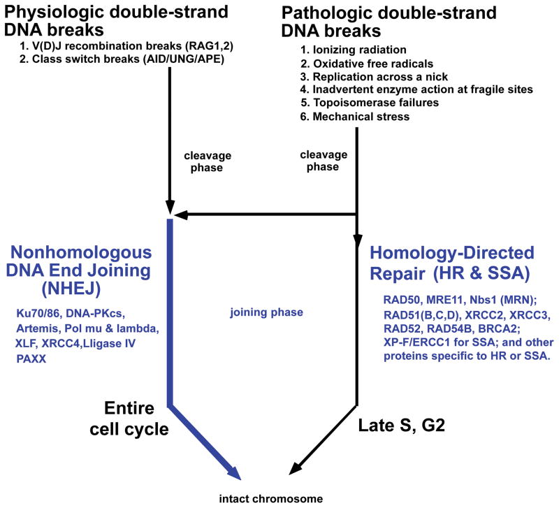
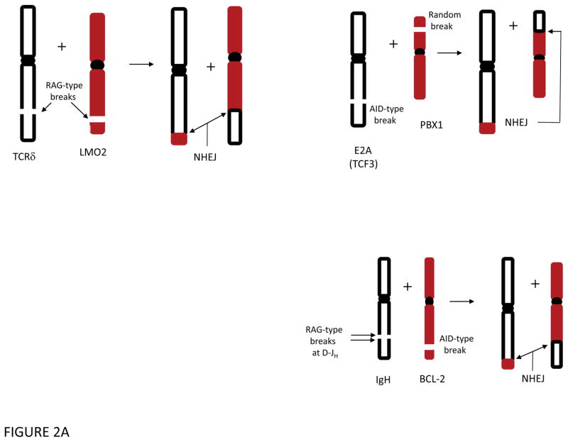
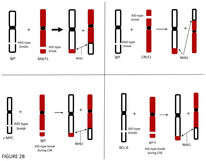
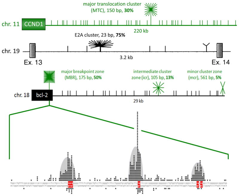
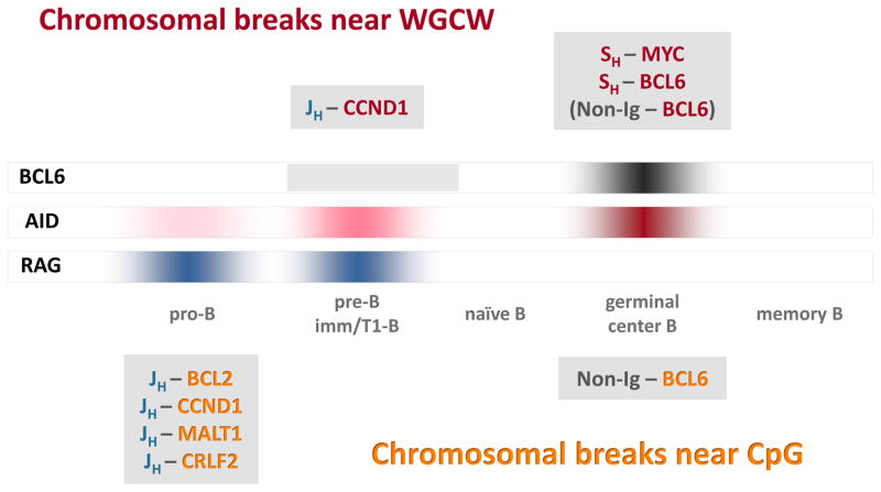
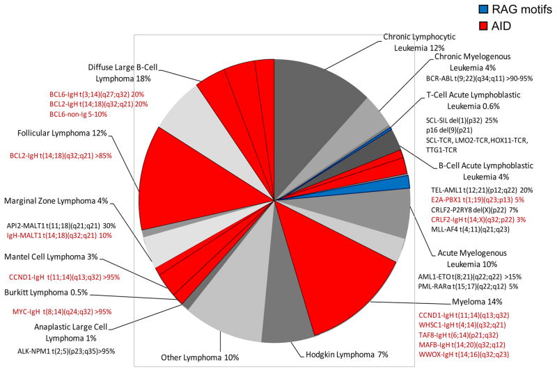
Analysis of chromosomal translocation sequence locations in human lymphomas has provided valuable clues about the mechanism of the translocations and when they occur. Biochemical analyses on the mechanisms of DNA breakage and rejoining permit formulation of detailed models of the human chromosomal translocation process in lymphoid neoplasms. Most human lymphomas are derived from B cells in which a DNA break at an oncogene is initiated by activation-induced deaminase (AID). The partner locus in many cases is located at one of the antigen receptor loci, and this break is generated by the recombination activating gene (RAG) complex or by AID. After breakage, the joining process typically occurs by non-homologous DNA end-joining (NHEJ). Some of the insights into this mechanism also apply to translocations that occur in non-lymphoid neoplasms.


Figures

Figure 1
Figure 1. Causes and Repair of Double-Strand DNA Breaks
Physiologic and pathologic causes of double-strand breaks in mammalian somatic cells are listed at the top. During S and G2 of the cell cycle, homology-directed repair is common because the two sister chromatids are in close proximity, providing a nearby homology donor. Homology-directed repair includes homologous recombination (HR) and single-strand annealing (SSA). At any time in the cell cycle, double-strand breaks can be repaired by nonhomologous DNA end joining (NHEJ). Proteins involved in the repair pathways are listed.
Figure 2
Figure 2. Common Chromosomal Translocations in T and B Cells
A. Some lymphoid translocations involve a DSB that is a RAG-type event at each of the two participating chromosomes (top left). Such RAG-type/RAG-type translocations are common in T cell lymphomas and a subset of B cell lymphomas. More commonly among B cell lymphomas, the initial translocation involves a RAG-type event at one chromosome, often the normal IgH locus [or one of the other antigen receptor loci, such as IgL (κ or λ) or one of the TCR loci] and an AID-type event at the other chromosome (bottom right). After exchange of the chromosomal arms, the DNA ends are joined by NHEJ to generate the two derivative chromosomes. Such events are the predominant mechanism found in most human B cell lymphomas. Centromeres are shown as a filled black oval. B. AID-Type events can cause translocations in early B cells or in germinal center B cells. In the upper left and right, an AID-type event and a RAG-type break give rise to translocations in pro-/pre-B cells. In the lower left and right, an AID-type event on one chromosome can give rise to a translocation involving a failed IgH class switch recombination (CSR) event, which is a physiologic AID-type event. Centromeres are shown as a filled black oval.
Figure 3
Figure 3. A High Proportion of Breakpoints on BCL-2, BCL-1, and E2A Fall into Fragile Regions that are Less than 600 bp
Schematics of the BCL-1 [near CCND1 gene], E2A (also called TCF3), and BCL-2 regions illustrate clustering of breakpoints within the various identified cluster regions [green or black starbursts]. The breakpoints that do not fall into cluster regions are plotted randomly for illustrative purposes [short vertical lines]. In the top line (green horizontal line), the MTC is located about 110 kb from the gene for the cyclin D1 oncoprotein. The 150 bp MTC contains about 30% of breakpoints, whereas the remaining 70% of events are distributed widely over the surrounding 340 kb as recently mapped and sequenced . The second line shows a diagram of intron 13 of the E2A gene, taken from , which showed that 75% of breakpoints occur in the 23 bp E2A cluster, while the surrounding 3 kb only account for 25%. The third line depicts relative proportions of breakpoints at the BCL-2 MBR, icr, and mcr cluster regions. The third exon of the BCL-2 gene, black box on left, contains the MBR [major breakpoint region (or zone)] within the 3′ UTR region, while the centromeric 29 kb contains the icr (intermediate cluster region/zone) and mcr (minor cluster region/zone). Short vertical lines in the gene diagram mark the approximate locations and relative abundance of patient breakpoints. The 175 bp MBR, 105 bp icr, and 561 bp mcr account for about 50%, 13%, and 5% of bcl-2 translocation breakpoints. Every CG sequence motif in each of the three fragile zones the ~29kb downstream of the BCL-2 gene (MBR, icr, mcr) is a hotspot for human translocation. The figure shows the location of nearly all published BCL-2 translocations with expanded detail of the MBR in the very bottom line of the figure. Fragile regions or zone for one of the oncogenes in this review, BCL-2, are shown, but the principles apply to the other fragile zones as well. Within each fragile zone, the actual translocations occur at DNA sequence motifs, consisting of either the sequence CG (CpG) or mCG (when methylated) or WGCW where W=A or T. Human lymphomas are clinically indistinguishable regardless of the position of the breakpoint within the 29 kb. We are trying to determine why these BCL-2 and related zones at other loci (such as BCL-1, MTC, E2A, MALT1, and CRLF2) are highly preferred for DNA breakage and translocation. In the expanded MBR DNA sequence diagram (bottom line of figure), each small black triangle marks the breakpoint of a single, patient translocation. The breakage frequency within this 175 bp region is 300-fold higher than what one would expect to occur at random. Patient breaks within the MBR are not uniformly distributed across the entire 175 bp, but rather are focused in 3 peaks (bell-shaped gray areas), and there are CG sequence motifs (red bases) located near the center of each peak . The p values for the proximity of the MBR breaks to CG sites are between 10−42 and 10−96 (highly significant). The importance of CG and WGCW as translocation sequence motifs applies not only to the BCL-2 gene, but also to most other human B cell neoplastic chromosomal translocations. For example, breaks at the major translocation cluster (MTC) of the BCL-1 gene are also located near CG sites (p values of 10−9 to 10−13), and breaks on the telomeric side of the MTC are significantly near WGCW sites. In the MALT1 gene translocations to IgH in human MALT lymphomas (Fig. 2B), the breaks are also located at CG sites (p = 0.002). The proximity of CG to breaks applies also to the E2A gene translocations (p = 10−4) and the CRLF2 gene translocations to IgH (p = 0.0004) in human pre-B ALL.
Figure 4
Figure 4. Timing of chromosomal translocations as a function of B-cell development
The three long rectangles in the middle of the figure (labeled BCL6, AID, and RAG) depict expression levels. Breakpoint motif analysis suggests that most lymphoma translocations occur either in germinal center B-cells, when AID and BCL-6 are highly expressed, or in pro-B/pre-B cells, when the RAG complex is highly expressed and AID is expressed at low levels (see text). The Ig breaks in most Ig-MYC and Ig-BCL6 translocations are in SH regions, which contain hundreds of WGCW repeats, and the MYC and BCL6 breaks occur near WGCW motifs scattered throughout these partner loci (red text in figure). The Ig breaks in most pro-B/pre-B cell translocations are generated at JH and DH segments by the RAG complex as part of the V(D)J recombination process (blue text). Most CpG breaks occur at Ig partner loci in pro-B/pre-B cell translocations but also in a subset of non-Ig–BCL6 rearrangements that probably occur in germinal center B-cells (orange text), .
Figure 5
Figure 5. Fraction of Human Hematopoietic Malignancies Explained by AID-Type Breaks
Each human hematopoietic malignancy is shown to reflect the fraction of all hematopoietic malignances. Numbers of events reflect the incidence of all events per year in the USA, including all ages (adults and children). The translocations and their percentage in each malignancy are estimated from Swerdlow et al . The portion in RED are translocations that involve at least one AID-type event, often at the oncogene. Some are AID-type/AID-type events, such as translocations between BCL-6 or c-MYC and the IgH switch regions. But most are AID-type/RAG-type events, such as BCL-1, BCL-2, MALT1, or CRLF2 to the IgH locus during failed DH to JH joining. The small fraction colored blue are RAG-type/RAG-type events. We note that many other translocations are not listed (e.g., BCR-ABL1 translocations also occur in B cell lineage ALL; and MLL-AF9 is seen in some cases of AML). This figure is not intended to be comprehensive, and readers are referred to current ACS statistics and Swerdlow et al. for further details.

References

    1. Tsai AG, et al. Human chromosomal translocations at CpG sites and a theoretical basis for their lineage and stage specificity. Cell. 2008;135:1130–42. The CpG sequence motif is decribed here as a site of translocation breakage. - PMC - PubMed
    1. Mahowald GK, Baron JM, Sleckman BP. Collateral damage from antigen receptor gene diversification. Cell. 2008;135:1009–12. - PubMed
    1. Gostissa M, Alt FW, Chiarle R. Mechanisms that promote and suppress chromosomal translocations in lymphocytes. Annu Rev Immunol. 2011;29:319–50. - PubMed
    1. Nussenzweig A, Nussenzweig MC. Origin of chromosomal translocations in lymphoid cancer. Cell. 2010;141:27–38. - PMC - PubMed
    1. Lieber MR. The Mechanism of Double-Strand DNA Break Repair by the Nonhomologous DNA End-Joining Pathway. Annu Rev Biochem. 2010;79:181–211. - PMC - PubMed

Substances