

Differential changes in Neuregulin-1 signaling in major brain regions in a lipopolysaccharide-induced neuroinflammation mouse model
- PMID: 27220549
- PMCID: PMC4918623
- DOI: 10.3892/mmr.2016.5325
Differential changes in Neuregulin-1 signaling in major brain regions in a lipopolysaccharide-induced neuroinflammation mouse model
Abstract
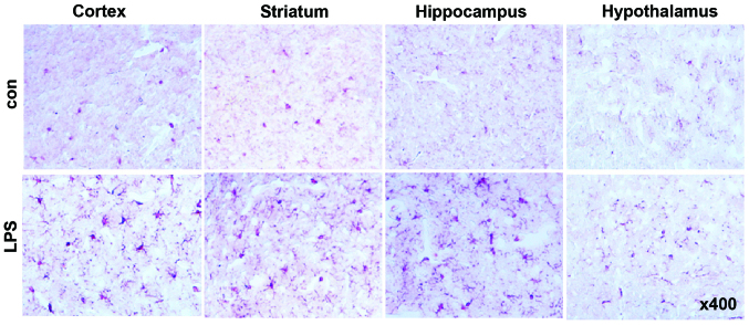
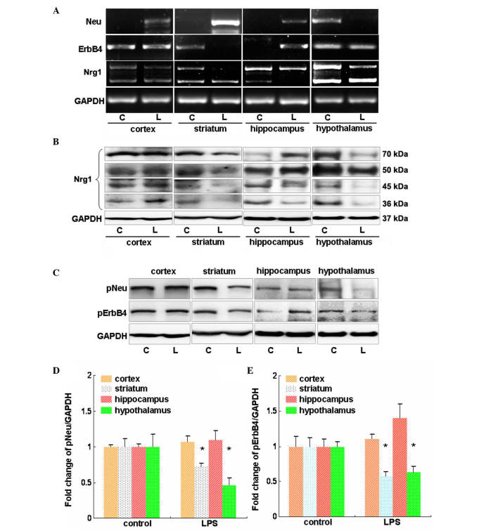
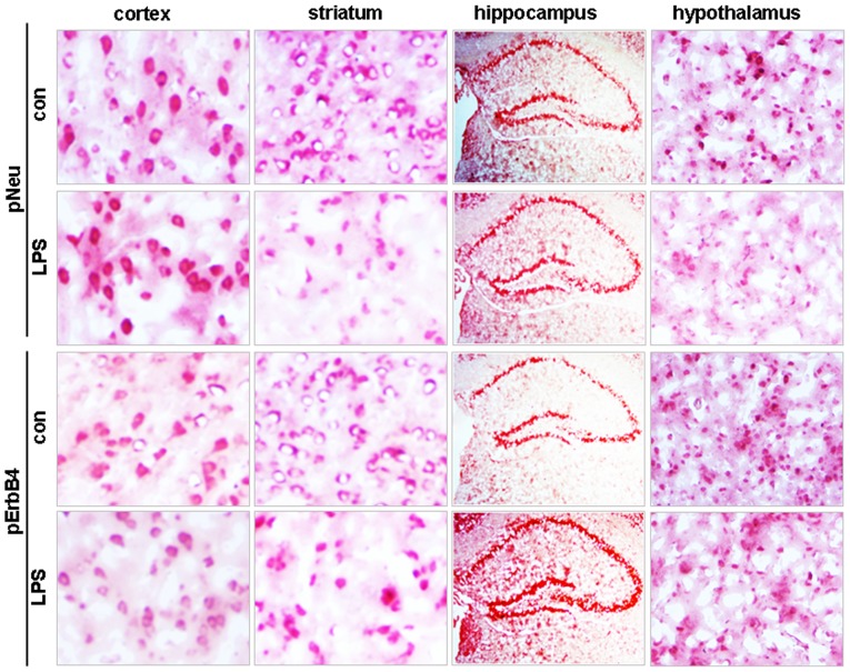
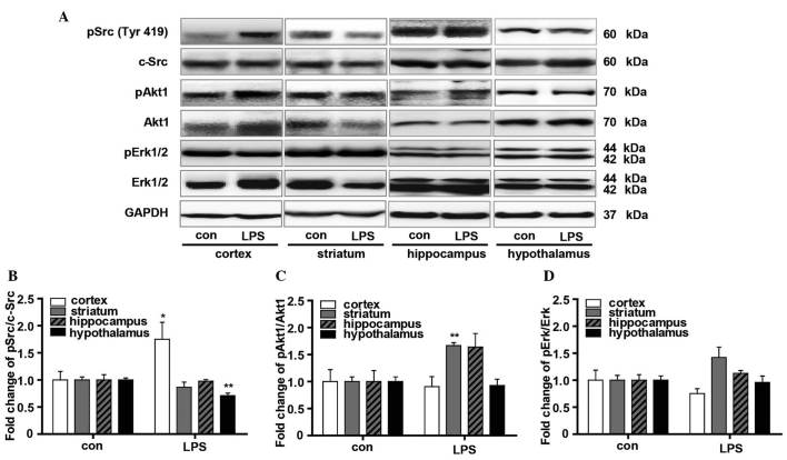
Neuregulin 1 (Nrg1) is involved in multiple biological processes in the nervous system. The present study investigated changes in Nrg1 signaling in the major brain regions of mice subjected to lipopolysaccharide (LPS)-induced neuroinflammation. At 24 h post‑intraperitoneal injection of LPS, mouse brain tissues, including tissues from the cortex, striatum, hippocampus and hypothalamus, were collected. Reverse transcription‑polymerase chain reaction was used to determine the expression of Nrg1 and its receptors, Neu and ErbB4, at the mRNA level. Western blotting was performed to determine the levels of these proteins and the protein levels of phosphorylated extracellular signal-regulated kinases (Erk)1/2 and Akt1. Immunohistochemical staining was utilized to detect the levels of pNeu and pErbB4 in these regions. LPS successfully induced sites of neuroinflammation in these regions, in which changes in Nrg1, Neu and ErbB4 at the mRNA and protein levels were identified compared with controls. LPS induced a reduction in pNeu and pErbB4 in the striatum and hypothalamus, although marginally increased pErbB4 levels were found in the hippocampus. LPS increased the overall phosphorylation of Src but this effect was reduced in the hypothalamus. Moreover, increased phosphorylation of Akt1 was found in the striatum and hippocampus. These data suggest diverse roles for Nrg1 signaling in these regions during the process of neuroinflammation.
Figures
Similar articles
-
Neuregulin-1 protects mouse cerebellum against oxidative stress and neuroinflammation.Brain Res. 2017 Sep 1;1670:32-43. doi: 10.1016/j.brainres.2017.06.012. Epub 2017 Jun 13. Brain Res. 2017. PMID: 28623147
-
Neuroinflammation-Induced Downregulation of Hippocampacal Neuregulin 1-ErbB4 Signaling in the Parvalbumin Interneurons Might Contribute to Cognitive Impairment in a Mouse Model of Sepsis-Associated Encephalopathy.Inflammation. 2017 Apr;40(2):387-400. doi: 10.1007/s10753-016-0484-2. Inflammation. 2017. PMID: 27913953
-
Spinal cord injury induced Neuregulin 1 signaling changes in mouse prefrontal cortex and hippocampus.Brain Res Bull. 2019 Jan;144:180-186. doi: 10.1016/j.brainresbull.2018.12.002. Epub 2018 Dec 7. Brain Res Bull. 2019. PMID: 30529367
-
The neuregulin-I/ErbB signaling system in development and disease.Adv Anat Embryol Cell Biol. 2007;190:1-65. Adv Anat Embryol Cell Biol. 2007. PMID: 17432114 Review.
-
Neuregulin 1-activated ERBB4 as a "dedicated" receptor for the Hippo-YAP pathway.Sci Signal. 2014 Dec 9;7(355):pe29. doi: 10.1126/scisignal.aaa2710. Sci Signal. 2014. PMID: 25492964 Review.
Cited by
-
Identification of novel biomarkers for prediction of neurological prognosis following cardiac arrest.Oncotarget. 2017 Mar 7;8(10):16144-16157. doi: 10.18632/oncotarget.14877. Oncotarget. 2017. PMID: 28147324 Free PMC article.
-
Nrg1 Intracellular Signaling Is Neuroprotective upon Stroke.Oxid Med Cell Longev. 2019 Sep 8;2019:3930186. doi: 10.1155/2019/3930186. eCollection 2019. Oxid Med Cell Longev. 2019. PMID: 31583038 Free PMC article.
-
Neuregulin in Health and Disease.Int J Brain Disord Treat. 2018;4(1):024. doi: 10.23937/2469-5866/1410024. Epub 2018 Nov 10. Int J Brain Disord Treat. 2018. PMID: 31032468 Free PMC article. No abstract available.
-
Dihydrolipoic acid protects against lipopolysaccharide-induced behavioral deficits and neuroinflammation via regulation of Nrf2/HO-1/NLRP3 signaling in rat.J Neuroinflammation. 2020 May 25;17(1):166. doi: 10.1186/s12974-020-01836-y. J Neuroinflammation. 2020. PMID: 32450903 Free PMC article.
-
Therapeutic efficacy of neuregulin 1-expressing human adipose-derived mesenchymal stem cells for ischemic stroke.PLoS One. 2019 Sep 27;14(9):e0222587. doi: 10.1371/journal.pone.0222587. eCollection 2019. PLoS One. 2019. PMID: 31560696 Free PMC article.
References
MeSH terms
Substances
LinkOut - more resources
Full Text Sources
Other Literature Sources
Medical
Research Materials
Miscellaneous
