

Effects of dexmedetomidine postconditioning on myocardial ischemia and the role of the PI3K/Akt-dependent signaling pathway in reperfusion injury
- PMID: 27221008
- PMCID: PMC4918562
- DOI: 10.3892/mmr.2016.5345
Effects of dexmedetomidine postconditioning on myocardial ischemia and the role of the PI3K/Akt-dependent signaling pathway in reperfusion injury
Abstract
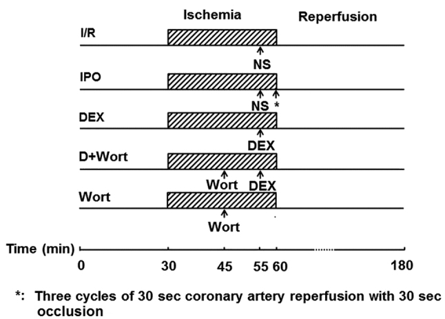
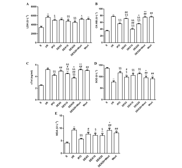
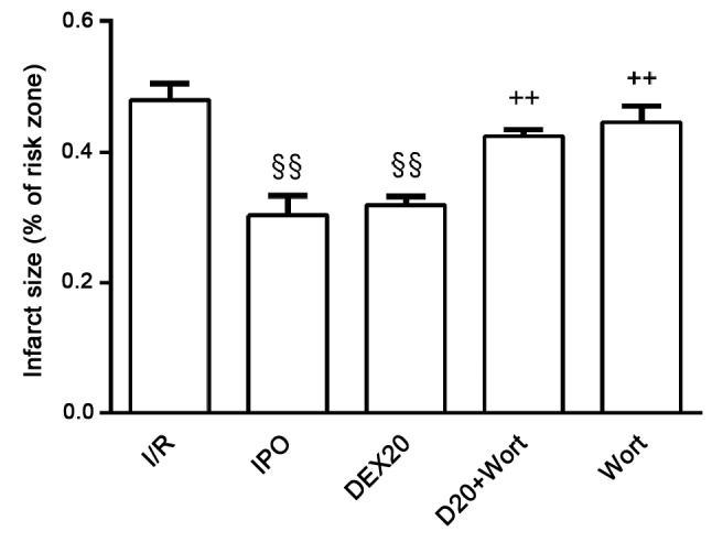
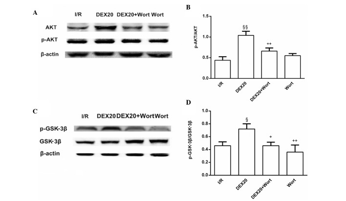
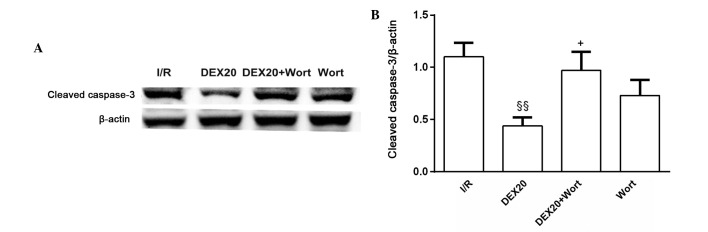
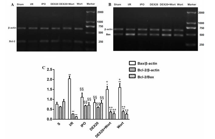
The present study aimed to determine whether post-ischemic treatment with dexmedetomidine (DEX) protected the heart against acute myocardial ischemia/reperfusion (I/R)‑induced injury in rats. The phosphatidylinositol‑3 kinase/protein kinase B(PI3K/Akt)‑dependent signaling pathway was also investigated. Male Sprague Dawley rats (n=64) were subjected to ligation of the left anterior descending artery (LAD), which produced ischemia for 25 min, followed by reperfusion. Following LAD ligation, rats were treated with DEX (5, 10 and 20 µg/kg) or underwent post‑ischemic conditioning, which included three cycles of ischemic insult. In order to determine the role of the PI3K/Akt signaling pathway, wortmannin (Wort), a PI3K inhibitor, was used to treat a group of rats that had also been treated with DEX (20 µg/kg). Post‑reperfusion, lactate dehydrogenase (LDH), cardiac troponin I (cTnI), creatine kinase isoenzymes (CK‑MB), superoxide dismutase (SOD) and malondialdehyde (MDA) serum levels were measured using an ultraviolet spectrophotometer. The protein expression levels of phosphorylated (p)‑Akt, Ser9‑p‑glycogen synthase kinase‑3β (p‑GSK‑3β) and cleaved caspase‑3 were detected in heart tissue by western blotting. The mRNA expression levels of B‑cell lymphoma 2 (Bcl‑2) and Bcl‑2‑associated X protein (Bax) were detected using reverse transcription‑polymerase chain reaction. At the end of the experiment, the hearts were removed and perfused in an isolated perfusion heart apparatus with Evans blue (1%) in order to determine the non‑ischemic areas. The risk and infarct areas of the heart were not dyed. As expected, I/R induced myocardial infarction, as determined by the increased serum levels of cTnI, CK‑MB and MDA, and the decreased levels of SOD. Post‑ischemic treatment with DEX increased the expression levels of p‑Akt and p‑GSK‑3β, whereas caspase‑3 expression was reduced following DEX treatment compared with in the I/R group. Compared with the I/R group, the ratio of Bcl‑2/Bax at the mRNA level was elevated in the DEX and ischemic post‑conditioning groups, whereas the expression levels of Bax were decreased. Conversely, the effects of DEX were attenuated by Wort. These results indicated that, similar to post‑ischemic conditioning, post‑ischemic treatment with DEX protects the heart against I/R via the PI3K/Akt‑dependent signaling pathway, possibly by activating GSK‑3β.
Figures
Similar articles
-
Effects of Dexmedetomidine Postconditioning on Myocardial Ischemia/Reperfusion Injury in Diabetic Rats: Role of the PI3K/Akt-Dependent Signaling Pathway.J Diabetes Res. 2018 Oct 8;2018:3071959. doi: 10.1155/2018/3071959. eCollection 2018. J Diabetes Res. 2018. PMID: 30402501 Free PMC article.
-
Remote ischemic postconditioning protects the heart by upregulating ALDH2 expression levels through the PI3K/Akt signaling pathway.Mol Med Rep. 2014 Jul;10(1):536-42. doi: 10.3892/mmr.2014.2156. Epub 2014 Apr 15. Mol Med Rep. 2014. PMID: 24736969
-
Effects of dexmedetomidine on myocardial ischemia-reperfusion injury through PI3K-Akt-mTOR signaling pathway.Eur Rev Med Pharmacol Sci. 2019 Aug;23(15):6736-6743. doi: 10.26355/eurrev_201908_18565. Eur Rev Med Pharmacol Sci. 2019. PMID: 31378917
-
Diabetic inhibition of preconditioning- and postconditioning-mediated myocardial protection against ischemia/reperfusion injury.Exp Diabetes Res. 2012;2012:198048. doi: 10.1155/2012/198048. Epub 2011 Aug 1. Exp Diabetes Res. 2012. PMID: 21822424 Free PMC article. Review.
-
The role of PI3K/AKT signaling pathway in myocardial ischemia-reperfusion injury.Int Immunopharmacol. 2023 Oct;123:110714. doi: 10.1016/j.intimp.2023.110714. Epub 2023 Jul 29. Int Immunopharmacol. 2023. PMID: 37523969 Review.
Cited by
-
The Effect of Dexmedetomidine on Oral Mucosal Blood Flow and the Absorption of Lidocaine.Anesth Prog. 2018 Fall;65(3):168-176. doi: 10.2344/anpr-65-03-02. Anesth Prog. 2018. PMID: 30235427 Free PMC article.
-
Influence of Hyperglycemia on Dexmedetomidine-Induced Cardioprotection in the Isolated Perfused Rat Heart.J Clin Med. 2020 May 13;9(5):1445. doi: 10.3390/jcm9051445. J Clin Med. 2020. PMID: 32413983 Free PMC article.
-
Dexmedetomidine alleviates cisplatin‑induced acute kidney injury by attenuating endoplasmic reticulum stress‑induced apoptosis via the α2AR/PI3K/AKT pathway.Mol Med Rep. 2020 Mar;21(3):1597-1605. doi: 10.3892/mmr.2020.10962. Epub 2020 Jan 24. Mol Med Rep. 2020. PMID: 32016445 Free PMC article.
-
Pre-cardiopulmonary bypass administration of dexmedetomidine decreases cardiac troponin I level following cardiac surgery with sevoflurane postconditioning.J Int Med Res. 2019 Aug;47(8):3623-3635. doi: 10.1177/0300060519856750. Epub 2019 Jun 24. J Int Med Res. 2019. PMID: 31234690 Free PMC article.
-
Remote ischemic post-conditioning protects against myocardial ischemia/reperfusion injury by inhibiting the Rho-kinase signaling pathway.Exp Ther Med. 2020 Jan;19(1):99-106. doi: 10.3892/etm.2019.8176. Epub 2019 Nov 8. Exp Ther Med. 2020. PMID: 31853278 Free PMC article.
References
-
- Cai Y, Xu H, Yan J, Zhang L, Lu Y. Molecular targets and mechanism of action of dexmedetomidine in treatment of ischemia/reperfusion injury. Mol Med Rep. 2014;9:1542–1550. - PubMed
-
- Ibacache M, Sanchez G, Pedrozo Z, Galvez F, Humeres C, Echevarria G, Duaso J, Hassi M, Garcia L, Díaz-Araya G, Lavandero S. Dexmedetomidine preconditioning activates pro-survival kinases and attenuates regional ischemia/reperfusion injury in rat heart. Biochim Biophys Acta. 18222012:537–545. - PubMed
MeSH terms
Substances
LinkOut - more resources
Full Text Sources
Other Literature Sources
Research Materials
Miscellaneous
