. 2016 May 25:6:26697.
doi: 10.1038/srep26697.

MicroRNA-146a suppresses ROCK1 allowing hyperphosphorylation of tau in Alzheimer's disease

Affiliations

MicroRNA-146a suppresses ROCK1 allowing hyperphosphorylation of tau in Alzheimer's disease

Gang Wang et al. Sci Rep. .

Abstract

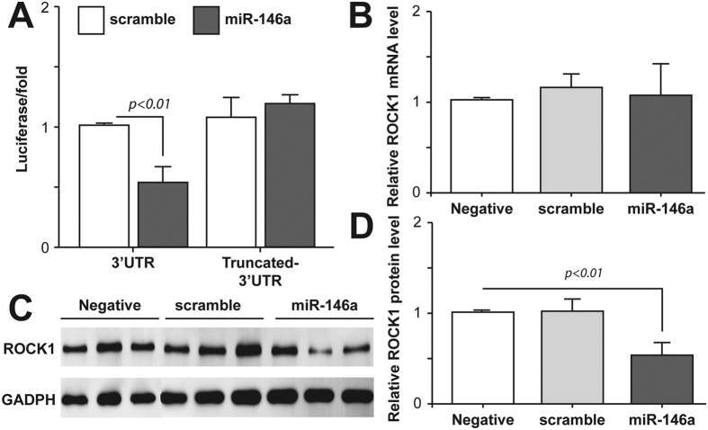
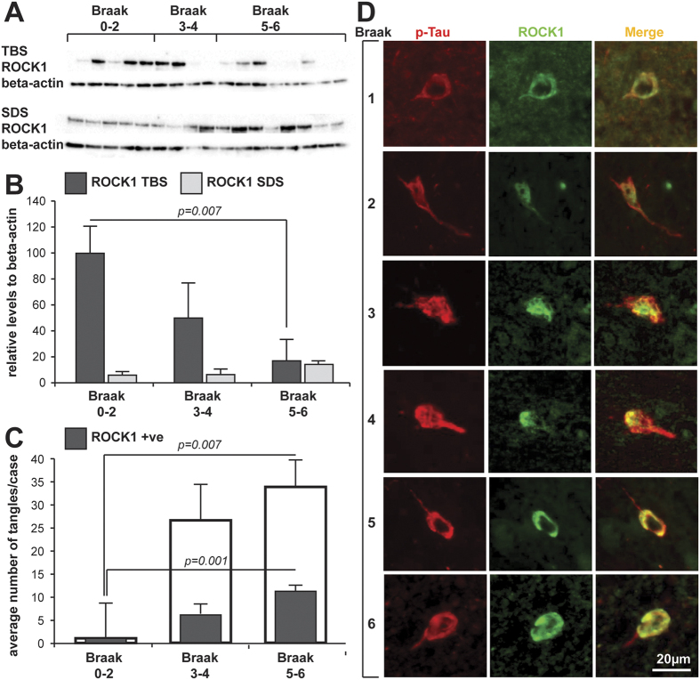
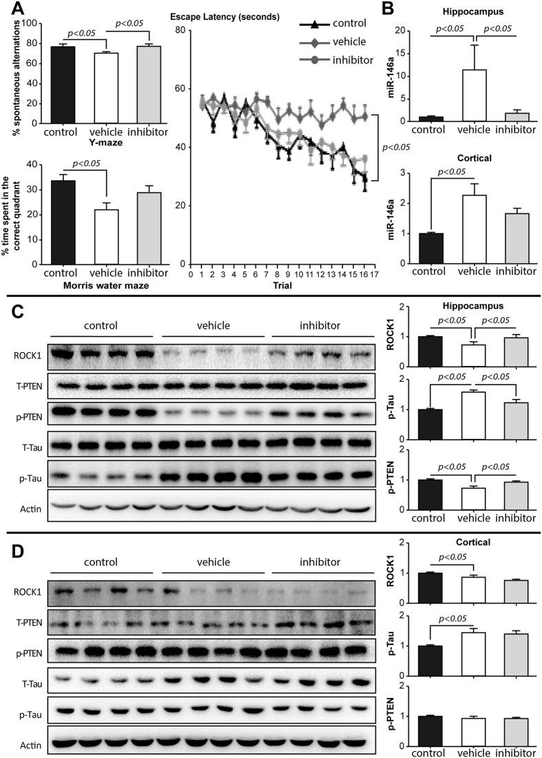
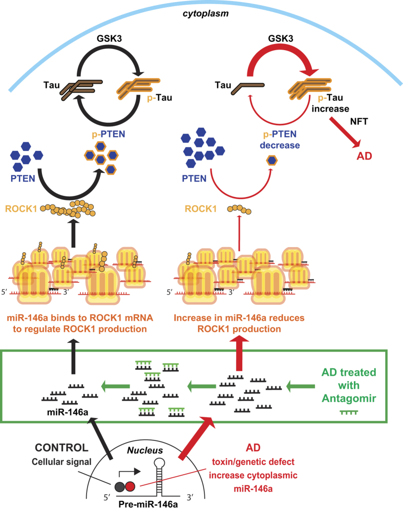
MicroRNA-146a is upregulated in the brains of patients with Alzheimer's disease (AD). Here, we show that the rho-associated, coiled-coil containing protein kinase 1 (ROCK1) is a target of microRNA-146a in neural cells. Knockdown of ROCK1 mimicked the effects of microRNA-146a overexpression and induced abnormal tau phosphorylation, which was associated with inhibition of phosphorylation of the phosphatase and tensin homolog (PTEN). The ROCK1/PTEN pathway has been implicated in the neuronal hyperphosphorylation of tau that occurs in AD. To determine the function of ROCK1 in AD, brain tissue from 17 donors with low, intermediate or high probability of AD pathology were obtained and analyzed. Data showed that ROCK1 protein levels were reduced and ROCK1 colocalised with hyperphosphorylated tau in early neurofibrillary tangles. Intra-hippocampal delivery of a microRNA-146a specific inhibitor (antagomir) into 5xFAD mice showed enhanced hippocampal levels of ROCK1 protein and repressed tau hyperphosphorylation, partly restoring memory function in the 5xFAD mice. Our in vitro and in vivo results confirm that dysregulation of microRNA-146a biogenesis contributes to tau hyperphosphorylation and AD pathogenesis, and inhibition of this microRNA could be a viable novel in vivo therapy for AD.


Figures

Figure 1
Figure 1. ROCK1 is a target of microRNA-146a in neural cells.
(A) The effect of microRNA-146a (miR-146a) on ROCK1 expression was assessed in neural SH-SY5Y cells using a luciferase reporter system. A vector expressing miR-146a and a psiCHECK-2 vector containing either the full length ROCK1 3′ UTR or a truncated 3′ UTR lacking the miR-146a binding sites were co-transfected into neural SH-SY5Y cells. In control cells the vector expressing miR-146a was replaced with a scrambled miR-146a vector (scramble). Fluorescence was measured 48 hours after transfection. MiR-146a bound to the full length ROCK1 3′ UTR and inhibited its activity, but did not bind to the truncated ROCK1 3′ UTR. (B) Real-time PCR was used to detect the mRNA expression level of ROCK1 in neural SH-SY5Y cells. Overexpression of miR-146a or scramble did not affect the relative ROCK1 mRNA level compared to neural cells without vectors (negative). Relative expression of ROCK1 mRNA was calculated using the ΔΔCT method. (C) Western blot was used to measure ROCK1 protein translation in neural cells. Overexpression of miR-146a significantly decreased expression of endogenous ROCK1 protein in neural SH-SY5Y cells compared to negative or scramble treated neural cells. (D) Overexpression of miR-146a significantly reduced the relative ROCK1 protein level compared to negative or scramble treated neural cells. All of the data are expressed as means ± SD of at least three independent experiments.
Figure 2
Figure 2. Molecular mechanisms of microRNA-146a associated with Alzheimer pathologies.
(A) Conditions inducing microRNA-146a (miR-146a) overexpression in neural SH-SY5Y cells included treatment with Aβ1–42 (5 μM) for 24 hours and stable overexpression of the APP Swedish mutation compared to untransfected control neural cells (means ± SD). (B,C) Overexpression of miR-146ain neural SH-SY5Y cells induces decreased levels of ROCK1 and tau hyperphosphorylation, as demonstrated in Western blots of total tau (T-Tau) and phosphorylated tau (Ser396, p-Tau) (means ± SD). Control neural cells expressed a scrambled miR-146a vector (scramble). (D,E) Knockdown of ROCK1 mRNA using siRNA in neural SH-SY5Y cells caused an increase in tau phosphorylation at Ser396 (p-Tau) compared to total tau (T-Tau) and decreased PTEN phosphorylation (p-PTEN) compared to total PTEN (T-PTEN) as detected in Western blots using an antibody specific for phosphorylation at Ser380/Thr382/Thr383. Scrambled ROCK1 siRNA (scramble) was used in control neural cell experiments. Quantified data are expressed as mean ± SD for at least three independent experiments.
Figure 3
Figure 3. ROCK1 in human brain tissue.
(A) Western blot of ROCK1 protein levels in the different tissue fractions (TBS = soluble fraction, SDS = SDS fraction, see methods) of the inferior temporal lobe of donors at different stages of AD pathology. (B) Relative quantitation of the ROCK1 Western blots in the different tissue fractions shown in (A). (C) Quantitation of the numbers of phospho-tau-immunoreactive tangles (open bars) and those colocalising ROCK1 (shaded bar) at different stages of AD. Significant increases in both types of tangles occur with increasing stages of AD. (D) Representative examples of double-labelled phospho-tau- (p-Tau) and ROCK1-immunoreactive tangles at the different stages of AD indicated at left. Brain tissue samples from healthy aged controls (N = 5 aged 89 ± 4 y, Braak neuritic stages 0–2), preclinical AD (N = 4 aged 85 ± 3 y, Braak neuritic stages 3–4) and clinical end-stage AD (N = 8 aged 86 ± 4 y, Braak neuritic stages 5–6). All data are expressed as mean ± SD with at least three replications of the Western blot data. Scale in (D) is equivalent for all micrographs.
Figure 4
Figure 4. Intra-hippocampal delivery of antagomir.
(A) The Y maze revealed that the percentage of spontaneous alternations was higher in 5xFAD mice treated with antagomir compared with Vehicle treated 5xFAD mice; and the Morris water maze showed shorter escape latency and higher percentage of time spent in the correct quadrant for 5xFAD mice treated with antagomir compared to Vehicle treated 5xFAD mice. (B) The relative expression of microRNA-146a (miR146a) was significantly inhibited in hippocampus, but not prefrontal cortex, after bilateral intra-hippocampal injections (n = 4). (C) The hippocampal protein levels of the related signaling pathway markers (ROCK1-pPTEN-pTau) were evaluated by Western blotting. A representative gel image is shown from four Control (wild-type), four Vehicle treated 5xFAD, and four 5xFAD with antagomir mice. (D) The prefrontal protein levels of the related signaling pathways (ROCK1-pPTEN-pTau) were evaluated by Western blotting. A representative gel image is shown from four Control (wild-type), four Vehicle treated 5xFAD, and four 5xFAD mice treated with antagomir mice. The data are shown as mean ± SE.
Figure 5
Figure 5. A model of the proposed inductive effects of microRNA-146a in Alzheimer’s disease (AD).
Normal cell signaling (such as by NF-κB) stimulates the generation of the primary microRNA-146a (miR-146a) transcript that is produced in the nucleus aspre-microRNA-146a (Pre-miR-146a), which is then shuttled into the cytoplasm and further processed. MiR-146a binds to complimentary sequences in the 3′ untranslated region (UTR) of its target mRNA transcript of ROCK1. This results in translational repression of ROCK1 protein. Tau phosphorylation (p-Tau) and dephosphorylation are maintained in a dynamic balance for normal cell functions. Protein kinases (such as GSK3 etc) phosphorylate tau, while phosphorylated PTEN (p-PTEN) dephosphorylates tau. The levels of p-PTEN are regulated by the levels of ROCK1 protein, indicating that ROCK1 plays an important role in tau phosphorylation. In AD (red pathway at right) neuronal cells produce more miR-146a that decreases the levels of ROCK1 protein and reduces the levels of p-PTEN, impeding tau dephosphorylation. Thus, p-tau accumulates in neurons to form neurofibrillary tangles (NFT), finally leadingto neuronal death in AD. Treatment with antagomir neutralizes miR-146a (green box) restoring ROCK1 protein levels and facilitating appropriate PTEN phosphorylation and tau dephosphorylation.

Similar articles

Cited by

References

    1. Hardy J. Amyloid, the presenilins and Alzheimer’s disease. Trends Neurosci. 20, 154–159 (1997). - PubMed
    1. Hutton M. & Hardy J. The presenilins and Alzheimer’s disease. Hum Mol Genet. 6, 1639–1646 (1997). - PubMed
    1. Scholz T. & Mandelkow E. Transport and diffusion of Tau protein in neurons. Cell Mol Life Sci. 71, 3139–3150, doi: 10.1007/s00018-014-1610-7 (2014). - DOI - PMC - PubMed
    1. Brunden K. R., Ballatore C., Lee V. M., Smith A. B. 3rd & Trojanowski J. Q. Brain-penetrant microtubule-stabilizing compounds as potential therapeutic agents for tauopathies. Biochem Soc Trans. 40, 661–666, doi: 10.1042/BST20120010 (2012). - DOI - PMC - PubMed
    1. Duan Y., Dong S., Gu F., Hu Y. & Zhao Z. Advances in the pathogenesis of Alzheimer’s disease: focusing on tau-mediated neurodegeneration. Transl Neurodegener. 1, 24, doi: 10.1186/2047-9158-1-24 (2012). - DOI - PMC - PubMed

Publication types