

Optic nerve head parameters of high-definition optical coherence tomography and Heidelberg retina tomogram in perimetric and preperimetric glaucoma
- PMID: 27221679
- PMCID: PMC4901845
- DOI: 10.4103/0301-4738.182938
Optic nerve head parameters of high-definition optical coherence tomography and Heidelberg retina tomogram in perimetric and preperimetric glaucoma
Abstract
Background: Heidelberg retina tomogram (HRT) and optical coherence tomography (OCT) are two widely used imaging modalities to evaluate the optic nerve head (ONH) in glaucoma.
Purpose: To compare the ONH parameters of HRT3 and high-definition OCT (HD-OCT) and evaluate their diagnostic abilities in perimetric and preperimetric glaucoma.
Design: Cross-sectional analysis.
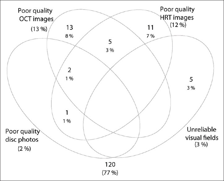
Methods: 35 control eyes (24 subjects), 21 preperimetric glaucoma eyes (15 patients), and 64 perimetric glaucoma eyes (44 patients) from the Longitudinal Glaucoma Evaluation Study underwent HRT3 and HD-OCT examinations.
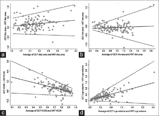
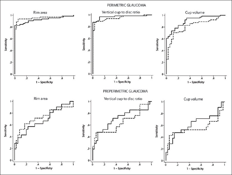
Statistical analysis: Agreement between the ONH parameters of HRT and HD-OCT were assessed using Bland-Altman plots. Diagnostic abilities of ONH parameters were evaluated using area under the receiver operating characteristic curves (AUCs), sensitivity at fixed specificity, and likelihood ratios (LR).
Results: Optic disc area, vertical cup to disc ratio, and cup volume with HD-OCT were larger than with HRT, while the rim area was smaller with HD-OCT (P < 0.001 for all comparisons). AUCs of all HD-OCT ONH parameters (0.90-0.97 in perimetric and 0.62-0.71 in preperimetric glaucoma) were comparable (P > 0.10) to the corresponding HRT ONH parameters (0.81-0.95 in perimetric and 0.55-0.72 in preperimetric glaucoma). LRs associated with diagnostic categorization of ONH parameters of both HD-OCT and HRT were associated with larger effects on posttest probability of perimetric compared to preperimetric glaucoma.
Conclusions: ONH measurements of HD-OCT and HRT3 cannot be used interchangeably. Though the diagnostic abilities of ONH parameters of HD-OCT and HRT in glaucoma were comparable, the same were significantly lower in preperimetric compared to perimetric glaucoma.
Figures
Similar articles
-
Ganglion cell-inner plexiform layer thickness of high definition optical coherence tomography in perimetric and preperimetric glaucoma.Invest Ophthalmol Vis Sci. 2014 Jul 11;55(8):4768-75. doi: 10.1167/iovs.14-14598. Invest Ophthalmol Vis Sci. 2014. PMID: 25015361
-
Optic disc imaging with spectral-domain optical coherence tomography: variability and agreement study with Heidelberg retinal tomograph.Ophthalmology. 2012 Sep;119(9):1852-7. doi: 10.1016/j.ophtha.2012.02.033. Epub 2012 May 8. Ophthalmology. 2012. PMID: 22572035
-
Glaucoma diagnosis optic disc analysis comparing Cirrus spectral domain optical coherence tomography and Heidelberg retina tomograph II.Jpn J Ophthalmol. 2013 Jan;57(1):41-6. doi: 10.1007/s10384-012-0205-9. Epub 2012 Oct 27. Jpn J Ophthalmol. 2013. PMID: 23104685
-
The role of clinical examination of the optic nerve head in glaucoma today.Curr Opin Ophthalmol. 2021 Mar 1;32(2):83-91. doi: 10.1097/ICU.0000000000000734. Curr Opin Ophthalmol. 2021. PMID: 33470671 Review.
-
The diagnostic value of optic nerve imaging in early glaucoma.Curr Opin Ophthalmol. 2001 Apr;12(2):100-4. doi: 10.1097/00055735-200104000-00004. Curr Opin Ophthalmol. 2001. PMID: 11224715 Review.
Cited by
-
Anterior Segment Optic Coherence Tomography Changes Before and After Phacoemulsification in Primary Open-Angle Glaucoma.Beyoglu Eye J. 2019 Aug 9;4(2):102-107. doi: 10.14744/bej.2019.39306. eCollection 2019. Beyoglu Eye J. 2019. PMID: 35187442 Free PMC article.
-
Comparison of the efficacy of latanoprost versus dorzolamide/timolol fixed combination therapy in patients with pseudoexfoliative glaucoma according to glaucoma stage.Arq Bras Oftalmol. 2022 Sep 23;87(1):0230. doi: 10.5935/0004-2749.2021-0230. eCollection 2022. Arq Bras Oftalmol. 2022. PMID: 36169430 Free PMC article.
-
Structural and functional changes to the retina and optic nerve following panretinal photocoagulation over a 2-year time period.Eye (Lond). 2017 Aug;31(8):1237-1244. doi: 10.1038/eye.2017.66. Epub 2017 Apr 28. Eye (Lond). 2017. PMID: 28452993 Free PMC article.
-
The use of deep learning technology for the detection of optic neuropathy.Quant Imaging Med Surg. 2022 Mar;12(3):2129-2143. doi: 10.21037/qims-21-728. Quant Imaging Med Surg. 2022. PMID: 35284277 Free PMC article. Review.
References
-
- Preferred practice pattern for primary open-angle glaucoma. San Francisco, California: American Academy of Ophthalmology; 2005.
-
- Varma R, Steinmann WC, Scott IU. Expert agreement in evaluating the optic disc for glaucoma. Ophthalmology. 1992;99:215–21. - PubMed
-
- Abrams LS, Scott IU, Spaeth GL, Quigley HA, Varma R. Agreement among optometrists, ophthalmologists, and residents in evaluating the optic disc for glaucoma. Ophthalmology. 1994;101:1662–7. - PubMed
-
- Azuara-Blanco A, Katz LJ, Spaeth GL, Vernon SA, Spencer F, Lanzl IM. Clinical agreement among glaucoma experts in the detection of glaucomatous changes of the optic disk using simultaneous stereoscopic photographs. Am J Ophthalmol. 2003;136:949–50. - PubMed
MeSH terms
LinkOut - more resources
Full Text Sources
Other Literature Sources
Medical
