Review
. 2016 Aug;7(4):531-9.
doi: 10.1007/s13244-016-0499-0. Epub 2016 May 24.

Idiopathic granulomatous mastitis: imaging update and review

Affiliations
Review

Idiopathic granulomatous mastitis: imaging update and review

Robert T Fazzio et al. Insights Imaging. 2016 Aug.

Abstract

Objectives: The purpose of this study was to review the imaging features of idiopathic granulomatous mastitis (IGM) with clinical and pathology correlation.

Methods: With institutional review board (IRB) approval, a retrospective search of the surgical pathology database from January 2000 to July 2015 was performed. Clinical, imaging and histology findings were reviewed. Cases of granulomatous mastitis without a known source, diagnosed with percutaneous or surgical biopsy, were included in our analysis.

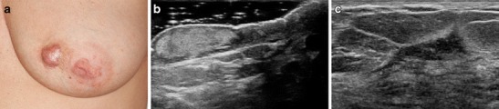
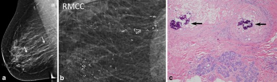
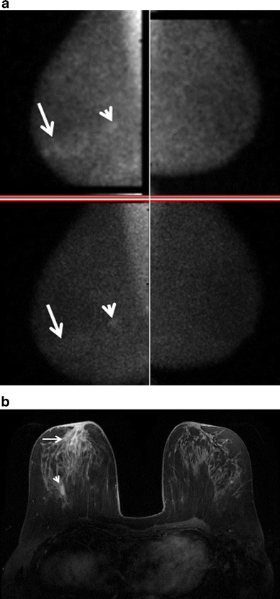
Results: Seventeen cases of IGM were identified with imaging available for review. The majority of patients presented with a palpable abnormality, whereas a minority were asymptomatic with an abnormal screening mammogram. At imaging, IGM most often demonstrated a focal asymmetry at mammography, a hypoechoic mass with irregular or angular margins at ultrasound, and robust enhancement with mixed progressive and plateau kinetics at magnetic resonance imaging (MRI). Axillary lymph nodes were reactive in appearance at ultrasound. Molecular breast imaging performed in one case showed mild focal asymmetric radiotracer uptake.

Conclusion: IGM is a rapidly progressive rare inflammatory condition of the breast resulting in non-necrotizing granuloma formation. Imaging features mimic breast carcinoma and diagnosis can be difficult. Radiologists' awareness of this condition is essential to prevent delayed or unnecessary treatment.

Teaching points: • Idiopathic granulomatous mastitis is rapidly progressive inflammatory condition. • Imaging features may mimic breast carcinoma or infection. • Ultrasound shows irregular hypoechoic masses with increased vascularity and sinus tracts. • MRI shows irregular, enhancing masses or non-mass enhancement with microabscesses. • MRI is useful for assessment of breast involvement and response to treatment.

Keywords: Breast; Breast ultrasound; Granulomatous mastitis; MRI; Mammography.


Figures

Fig. 1
Fig. 1
A 27-year-old woman with an enlarging palpable right breast mass admitted to the hospital for intractable joint pain. An erythematous rash was noted on her right breast and extremities. She was 8 months post-partum, breastfeeding with difficulty on the right. Bilateral mediolateral oblique mammogram (a) demonstrates regional asymmetry in the middle depth upper breast (arrows). b Clinical photograph of the patient’s forearm shows erythematous areas (arrows), representing erythema nodosum. c Photomicrograph (original magnification ×200; haematoxylin-eosin [H-E] stain) showing non-necrotizing granulomatous inflammation (arrow) composed of histiocytes and giant cells
Fig. 2
Fig. 2
A 24-year-old Albanian mother of a 4-year-old (breastfed for 1 year) presented with a left breast lump, pain, erythema and drainage from multiple sites. Clinical photograph (a) of the left breast shows four eschars (arrows), two of which in the upper breast were draining cloudy fluid. Also shown is nipple inversion. b Ultrasound (US) image of the upper left breast shows a hypoechoic mass with indistinct margins with tract extending into adjacent tissue and to the skin surface (arrows) to the uppermost eschars noted the clinical photograph (a). c Axial T1-weighted post-contrast subtraction axial MRI image shows an irregular, intensely enhancing mass in the left breast extending to the chest wall with central areas of non-enhancement consistent with abscess formation. Ultrasound biopsy revealed IGM
Fig. 3
Fig. 3
A 50-year-old woman presenting with painful, rapidly enlarging areolar skin lesion and breast mass. Clinical photograph (a) of the left breast shows a raised red skin lesion at the 9 o’clock position of the areola. Skin biopsy showed non-necrotizing granulomatous inflammation. b US image of the left areola and nipple show a hyperechoic area of skin thickening, which involves the dermis but does not connect to the underlying breast tissue. c US of the palpable breast mass demonstrates an irregular hypoechoic mass with angular margins and hyperechoic rim which did not connect to the areolar skin lesion. Percutaneous biopsy showed non-necrotizing granulomatous inflammation. Special stains for microorganisms were negative
Fig. 4
Fig. 4
A 36-year-old female mother of two with a left breast lump and bilateral breast pain; IGM affecting both breasts. This patient breastfed both children; the last time was 2 years prior to presentation. a Bilateral mediolateral oblique mammograms show heterogeneously dense breast tissue but no abnormality in the left upper breast in the region of the palpable mass. b US image of the left upper outer breast shows a mixed echogenicity shadowing mass with indistinct margins (arrows), corresponding to a region of patient concern. c Axial T1 weighted post-contrast MIP with color kinetic analysis demonstrates bilateral heterogeneous enhancement, left greater than right, showing extent of involvement in both breasts (blue represents progressive enhancement and green plateau enhancement). d Sagittal diffusion-weighted (b = 800) image (left), corresponding ADC map (centre; 0.9 × 10−3 mm2/s), and time intensity enhancement curve (right) of the right breast demonstrates moderate restricted water diffusion with lower mean ADC values than what is observed in normal breast tissue
Fig. 5
Fig. 5
A 37-year-old asymptomatic mother of three who finished breastfeeding her third child 4 months prior to scheduled screening mammogram. a Unilateral right mediolateral oblique and b magnified craniocaudal mammograms demonstrate abundant segmental coarse heterogeneous calcifications not present on a prior comparison study performed 2 years earlier (not shown). c Photomicrograph (original magnification × 100; H-E stain) of stereotactic biopsy specimen demonstrates calcifications (arrows) in the background of histiocytic inflammation within a breast lobule
Fig. 6
Fig. 6
A 39-year-old woman treated with multiple prior incision and drainages of the lateral lower left breast, presenting with new pain and palpable thickening left inner upper breast. a Post-contrast, vibrant, T1-weighted, fat-saturated axial MRI image showing extensive rapid enhancement of the left inner breast with central areas of non-enhancement consistent with microabscess formation. b US of the same area shows multiple serpiginous hypoechoic areas with increased vascularity noted on color Doppler evaluation
Fig. 7
Fig. 7
A 50-year-old woman with right breast pain and redness not resolving with antibiotics. Biopsy showed IGM. a Dual head prototype MLO bilateral molecular breast imaging (MBI) scanned prior to commercially available MBI demonstrates mild unilateral right non-mass subareolar uptake (arrows) best seen on the superior detector (upper left image). The posterior area of mass enhancement (arrowhead) was a biopsy proven fibroadenoma. Left breast is negative. b Axial T1 weighted post gadolinium fat saturated MRI image shows non-mass enhancement in the right subareolar region corresponding to the focal uptake on MBI (arrows); MR-guided biopsy revealed IGM. The enhancing fibroadenoma is noted posterior depth right breast (arrowhead)
Fig. 8
Fig. 8
A 34-year-old pregnant patient with area of redness and pain left breast and associated nipple inversion. One of many aspirations grew Corynebacterium Kroppenstedtii. a Clinical photograph shows nipple retraction and a large area of inflammation involving the lateral left breast. b Ultrasound image of the left lateral breast demonstrates multiple hypoechoic masses with interconnecting tracts and marked parenchymal vascularity as seen on color Doppler evaluation, very similar to ultrasound findings in Fig. 6

References

    1. Kessler E, Wolloch Y. Granulomatous mastitis: a lesion clinically simulating carcinoma. Am J Clin Pathol. 1972;58:642–6. doi: 10.1093/ajcp/58.6.642. - DOI - PubMed
    1. Erhan Y, Veral A, Kara E, et al. A clinicopthologic study of a rare clinical entity mimicking breast carcinoma: idiopathic granulomatous mastitis. Breast. 2000;9:52–6. doi: 10.1054/brst.1999.0072. - DOI - PubMed
    1. Akbulut S, Yilmaz D, Bakir S. Methotrexate in the management of idiopathic granulomatous mastitis: review of 108 published cases and report of four cases. Breast J. 2010;17:661–8. doi: 10.1111/j.1524-4741.2011.01162.x. - DOI - PubMed
    1. Boufettal H, Essodegui F, Noun M, et al. Idiopathic granulomatous mastitis: a report of twenty cases. Diagn Interv Imaging. 2012;93:586–96. doi: 10.1016/j.diii.2012.04.028. - DOI - PubMed
    1. Lin C-H, Hsu C-W, Tsao T-Y, Chou J. Idiopathic granulomatous mastitis associated with risperidone-induced hyperprolactinemia. Diagn Pathol. 2012;7:2. doi: 10.1186/1746-1596-7-2. - DOI - PMC - PubMed

LinkOut - more resources