. 2016 May 25:6:26612.
doi: 10.1038/srep26612.

Polyhydroxyalkanoate (PHA) Granules Have no Phospholipids

Affiliations

Polyhydroxyalkanoate (PHA) Granules Have no Phospholipids

Stephanie Bresan et al. Sci Rep. .

Abstract

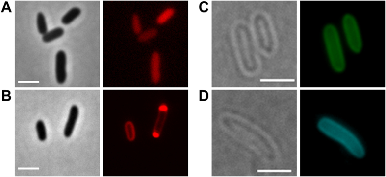
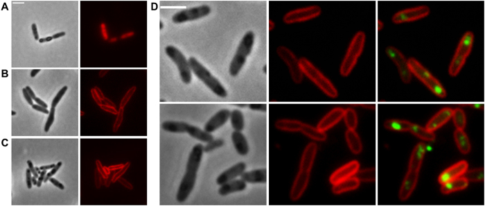
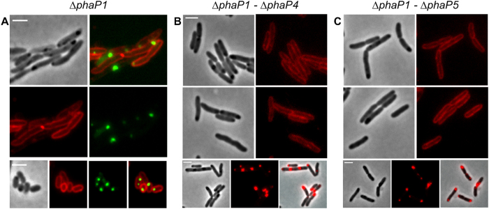
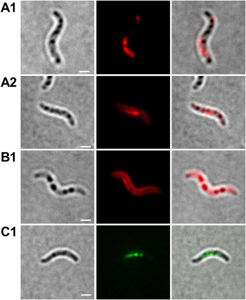
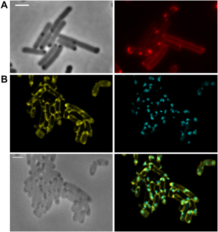
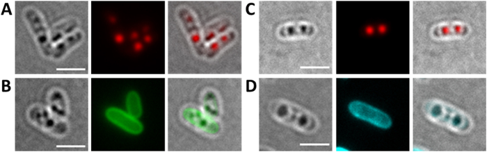
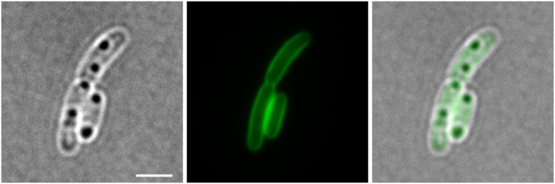
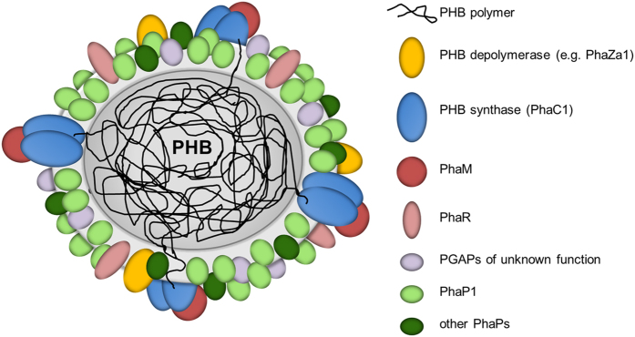
Polyhydroxybutyrate (PHB) granules, also designated as carbonosomes, are supra-molecular complexes in prokaryotes consisting of a PHB polymer core and a surface layer of structural and functional proteins. The presence of suspected phospholipids in the surface layer is based on in vitro data of isolated PHB granules and is often shown in cartoons of the PHB granule structure in reviews on PHB metabolism. However, the in vivo presence of a phospholipid layer has never been demonstrated. We addressed this topic by the expression of fusion proteins of DsRed2EC and other fluorescent proteins with the phospholipid-binding domain (LactC2) of lactadherin in three model organisms. The fusion proteins specifically localized at the cell membrane of Ralstonia eutropha but did not co-localize with PHB granules. The same result was obtained for Pseudomonas putida, a species that accumulates another type of polyhydroxyalkanoate (PHA) granules related to PHB. Notably, DsRed2EC-LactC2 expressed in Magnetospirillum gryphiswaldense was detected at the position of membrane-enclosed magnetosome chains and at the cytoplasmic membrane but not at PHB granules. In conclusion, the carbonosomes of representatives of α-proteobacteria, β-proteobacteria and γ-proteobacteria have no phospholipids in vivo and we postulate that the PHB/PHA granule surface layers in natural producers generally are free of phospholipids and consist of proteins only.


Figures

Figure 1
Figure 1. Expression of fluorescent proteins in E. coli.
(A) expression of DsRed2EC alone (phase contrast/red channel), fluorescence visible in the cytoplasm, (B) Expression of DsRed2EC-LactC2 fusion (phase contrast/red channel), note, uniform fluorescence of the cell membrane and additional fluorescent foci at the cell poles in some cells, (C) Expression of sfGFP-LactC2 fusion (bright field/green channel), (D) Expression of mTurquoise2-LactC2 (bright filed/blue channel). Scale bars correspond to 2 μm. Since E. coli is not able to synthesize PHB no granules are visible.
Figure 2
Figure 2. Expression of DsRed2EC and DsRed2EC-LactC2 in R. eutropha H16.
Expression of DsRed2EC alone in wild type (A). Expression of DsRed2EC-LactC2 in ∆phaC mutant (B) and in wild type (C). Phase contrast (left) and red channel (right) in (A–C). In (D), DsRed2EC-LactC2 was co-expressed with eYFP-PhaC (C319A) in R. eutropha wild type (from left to right: phase contrast, red channel, merge of red and green channels). Scale bars correspond to 2 μm.
Figure 3
Figure 3. Expression of DsRed2EC-LactC2 in R. eutropha phasin mutants.
Co-expression of DsRed2EC-LactC2 and eYFP-PhaC in ∆phaP1 mutant (A). Expression of DsRed2EC-LactC2 in ∆phaP1-phaP4 mutant (B) and in ∆phaP1-phaP5 mutant (C). Phase contrast and fluorescent images are shown. In the bottom rows of (B,C) cells were additionally stained with Nile red to indicate the position of PHB granules more clearly than in phase contrast images (phase contrast, red channel, merge). Scale bars correspond to 2 μm.
Figure 4
Figure 4. Expression fusion proteins in M. gryphiswaldense.
Cells expressing DsRed2EC-LactC2 focussed to filament-like fluorescence representing magnetosome-filaments (A1 and A2). Cell expressing DsRed2EC-LactC2 focussed to cell membrane fluorescence (B1). Cell expressing MamC-GFP (C1). Note, presence of several globular inclusions in all images (PHB granules) that do not co-localize with DsRed2EC-LactC2 or with MamC-eGFP. Individual magnetosomes are too small (≈35 nm) to be visible in bright field. From left to right: bright field, fluorescence channel, merge. Scale bars correspond to 2 μm.
Figure 5
Figure 5. Expression of DsRed2EC-LactC2 and Venus-LactC2 in recombinant PHB accumulating E. coli.
E. coli HMS174 cells co-expressing the phaCAB genes of R. eutropha (pJM9238) and DsRed2EC-LactC2 are shown in (A) phase contrast left, red channel right. Microscopic images of E. coli BL21(DE3) co-expressing the phaC-Cerulean-phaAB operon and Venus-LactC2 from the pETDuet-vector in (B) Venus channel middle left, Cerulean channel middle right, phase contrast bottom left, merge in bottom right. Scale bars correspond to 2 μm.
Figure 6
Figure 6. Expression of LactC2 fusion proteins in P. putida.
Cells of P. putida expressing sfGFP-LactC2 (A,B) or mTurquoise-LactC2 (C,D) were grown in mineral salts medium with sodium octanoate to promote PHA granule formation. Cells were stained with Nile red in (A,C) to visualize PHA granules. From left to right: bright field, red (top row) or green/turquoise (bottom row) channel and overlay images. Note, formation of globular inclusions visible in bright field that are stained by Nile-red (PHA granules) but that show no sfGFP-LactC2 or mTurquoise-LactC2 fluorescence. Scale bars correspond to 2 μm.
Figure 7
Figure 7. Expression of eYFP-Psd (phosphatidyl-serine decarboxylase) in R. eutropha H16.
From left to right: bright field, green channel, merge). eYFP-Psd co-localizes with the cell membrane but not with globular structures (PHB granules) that are visible in bright field. Scale bar corresponds to 2 μm.
Figure 8
Figure 8. Model of an in vivo PHB granule in R. eutropha H16.
The surface layer is free of phospholipids and consists of proteins only. The presently known PHB granule associated proteins (PGAPs) are symbolised by differentially coloured globules. All proteins in this model had been previously shown to be bound to PHB granules in vivo by expression of appropriate fusions with fluorescent proteins. For details and overview see references. The dimension of the surface layer is enlarged relative to the polymer core for better visibility.

References

    1. Anderson A. J. & Dawes E. A. Occurrence, metabolism, metabolic role, and industrial uses of bacterial polyhydroxyalkanoates. Microbiol. Rev. 54, 450–472 (1990). - PMC - PubMed
    1. Madison L. L. & Huisman G. W. Metabolic engineering of poly(3-hydroxyalkanoates): from DNA to plastic. Microbiol. Mol. Biol. Rev. 63, 21–53 (1999). - PMC - PubMed
    1. Stubbe J. et al. Nontemplate-dependent polymerization processes: polyhydroxyalkanoate synthases as a paradigm. Annu. Rev. Biochem. 74, 433–480 (2005). - PubMed
    1. Pötter M. & Steinbüchel A. Biogenesis and Structure of polyhydroxyalkanoate granules. Microbiol. Monogr. 1, 110–136 (2006).
    1. Grage K. et al. Bacterial polyhydroxyalkanoate granules: biogenesis, structure, and potential use as nano-/micro-beads in biotechnological and biomedical applications. Biomacromolecules 10, 660–669 (2009). - PubMed

Publication types

MeSH terms

LinkOut - more resources