Shox2 influences mesenchymal stem cell fate in a co-culture model in vitro
- PMID: 27222368
- PMCID: PMC4918598
- DOI: 10.3892/mmr.2016.5306
Shox2 influences mesenchymal stem cell fate in a co-culture model in vitro
Abstract
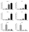
Sinoatrial node (SAN) dysfunction is a common cardiovascular problem, and the development of a cell sourced biological pacemaker has been the focus of cardiac electrophysiology research. The aim of biological pacemaker therapy is to produce SAN-like cells, which exhibit spontaneous activity characteristic of the SAN. Short stature homeobox 2 (Shox2) is an early cardiac transcription factor and is crucial in the formation and differentiation of the sinoatrial node (SAN). The present study aimed to improve pacemaker function by overexpression of Shox2 in canine mesenchymal stem cells (cMSCs) to induce a phenotype similar to native pacemaker cells. To achieve this objective, the cMSCs were transfected with lentiviral pLentis‑mShox2‑red fluorescent protein, and then co‑cultured with rat neonatal cardiomyocytes (RNCMs) in vitro for 5-7 days. The feasibility of regulating the differentiation of cMSCs into pacemaker‑like cells by Shox2 overexpression was investigated. Reverse transcription-quantitative polymerase chain reaction and western blotting showed that Shox2‑transfected cMSCs expressed high levels of T box 3, hyperpolarization-activated cyclic nucleotide‑gated cation channel and Connexin 45 genes, which participate in SAN development, and low levels of working myocardium genes, Nkx2.5 and Connexin 43. In addition, Shox2‑transfected cMSCs were able to pace RNCMs with a rate faster than the control cells. In conclusion, these data indicate that overexpression of Shox2 in cMSCs can greatly enhance the pacemaker phenotype in a co-culture model in vitro.
Figures


Similar articles
-
Transcription factor Tbx18 induces the differentiation of c-kit+ canine mesenchymal stem cells (cMSCs) into SAN-like pacemaker cells in a co-culture model in vitro.Am J Transl Res. 2018 Aug 15;10(8):2511-2528. eCollection 2018. Am J Transl Res. 2018. PMID: 30210689 Free PMC article.
-
A common Shox2-Nkx2-5 antagonistic mechanism primes the pacemaker cell fate in the pulmonary vein myocardium and sinoatrial node.Development. 2015 Jul 15;142(14):2521-32. doi: 10.1242/dev.120220. Epub 2015 Jul 2. Development. 2015. PMID: 26138475 Free PMC article.
-
Exogenous Nkx2.5- or GATA-4-transfected rabbit bone marrow mesenchymal stem cells and myocardial cell co-culture on the treatment of myocardial infarction in rabbits.Mol Med Rep. 2015 Aug;12(2):2607-21. doi: 10.3892/mmr.2015.3775. Epub 2015 May 12. Mol Med Rep. 2015. PMID: 25975979 Free PMC article.
-
Shox2: The Role in Differentiation and Development of Cardiac Conduction System.Tohoku J Exp Med. 2018 Mar;244(3):177-186. doi: 10.1620/tjem.244.177. Tohoku J Exp Med. 2018. PMID: 29503396 Review.
-
On the Evolution of the Cardiac Pacemaker.J Cardiovasc Dev Dis. 2017 Apr 27;4(2):4. doi: 10.3390/jcdd4020004. J Cardiovasc Dev Dis. 2017. PMID: 29367536 Free PMC article. Review.
Cited by
-
Transcription factor Tbx18 induces the differentiation of c-kit+ canine mesenchymal stem cells (cMSCs) into SAN-like pacemaker cells in a co-culture model in vitro.Am J Transl Res. 2018 Aug 15;10(8):2511-2528. eCollection 2018. Am J Transl Res. 2018. PMID: 30210689 Free PMC article.
-
Conversion of Unmodified Stem Cells to Pacemaker Cells by Overexpression of Key Developmental Genes.Cells. 2023 May 13;12(10):1381. doi: 10.3390/cells12101381. Cells. 2023. PMID: 37408215 Free PMC article.
-
SHOX2 is a Potent Independent Biomarker to Predict Survival of WHO Grade II-III Diffuse Gliomas.EBioMedicine. 2016 Nov;13:80-89. doi: 10.1016/j.ebiom.2016.10.040. Epub 2016 Oct 28. EBioMedicine. 2016. PMID: 27840009 Free PMC article.
-
Nkx2.5: a crucial regulator of cardiac development, regeneration and diseases.Front Cardiovasc Med. 2023 Dec 6;10:1270951. doi: 10.3389/fcvm.2023.1270951. eCollection 2023. Front Cardiovasc Med. 2023. PMID: 38124890 Free PMC article. Review.
References
MeSH terms
Substances
LinkOut - more resources
Full Text Sources
Other Literature Sources
Research Materials

