. 2016 May 24;7(3):e00538-16.
doi: 10.1128/mBio.00538-16.

Staphylococcus aureus Protein A Mediates Interspecies Interactions at the Cell Surface of Pseudomonas aeruginosa

Affiliations

Staphylococcus aureus Protein A Mediates Interspecies Interactions at the Cell Surface of Pseudomonas aeruginosa

Catherine R Armbruster et al. mBio. .

Abstract

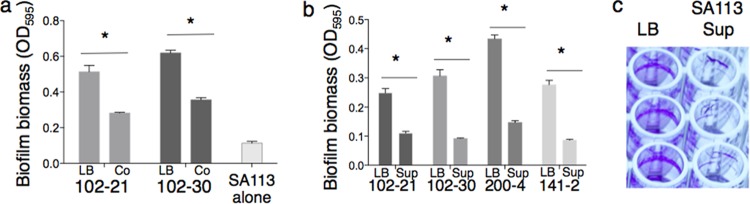
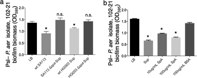
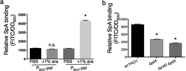
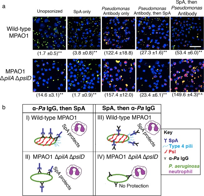
While considerable research has focused on the properties of individual bacteria, relatively little is known about how microbial interspecies interactions alter bacterial behaviors and pathogenesis. Staphylococcus aureus frequently coinfects with other pathogens in a range of different infectious diseases. For example, coinfection by S. aureus with Pseudomonas aeruginosa occurs commonly in people with cystic fibrosis and is associated with higher lung disease morbidity and mortality. S. aureus secretes numerous exoproducts that are known to interact with host tissues, influencing inflammatory responses. The abundantly secreted S. aureus staphylococcal protein A (SpA) binds a range of human glycoproteins, immunoglobulins, and other molecules, with diverse effects on the host, including inhibition of phagocytosis of S. aureus cells. However, the potential effects of SpA and other S. aureus exoproducts on coinfecting bacteria have not been explored. Here, we show that S. aureus-secreted products, including SpA, significantly alter two behaviors associated with persistent infection. We found that SpA inhibited biofilm formation by specific P. aeruginosa clinical isolates, and it also inhibited phagocytosis by neutrophils of all isolates tested. Our results indicate that these effects were mediated by binding to at least two P. aeruginosa cell surface structures-type IV pili and the exopolysaccharide Psl-that confer attachment to surfaces and to other bacterial cells. Thus, we found that the role of a well-studied S. aureus exoproduct, SpA, extends well beyond interactions with the host immune system. Secreted SpA alters multiple persistence-associated behaviors of another common microbial community member, likely influencing cocolonization and coinfection with other microbes.

Importance: Bacteria rarely exist in isolation, whether on human tissues or in the environment, and they frequently coinfect with other microbes. However, relatively little is known about how microbial interspecies interactions alter bacterial behaviors and pathogenesis. We identified a novel interaction between two bacterial species that frequently infect together-Staphylococcus aureus and Pseudomonas aeruginosa We show that the S. aureus-secreted protein staphylococcal protein A (SpA), which is well-known for interacting with host targets, also binds to specific P. aeruginosa cell surface molecules and alters two persistence-associated P. aeruginosa behaviors: biofilm formation and uptake by host immune cells. Because S. aureus frequently precedes P. aeruginosa in chronic infections, these findings reveal how microbial community interactions can impact persistence and host interactions during coinfections.


Figures

FIG 1
FIG 1
(a) Clonally related P. aeruginosa isolates from patient 102 exhibited inhibition of biofilm formation when grown in coculture with S. aureus SA113, compared to monoculture in LB. All experiments shown are from crystal violet-stained biofilms after 4 h of growth. The pairs of bars in the graph represent results for P. aeruginosa cells grown in monoculture in LB (left) or results for coculture with S. aureus S113 (right). (b) P. aeruginosa respiratory isolates from multiple CF patients, including patient 102, displayed biofilm inhibition when grown in S. aureus SA113 supernatant, compared with growth in LB. Images are from experiments using crystal violet-stained biofilms after 4 h of growth. Each pair of bars in the graph represent results for growth in LB (left) and growth in SA113 supernatant (right). For panels a and b, an asterisk indicates that the biofilm biomass of patient 102 P. aeruginosa isolates grown in SA113 supernatant differed significantly from that of each strain grown in LB (Student’s t test; P < 0.05). (c) A representative image of the biofilm inhibition phenotype in the crystal violet assay. The isolate shown is P. aeruginosa 102-21, after 4 h of growth in buffered medium (LB-MOPS) or cell-free SA113 supernatant after growth in the same medium.
FIG 2
FIG 2
(a) Inhibition of biofilm formation by P. aeruginosa isolate 102-21 is lost in the absence of SpA. Culture supernatant from S. aureus lab strain HG003 exhibited biofilm inhibitory activity against isolate 102-21, but HG003 Δspa supernatant did not. An asterisk indicates a significant difference in biofilm biomass compared to the LB control (P < 0.05); n.s., not statistically significant compared to the LB control (P > 0.05). (b) Purified SpA inhibited biofilm formation by Psl P. aeruginosa in a 4-h crystal violet assay. An asterisk indicates a significant difference in biofilm biomass compared to that of the LB control (P < 0.001).
FIG 3
FIG 3
(a) SpA binds to P. aeruginosa Psl. Shown are fluorescence levels of FITC-SpA after incubation with the indicated strains, followed by washing. The asterisk indicates a significant difference in relative FITC fluorescence compared to the PBS control (P < 0.05); n.s., not statistically significant compared to the PBS control (P > 0.05). (b) FITC-SpA binding assay (also shown in Fig. S5a in the supplemental material) with the indicated strains, suggesting that SpA binds to P. aeruginosa surface type IV pili. Asterisks indicate a significant difference in relative FITC fluorescence compared to that of wild-type PAO1 (P < 0.05).
FIG 4
FIG 4
(a) SpA protects P. aeruginosa from neutrophil phagocytosis, as shown in these representative images from neutrophil phagocytosis experiments of the indicated P. aeruginosa strains, with and without the addition of SpA and/or antipseudomonal antibody. Blue, nuclei; green/yellow, extracellular P. aeruginosa; red, internalized P. aeruginosa. Asterisks indicate a significant difference in phagocytosis compared to the addition of Pseudomonas antibody only (*, P < 0.05; **, P < 0.001; n.s., no significant difference relative to Pseudomonas antibody only). Bar, 20 µm. Magnification (oil objective), ×60. The numbers (means ± standard deviations) of internalized bacteria are indicated in parentheses and are representative of 100 infected cells (neutrophils), examined from triplicate coverslips. (b) Model of two mechanisms of SpA protection of P. aeruginosa from IgG-mediated opsonophagocytosis. (I and II) Broad protection for both wild-type MPAO1 and MPAO1 ΔpilA ΔpslD due to SpA binding to the exposed Fc region of the antipseudomonal (α-Pa) IgG. (III) Specific protection of wild-type MPAO1 via binding of SpA to two receptors on the P. aeruginosa cell surface (Psl and type 4 pili) prior to opsonization with IgG. (IV) When preincubated with SpA prior to opsonization by IgG, MPAO1 ΔpilA ΔpslD is not protected from neutrophil phagocytosis, because it is unable to bind SpA on its cell surface.

References

    1. Donlan RM. 2001. Biofilms and device-associated infections. Emerg Infect Dis 7:277–281. doi:10.3201/eid0702.700277. - DOI - PMC - PubMed
    1. Bongiorni MG, Tascini C, Tagliaferri E, Di Cori A, Soldati E, Leonildi A, Zucchelli G, Ciullo I, Menichetti F. 2012. Microbiology of cardiac implantable electronic device infections. Europace 14:1334–1339. doi:10.1093/europace/eus044. - DOI - PubMed
    1. Yamane K, Hirose H, Mather PJ, Silvestry SC. 2011. Mycotic pseudoaneurysm of the ascending aorta after heart transplantation: case report. Transplant Proc 43:2055–2058. doi:10.1016/j.transproceed.2010.12.056. - DOI - PubMed
    1. Sousa C, Botelho C, Rodrigues D, Azeredo J, Oliveira R. 2012. Infective endocarditis in intravenous drug abusers: an update. Eur J Clin Microbiol Infect Dis 31:2905–2910. doi:10.1007/s10096-012-1675-x. - DOI - PubMed
    1. Hauser AR, Jain M, Bar-Meir M, McColley SA. 2011. Clinical significance of microbial infection and adaptation in cystic fibrosis. Clin Microbiol Rev 24:29–70. doi:10.1128/CMR.00036-10. - DOI - PMC - PubMed

MeSH terms

Substances