

Case Reports
doi: 10.5826/dpc.0602a02.
eCollection 2016 Apr.
Development of poorly differentiated invasive squamous cell carcinoma in giant Bowen's disease: a case report with dermatoscopy
Affiliations
- PMID: 27222765
- PMCID: PMC4866620
- DOI: 10.5826/dpc.0602a02
Item in Clipboard
Case Reports
Development of poorly differentiated invasive squamous cell carcinoma in giant Bowen's disease: a case report with dermatoscopy
Dermatol Pract Concept.
.
Abstract
Bowen's disease (BD) is an in situ form of squamous cell carcinoma (SCC), often occurring in the chronically UV-damaged skin of elderly people. The risk of progression of BD to invasive SCC varies between 3% and 5%, and one-third of invasive tumors may metastasize. Herein we discuss the dermatoscopic findings of a case of giant Bowen's disease, which progressed to poorly differentiated invasive SCC.
Keywords: Bowen’s disease squamous cell carcinoma; dermatoscopy; dermoscopy.
Figures
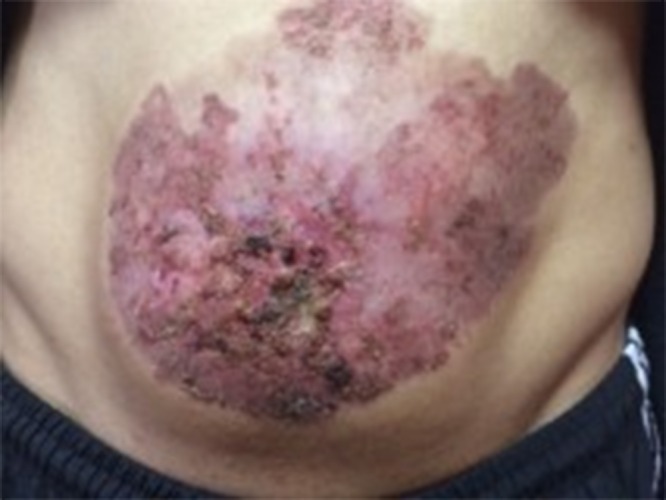
Clinical image of the patient showed a 35×25 cm erythematous hyperkeratotic, crusted, and ulcerated plaque on the abdominal region. [Copyright: ©2016 Akay et al.]
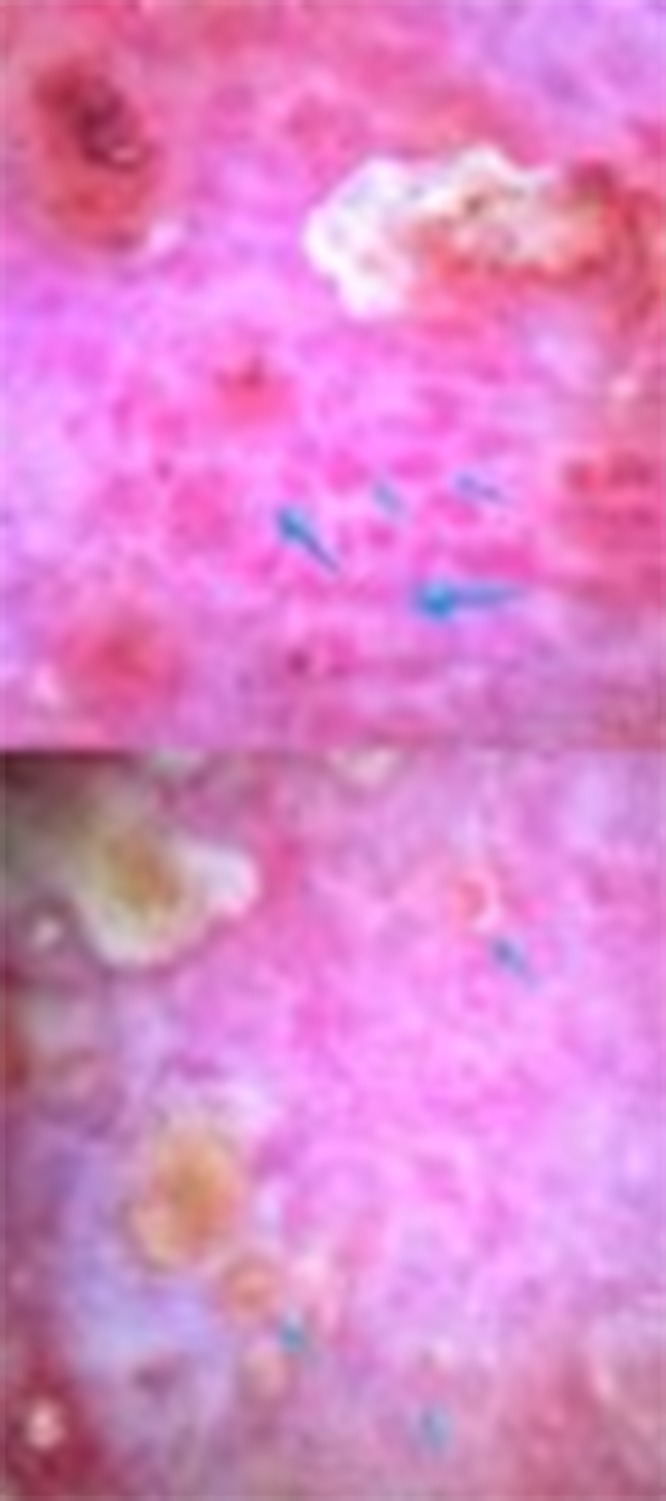
Dermatoscopy of the central region. (A) Linear arrangement of coiled vessels becoming convoluted and larger in size (blue arrows) intermingled with ulceration and adherent fibrin crusts. (B) White circles (blue arrows). [Copyright: ©2016 Akay et al.]
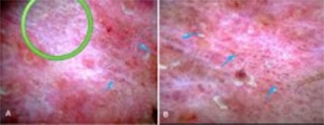
Dermatoscopy the peripheral region. (A, B) White and pink structureless areas, white lines (green circle) and a linear arrangement of gray dots and coiled vessels (blue arrows). No ulceration or white circles are seen. [Copyright: ©2016 Akay et al.]
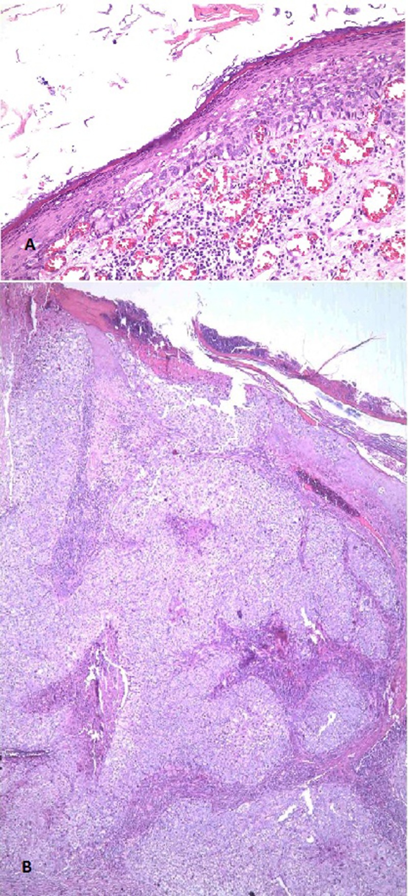
(A) Histopathology of the peripheral part which is consistent with BD: An acanthotic epidermis shows full-thickness epidermal replacement by crowded keratinocytes that demonstrate disordered dyspolarity, lack of maturation, atypical mitotic figures and nuclear pleomorphism with hyperchromasia. A band-like lymphocytic infiltrate is apparent in the papillary dermis. (B) Histopathology of the central region shows invasive part of tumor which extends as invasive broad tongues into the dermis. This part of the tumor composed of pleomorphic atypical epithelioid cells with high nucleocytoplasmic ratio and frequent mitosis. Squamous differentiation features, such as squamous pearls or single cell keratinization, are not obvious. Foci of necrosis can be noted at the right part of the figure. [Copyright: ©2016 Akay et al.]
Similar articles
-
Dermatoscopic findings and dermatopathological correlates in clinical variants of actinic keratosis, Bowen's disease, keratoacanthoma, and squamous cell carcinoma.Dermatol Ther. 2021 May;34(3):e14877. doi: 10.1111/dth.14877. Epub 2021 Mar 7. Dermatol Ther. 2021. PMID: 33583118
-
High-risk human papillomavirus in a child with digital pigmented Bowen's disease: Case report and dermoscopic findings.Pediatr Dermatol. 2018 Sep;35(5):e265-e267. doi: 10.1111/pde.13552. Epub 2018 Jun 22. Pediatr Dermatol. 2018. PMID: 29931706
-
Cyclin A and beta-catenin expression in actinic keratosis, Bowen's disease and invasive squamous cell carcinoma of the skin.Br J Dermatol. 2005 Dec;153(6):1166-75. doi: 10.1111/j.1365-2133.2005.06898.x. Br J Dermatol. 2005. PMID: 16307653
-
Bowen's Disease in Dermoscopy.Acta Dermatovenerol Croat. 2018 Jun;26(2):157-161. Acta Dermatovenerol Croat. 2018. PMID: 29989873 Review.
-
Race-Specific and Skin of Color Dermatoscopic Characteristics of Skin Cancer: A Literature Review.Dermatol Pract Concept. 2023 Oct 1;13(4 S1):e2023311S. doi: 10.5826/dpc.1304S1a311S. Dermatol Pract Concept. 2023. PMID: 37874992 Free PMC article. Review.
Cited by
-
Giant Bowen's Disease on the Face: Case Report and Review of the Literature.Open Access Maced J Med Sci. 2019 Feb 22;7(4):606-609. doi: 10.3889/oamjms.2019.190. eCollection 2019 Feb 28. Open Access Maced J Med Sci. 2019. PMID: 30894921 Free PMC article.
References
-
- Kossard S, Rosen R. Cutaneous Bowen’s disease. An analysis of 1001 cases according to age, sex, and site. J Am Acad Dermatol. 1992;27:406–10. - PubMed
-
- Reizner GT, Chuang TY, Elpern DJ, Stone JL, Farmer ER. Bowen’s disease (squamous cell carcinoma in situ) in Kauai, Hawaii. A population-based incidence report. J Am Acad Dermatol. 1994;31:596–600. - PubMed
Publication types
LinkOut - more resources
Full Text Sources
Other Literature Sources
Research Materials