

Spitz/Reed nevi: a review of clinical-dermatoscopic and histological correlation
- PMID: 27222770
- PMCID: PMC4866625
- DOI: 10.5826/dpc.0602a07
Spitz/Reed nevi: a review of clinical-dermatoscopic and histological correlation
Abstract
Background: Spitz/Reed nevi are melanocytic lesions that may mimic melanoma at clinical, dermatoscopic and histopathological levels. Management strategies of these lesions remain controversial.
Objectives: We aim a correlation among clinical-dermatoscopic and histological features of a series of Spitz/Reed nevi diagnosed during 7 years at the Department of Dermatology.
Methods: Clinical, dermatoscopic and histological features of Spitz/Reed nevi diagnosed at our tertiary hospital from 2008 to 2014 were reviewed in order to seek correlation.
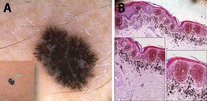
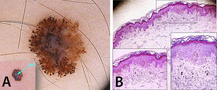
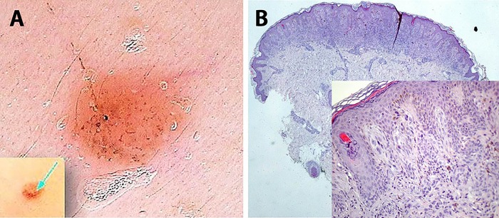
Results: All described dermatoscopic patterns for Spitz/Reed nevi were found among the 47 enrolled patients; starburst and atypical/multicomponent patterns prevailed (57.4%). Reticular pattern predominated among children younger than 12 years, whereas homogeneous pattern was more frequent in patients older than 12 years, although these differences were not statistically significant (P=0.785). Among histological atypical lesions, all dermatoscopic patterns were represented, but the atypical/multicomponent predominated (56.3%). Two out of 11 dermatoscopically atypical lesions did not show histopathological counterpart.
Conclusions: The excision of Spitz/Reed nevi in adults is supported, given the inability to accurately predict those with histopathological atypia, based on clinical and dermatoscopic features, which may raise concern about malignancy.
Keywords: Reed nevus; Spitz nevus; dermatoscopy; spindle and/or epithelioid cell nevus.
Figures
Similar articles
-
Differentiation of pigmented Spitz nevi and Reed nevi by integration of dermatopathologic and dermatoscopic findings.Dermatol Pract Concept. 2012 Jan 31;2(1):13-24. doi: 10.5826/dpc.0201a03.. eCollection 2012 Jan. Dermatol Pract Concept. 2012. PMID: 24765545 Free PMC article.
-
[Spitz nevus and Reed nevus: simulating melanoma in adults].Pathologe. 1998 Nov;19(6):403-11. doi: 10.1007/s002920050304. Pathologe. 1998. PMID: 9885003 Review. German.
-
Clinical and dermoscopic features of small Reed nevus (<6 mm).J Eur Acad Dermatol Venereol. 2013 Jul;27(7):919-21. doi: 10.1111/j.1468-3083.2012.04457.x. Epub 2012 Feb 10. J Eur Acad Dermatol Venereol. 2013. PMID: 22324638
-
Spitz nevi: beliefs, behaviors, and experiences of pediatric dermatologists.JAMA Dermatol. 2013 Mar;149(3):283-91. doi: 10.1001/jamadermatol.2013.1124. JAMA Dermatol. 2013. PMID: 23553063
-
Role of In Vivo Reflectance Confocal Microscopy in the Analysis of Melanocytic Lesions.Acta Dermatovenerol Croat. 2018 Apr;26(1):64-67. Acta Dermatovenerol Croat. 2018. PMID: 29782304 Review.
Cited by
-
Eruptive Disseminated Spitz Nevi With Architectural Features of Clark/Dysplastic Nevus.Dermatol Pract Concept. 2025 Jul 31;15(3):5323. doi: 10.5826/dpc.1503a5323. Dermatol Pract Concept. 2025. PMID: 40790444 Free PMC article. No abstract available.
-
Pigmented Spindle Cell Nevus of Reed of the Eyelid.Ocul Oncol Pathol. 2017 Sep;3(3):176-180. doi: 10.1159/000454864. Epub 2017 Jan 27. Ocul Oncol Pathol. 2017. PMID: 29134183 Free PMC article.
-
THE FAST CLINICAL EVOLUTION OF A SPITZ NEVUS: THREE-YEAR FOLLOW-UP OF A CHILD.Rev Paul Pediatr. 2017 Oct-Dec;35(4):476-479. doi: 10.1590/1984-0462/;2017;35;4;00016. Epub 2017 Sep 21. Rev Paul Pediatr. 2017. PMID: 28977136 Free PMC article.
References
-
- Reed RJ, Ichinose H, Clark WH, Jr, Mihm MC., Jr Common and uncommon melanocytic nevi and borderline melanomas. Semin Oncol. 1975;2:119–47. - PubMed
-
- Argenziano G, Soyer HP, Ferrara G, et al. Superficial black network: an additional dermoscopic clue for the diagnosis of pigmented spindle and/or epithelioid cell nevus. Dermatology. 2001;203:333–5. - PubMed
Publication types
LinkOut - more resources
Full Text Sources
Other Literature Sources