

Sclerosing hemangioma of the lung showing strong FDG avidity on PET scan: Case report and review of the current literature
- PMID: 27222778
- PMCID: PMC4821325
- DOI: 10.1016/j.rmcr.2015.12.005
Sclerosing hemangioma of the lung showing strong FDG avidity on PET scan: Case report and review of the current literature
Abstract
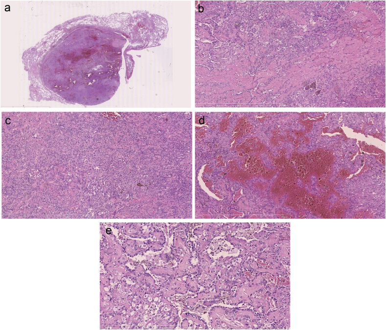
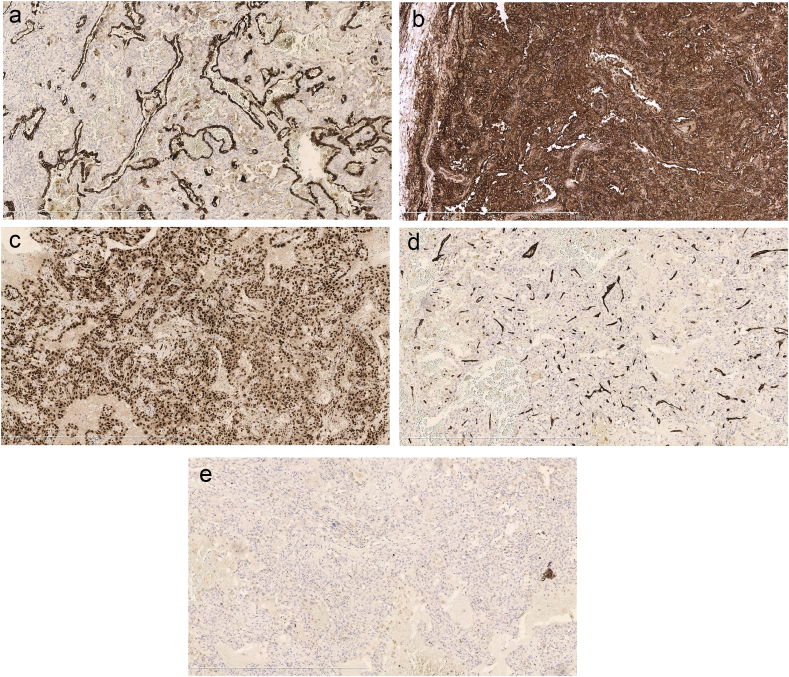
Sclerosing Hemangioma is a rare lung tumor with polymorphic histologic features that usually occurs in middle aged women. Based on many immunohistochemical and ultrastructural studies, it is most probably derived from undifferentiated respiratory epithelial cells. Symptoms are usually due to enlargement of the tumor and compression of the surrounding tissues. Occurrence of multiple lesions or metastasis is extremely rare although some authors consider sclerosing hemangioma as a potentially low grade malignancy tumor. It usually presents with low to moderate uptake on FDG PET imaging. We present a case of sclerosing hemangioma with strong FDG avidity on PET scan in a 41 year old lady with history of haemoptysis. A full review of the literature on this topic was performed.
Keywords: Lung; PET avidity; Sclerosing hemangioma.
Figures
Similar articles
-
[The fluorodeoxyglucose-positron emission tomography (FDG-PET) findings and surgical strategy for pulmonary sclerosing hemangioma].Kyobu Geka. 2010 Aug;63(9):769-73. Kyobu Geka. 2010. PMID: 20715456 Japanese.
-
More advantages in detecting bone and soft tissue metastases from prostate cancer using 18F-PSMA PET/CT.Hell J Nucl Med. 2019 Jan-Apr;22(1):6-9. doi: 10.1967/s002449910952. Epub 2019 Mar 7. Hell J Nucl Med. 2019. PMID: 30843003
-
18F-FDG PET/CT features of pulmonary sclerosing hemangioma.Acta Radiol. 2013 Feb 1;54(1):24-9. doi: 10.1258/ar.2011.110474. Epub 2012 Jan 30. Acta Radiol. 2013. PMID: 22291338
-
So-called sclerosing hemangioma of lung: current concept.Ann Diagn Pathol. 2010 Feb;14(1):60-7. doi: 10.1016/j.anndiagpath.2009.07.002. Epub 2009 Dec 11. Ann Diagn Pathol. 2010. PMID: 20123460 Review.
-
Diversity of imaging features of ovarian sclerosing stromal tumors on MRI and PET-CT: a case report and literature review.J Ovarian Res. 2018 Dec 20;11(1):101. doi: 10.1186/s13048-018-0473-1. J Ovarian Res. 2018. PMID: 30572921 Free PMC article. Review.
References
-
- Sugio K., Yohohama H., Kaneko S., Ishida T., Sugimachi K. Sclerosing hemangioma of the lung: radiographic and pathological study. Ann. Thorac. Surg. 1992;53:295–300. - PubMed
-
- Miyagawa-Hayashino A., Tazelaar H.D., Langel D., Colby T. Pulmonary sclerosing hemangioma with lymph node metastases. Arch. Pathol. Lab. Med. 2003;127:321–325. - PubMed
-
- Hanaoka J., Ohuchi M., Inoue S., Sawai S., Tezuka N., Fujino S. Bilateral multiple sclerosing hemangioma. Jpn. J. Thorac. Cardiovasc Surg. 2005;53:157–161. - PubMed
-
- Liebow A.A., Hubbell D.S. Sclerosing hemangioma (histiocytoma, xanthoma) of the lung. Cancer. 1956;9:53–75. - PubMed
Publication types
LinkOut - more resources
Full Text Sources
Other Literature Sources
