

Predominant diffuse ground glass opacity in both lung fields: A case of sarcoidosis with atypical CT findings
- PMID: 27222788
- PMCID: PMC4821339
- DOI: 10.1016/j.rmcr.2016.01.007
Predominant diffuse ground glass opacity in both lung fields: A case of sarcoidosis with atypical CT findings
Abstract
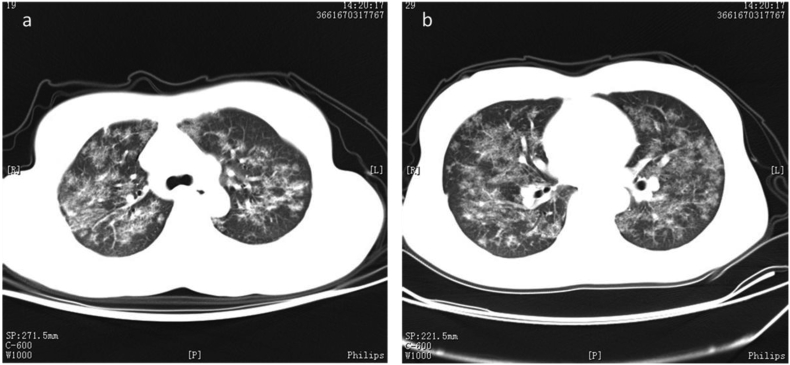
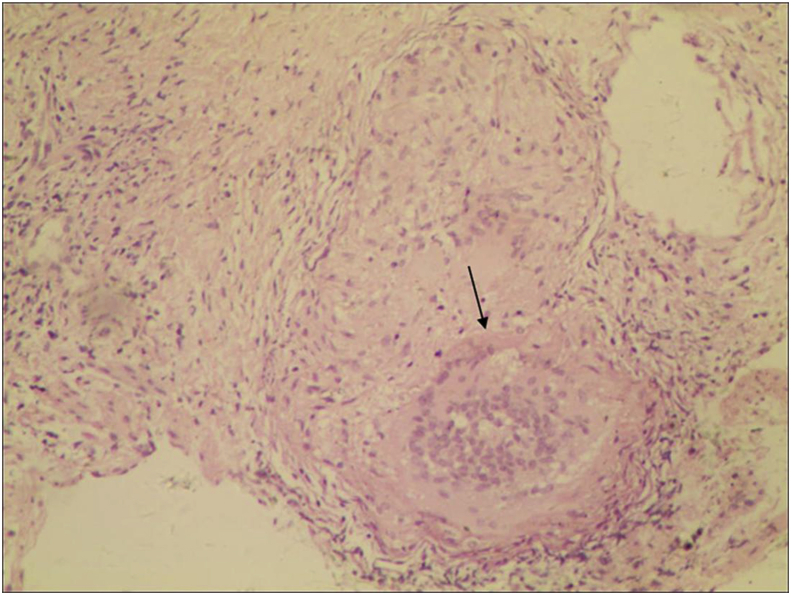
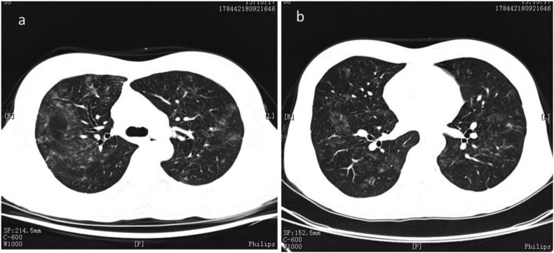
Sarcoidosis can cause fatal diffuse lung fibrosis in the end stage, so its early diagnosis and treatment can prevent the progression of fibrosis. Predominant ground glass opacity on high-resolution CT (HRCT) scans is a rare presentation of sarcoidosis. We report the case of a patient who presented with very few symptoms and signs of sarcoidosis; HRCT revealed large-scale ground glass opacity and minor lymphadenopathy. Bronchoalveolar lavage fluid contained turbid liquid. Sarcoidosis could be confirmed only based on pathological examination of the resected tissue. The patient was administrated prednisone at 40 mg/d orally with tapering of the dose. Lung HRCT scans taken 6 months after the prednisone treatment showed ablation of the ground glass opacity. This case report sheds light on an atypical HRCT presentation of sarcoidosis; the findings here will be useful for the early diagnosis of sarcoidosis and prevention of fatal complications.
Keywords: Ground glass opacity; HRCT; Sarcoidosis.
Figures
Similar articles
-
[A case of pulmonary sarcoidosis demonstrating panlobular ground-glass opacity with mosaic distribution].Nihon Kokyuki Gakkai Zasshi. 2009 Mar;47(3):212-7. Nihon Kokyuki Gakkai Zasshi. 2009. PMID: 19348268 Japanese.
-
[Diffuse ground-glass opacity of the lung. A guide to interpreting the high-resolution computed tomographic (HRCT) picture].Radiol Med. 1994 Nov;88(5):576-81. Radiol Med. 1994. PMID: 7824771 Review. Italian.
-
[Granulomatous diseases and pathogenic microorganism].Kekkaku. 2008 Feb;83(2):115-30. Kekkaku. 2008. PMID: 18326339 Japanese.
-
Thoracic Sarcoidosis: Imaging with High Resolution Computed Tomography.J Clin Diagn Res. 2017 Feb;11(2):TC15-TC18. doi: 10.7860/JCDR/2017/24165.9459. Epub 2017 Feb 1. J Clin Diagn Res. 2017. PMID: 28384959 Free PMC article.
-
Recent advances in radiology of the interstitial lung disease.Curr Opin Pulm Med. 1998 Sep;4(5):281-7. doi: 10.1097/00063198-199809000-00007. Curr Opin Pulm Med. 1998. PMID: 10813203 Review.
Cited by
-
Calcium Chaos in Sarcoidosis: A Tale of Severe Hypercalcemia's Diagnostic Challenge.Cureus. 2024 Mar 16;16(3):e56271. doi: 10.7759/cureus.56271. eCollection 2024 Mar. Cureus. 2024. PMID: 38623131 Free PMC article.
-
Mass-Like Ground-Glass Opacities in Sarcoidosis: A Rare Presentation Not Previously Described.Case Rep Radiol. 2018 Aug 14;2018:5686915. doi: 10.1155/2018/5686915. eCollection 2018. Case Rep Radiol. 2018. PMID: 30186655 Free PMC article.
-
Comparison of typical and atypical computed tomography patterns regarding reversibility and fibrosis in pulmonary sarcoidosis.Ann Thorac Med. 2021 Jan-Mar;16(1):118-125. doi: 10.4103/atm.ATM_187_20. Epub 2021 Jan 14. Ann Thorac Med. 2021. PMID: 33680132 Free PMC article.
References
-
- Aikins A., Kanne J.P., Chung J.H. Galaxy sign. J. Thorac. Imaging. 2012;27(6):W164. - PubMed
-
- Polverosi R., Russo R., Coran A. Typical and atypical pattern of pulmonary sarcoidosis at high-resolution CT: relation to clinical evolution and therapeutic procedures. Radiol. Med. 2014;119(6):384–392. - PubMed
-
- Kumazoe H., Matsunaga K., Nagata N. “Reversed halo sign” of high-resolution computed tomography in pulmonary sarcoidosis. J. Thorac. Imaging. 2009;24(1):66–68. - PubMed
-
- Marchiori E., Zanetti G., Barreto M.M. Atypical distribution of small nodules on high resolution CT studies: patterns and differentials. Respir. Med. 2011;105(9):1263–1267. - PubMed
Publication types
LinkOut - more resources
Full Text Sources
Other Literature Sources
