

The volumetric and shape changes of the putamen and thalamus in first episode, untreated major depressive disorder
- PMID: 27222797
- PMCID: PMC4873692
- DOI: 10.1016/j.nicl.2016.04.008
The volumetric and shape changes of the putamen and thalamus in first episode, untreated major depressive disorder
Abstract
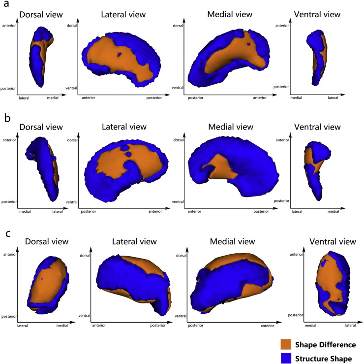
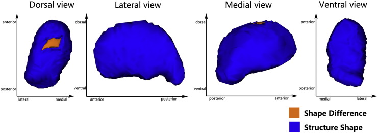
Previous MRI studies confirmed abnormalities in the limbic-cortical-striatal-pallidal-thalamic (LCSPT) network or limbic-cortico-striatal-thalamic-cortical (LCSTC) circuits in patients with major depressive disorder (MDD), but few studies have investigated the subcortical structural abnormalities. Therefore, we sought to determine whether focal subcortical grey matter (GM) changes might be present in MDD at an early stage. We recruited 30 first episode, untreated patients with major depressive disorder (MDD) and 26 healthy control subjects. Voxel-based morphometry was used to evaluate cortical grey matter changes, and automated volumetric and shape analyses were used to assess volume and shape changes of the subcortical GM structures, respectively. In addition, probabilistic tractography methods were used to demonstrate the relationship between the subcortical and the cortical GM. Compared to healthy controls, MDD patients had significant volume reductions in the bilateral putamen and left thalamus (FWE-corrected, p < 0.05). Meanwhile, the vertex-based shape analysis showed regionally contracted areas on the dorsolateral and ventromedial aspects of the bilateral putamen, and on the dorsal and ventral aspects of left thalamus in MDD patients (FWE-corrected, p < 0.05). Additionally, a negative correlation was found between local atrophy in the dorsal aspects of the left thalamus and clinical variables representing severity. Furthermore, probabilistic tractography demonstrated that the area of shape deformation of the bilateral putamen and left thalamus have connections with the frontal and temporal lobes, which were found to be related to major depression. Our results suggested that structural abnormalities in the putamen and thalamus might be present in the early stages of MDD, which support the role of subcortical structure in the pathophysiology of MDD. Meanwhile, the present study showed that these subcortical structural abnormalities might be the potential trait markers of MDD.
Keywords: Major depressive disorder; Putamen; Shape analysis; Thalamus; Volumetric analysis.
Figures


Similar articles
-
Gray Matter Abnormalities in Non-comorbid Medication-naive Patients with Major Depressive Disorder or Social Anxiety Disorder.EBioMedicine. 2017 Jul;21:228-235. doi: 10.1016/j.ebiom.2017.06.013. Epub 2017 Jun 15. EBioMedicine. 2017. PMID: 28633986 Free PMC article.
-
Gray matter structural alterations in first-episode drug-naïve adolescents with major depressive disorder: a comprehensive morphological analysis study.Psychol Med. 2025 Apr 11;55:e113. doi: 10.1017/S0033291725000790. Psychol Med. 2025. PMID: 40211094 Free PMC article.
-
Longitudinal brain volume changes in major depressive disorder.J Neural Transm (Vienna). 2018 Oct;125(10):1433-1447. doi: 10.1007/s00702-018-1919-8. Epub 2018 Aug 27. J Neural Transm (Vienna). 2018. PMID: 30167933
-
Grey matter volume abnormalities in the first depressive episode of medication-naïve adult individuals: a systematic review of voxel based morphometric studies.Int J Psychiatry Clin Pract. 2021 Nov;25(4):407-420. doi: 10.1080/13651501.2020.1861632. Epub 2020 Dec 22. Int J Psychiatry Clin Pract. 2021. PMID: 33351672
-
Meta-analysis of volumetric abnormalities in cortico-striatal-pallidal-thalamic circuits in major depressive disorder.Psychol Med. 2012 Apr;42(4):671-81. doi: 10.1017/S0033291711001668. Epub 2011 Sep 13. Psychol Med. 2012. PMID: 21910935 Review.
Cited by
-
Putamen gray matter volumes in neuropsychiatric and neurodegenerative disorders.World J Psychiatry Ment Health Res. 2019;3(1):1020. Epub 2019 May 30. World J Psychiatry Ment Health Res. 2019. PMID: 31328186 Free PMC article.
-
Body-mind relaxation meditation modulates the thalamocortical functional connectivity in major depressive disorder: a preliminary resting-state fMRI study.Transl Psychiatry. 2021 Oct 23;11(1):546. doi: 10.1038/s41398-021-01637-8. Transl Psychiatry. 2021. PMID: 34689151 Free PMC article.
-
Adaptive Whole-Brain Dynamics Predictive Method: Relevancy to Mental Disorders.Research (Wash D C). 2025 Apr 5;8:0648. doi: 10.34133/research.0648. eCollection 2025. Research (Wash D C). 2025. PMID: 40190349 Free PMC article.
-
Cortisol effects on brain functional connectivity during emotion processing in women with depression.J Affect Disord. 2021 May 15;287:247-254. doi: 10.1016/j.jad.2021.03.034. Epub 2021 Mar 19. J Affect Disord. 2021. PMID: 33799044 Free PMC article. Clinical Trial.
-
Identification and discovery of imaging genetic patterns using fusion self-expressive network in major depressive disorder.Front Neurosci. 2023 Nov 21;17:1297155. doi: 10.3389/fnins.2023.1297155. eCollection 2023. Front Neurosci. 2023. PMID: 38075264 Free PMC article.
References
-
- Alexander G.E., DeLong M.R., Strick P.L. Parallel organization of functionally segregated circuits linking basal ganglia and cortex. Annu. Rev. Neurosci. 1986;9:357–381. - PubMed
-
- Angst J. Major Depression in 1998: Are We Providing Optimal Therapy? J. Clin. Psychiatry. 1999 - PubMed
-
- Association, A.P. Practice guideline for the treatment of patients with major depressive disorder (revision) Am. J. Psychiatry. 2000;157:1–45. - PubMed
-
- Baumann B., Danos P., Krell D., Diekmann S., Leschinger A., Stauch R., Wurthmann C., Bernstein H.G., Bogerts B. Reduced volume of limbic system-affiliated basal ganglia in mood disorders: preliminary data from a postmortem study. J. Neuropsychiatry Clin. Neurosci. 1999;11:71–78. - PubMed
Publication types
MeSH terms
LinkOut - more resources
Full Text Sources
Other Literature Sources
