. 2017 Apr 25;8(17):27725-27739.
doi: 10.18632/oncotarget.9486.

Platelet factor 4 is produced by subsets of myeloid cells in premetastatic lung and inhibits tumor metastasis

Affiliations

Platelet factor 4 is produced by subsets of myeloid cells in premetastatic lung and inhibits tumor metastasis

Jiang Jian et al. Oncotarget. .

Abstract

Bone marrow-derived myeloid cells can form a premetastatic niche and provide a tumor-promoting microenvironment. However, subsets of myeloid cells have also been reported to have anti-tumor properties. It is not clear whether there is a transition between anti- and pro- tumor function of these myeloid cells, and if so, what are the underlying molecular mechanisms. Here we report platelet factor 4 (PF4), or CXCL4, but not the other family members CXCL9, 10, and 11, was produced at higher levels in the normal lung and early stage premetastatic lungs but decreased in later stage lungs. PF4 was mostly produced by Ly6G+CD11b+ myeloid cell subset. Although the number of Ly6G+CD11b+ cells was increased in the premetastatic lungs, the expression level of PF4 in these cells was decreased during the metastatic progression. Deletion of PF4 (PF4 knockout or KO mice) led an increased metastasis suggesting an inhibitory function of PF4. There were two underlying mechanisms: decreased blood vessel integrity in the premetastatic lungs and increased production of hematopoietic stem/progenitor cells (HSCs) and myeloid derived suppressor cells (MDSCs) in tumor-bearing PF4 KO mice. In cancer patients, PF4 expression levels were negatively correlated with tumor stage and positively correlated with patient survival. Our studies suggest that PF4 is a critical anti-tumor factor in the premetastatic site. Our finding of PF4 function in the tumor host provides new insight to the mechanistic understanding of tumor metastasis.

Keywords: PF4; metastasis; myeloid cells; premetastatic lung.


Conflict of interest statement

CONFLICTS OF INTEREST

The authors declare no competing financial interest.

The research work was supported by NCI intramural funding to Drs. Li Yang, and the Collaborative Research of National Institutes of Health-Natural Science Foundation of China (Nos.81361120399) to Xinhua Liang.

Figures
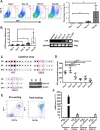
Figure 1
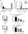
Figure 1. PF4 was induced in premetastatic lung of mice bearing tumors
(A) Flow cytometry analysis of 4T1-GFP+ tumor cells from single cell suspension of lungs from nude mice received tumor cell injection into the mammary fat pad (n = 5–8 mice per time point). Quantitative data are on the right. (B) GFP-PCR of RNA extraction from circulating nucleated cells in blood of mice received 4T1-GFP injections at different days. The gel electrophoresis is on the right. (C) Upper and lower left panels: cytokine array detecting the expression of PF4, CXCL9, and CXCL11 in the premetastatic lung of 4T1 tumor bearing mice at different days after tumor injection (D5, D10) with D 0 as control. Lower right panel: Western blot detecting CXCL10 expression. Each sample was a pool from 3–5 mice. (D) PF4 ELISA of lung protein extraction from mice received 4T1 injection at different days indicated (n = 3 mice per group). (E) Pre and post-sorting of Gr-1+CD11b+ cells by FACS from lungs of 4T1 tumor-bearing mice. (F) Q-PCR of PF4, CXCL9, CXCL10, and CXCL11 in sorted Gr-1+CD11b+ myeloid cells from day 10 lungs as well as B16F10 and 4T1 tumor cells. Shown is one of the 3 experiments performed. Data are presented as Mean +/− SEM. ***P < 0.001.
Figure 2
Figure 2. PF4 production was decreased in myeloid cells sorted from the premetastatic lungs during metastatic progression
(A) PF4 ELISA of immune cell subtypes from lungs of normal mice. The cells were sorted from single cell suspension of lungs. (B) Percentage of myeloid cell subsets and megakaryocytes from lungs of normal or 4T1 tumor-bearing mice at different days after tumor cell injection, by flow cytometry analysis of single cell suspension of lungs. (C) PF4 production level in Ly6G+CD11b+ cells from normal lungs normalized to the fold changes of cell numbers, Q-PCR in upper panel, and PF4 ELISA in lower panel. (D) PF4 Q-PCR (upper panel) or ELISA (lower panel) of Ly6G+CD11b+ cells from lungs of normal or 4T1 tumor bearing mice at different days after injection. Each sample was a pool from 3–5 mice. Data are presented as Mean +/− SEM. *P < 0.05, **P < 0.01, ***P < 0.001.
Figure 3
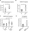
Figure 3. PF4 inhibits tumor growth and metastasis of B16-F10 melanoma
(A) Tumor volume of B16-F10 in wild type and PF4 KO mice (n = 5–6 mice per group). (B) Number of lung metastasis of B16-F10 in wild type and PF4 KO mice. The tumor cells were injected through tail vein (n = 7–11 mice per group). (C) Fold changes in metastasis for mice received both intradermal (to precondition the lungs) and tail vein injection of B16-F10 tumor cells, labeled as ID + TVI; the metastasis number from mice received TVI alone were divided by the average cross each genotype group, labeled as TVI. N = 14–16 mice per group. (D) Lung metastasis of B16-F10 when co-injected with wild type or PF4 deficient Gr-1+CD11b+ cells (n = 8 mice per group). Data are presented as Mean +/− SEM. *P < 0.05, **P < 0.01.
Figure 4
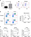
Figure 4. Increased hematopoietic progenitor cells and Gr-1+CD11b+ cells in PF4 KO mice and WT control animals
(A) Percentage of Sca1+CD117-Lin- HSCs in 7AAD- cells from bone marrow of normal or tumor-bearing PF4 KO mice (n = 8 mice per group). Left panel: quantitative data, right panel: representative plots. (B) Gr-1+CD11b+ cells in BM (n = 3–8 mice per group). Left panel: representative plots, right panel: quantitative data. (C) Gr-1+CD11b+ cells in spleen, peripheral blood, and lung of normal or tumor-bearing PF4 KO mice (n = 3–4 mice per group). Data are presented as Mean +/− SEM. *P < 0.05, **P < 0.01, ***P < 0.001.
Figure 5
Figure 5. Blood vessel leakage and CD31+ cells are increased in the premetastatic lungs of PF4 KO mice
(A) Blood vessel leakage in WT and PF4 KO mice with or without B16-F10 tumor bearing (n = 3 mice per group) showing by infusing Evans Blue. (B) Quantitative data from A. (C) Immunofluorescence staining of CD31+ in lung sections of WT and PF4 KO mice with or without B16-F10 tumor injection (n = 3 mice per group). The red pixel counts were obtained from 5 representative pictures for each group using Image J. The signals were divided by the average from normal WT, and plotted as relative density. (D) Percentage of CD31+ cells in the CD45-Lin- cells of single cell suspension from the lungs (n = 3 mice per group). Left panel: quantitative data, right panel: representative plots. Data are presented as Mean +/− SEM. *P < 0.05, **P < 0.01, ***P < 0.001.
Figure 6
Figure 6. PF4 expression levels negatively correlate with human cancer progression
(A) PF4 expression levels with breast cancer patient survival (left panel), and different stages of tumor progression (right panel). The TCGA breast cancer dataset is from “The Cancer Genome Atlas” (TCGA) (www.cancergenome.nih.gov), Oncomine. (B) PF4 expression levels in different stages of human colorectal cancers (left panel), and in metastasis compared with primary tumors (right panel). Bittner colorectal cohort from Oncomine (https://www.oncomine.org) was analyzed. (C) PF4 expression levels in different stages of human lung cancers, Bittner dataset, Oncomine. All data sets were analyzed by Genespring GX 10.0 software (Agilent Technologies). Data are presented as Mean +/− SEM. *P < 0.05, **P < 0.01, ***P < 0.001.

References

    1. Coussens LM, Werb Z. Inflammation and cancer. Nature. 2002;420:860–867. - PMC - PubMed
    1. Joyce JA, Pollard JW. Microenvironmental regulation of metastasis. Nat Rev Cancer. 2009;9:239–252. - PMC - PubMed
    1. Gabrilovich DI, Nagaraj S. Myeloid-derived suppressor cells as regulators of the immune system. Nat Rev Immunol. 2009;9:162–174. - PMC - PubMed
    1. Pollard JW. Trophic macrophages in development and disease. Nat Rev Immunol. 2009;9:259–270. - PMC - PubMed
    1. Mantovani A. Cancer: Inflaming metastasis. Nature. 2009;457:36–37. - PubMed

MeSH terms