Review
. 2016 May;7(1):10.1128/ecosalplus.ESP-0002-2016.
doi: 10.1128/ecosalplus.ESP-0002-2016.

Aminoacyl-tRNA Synthetases in the Bacterial World

Affiliations
Review

Aminoacyl-tRNA Synthetases in the Bacterial World

Richard Giegé et al. EcoSal Plus. 2016 May.

Abstract

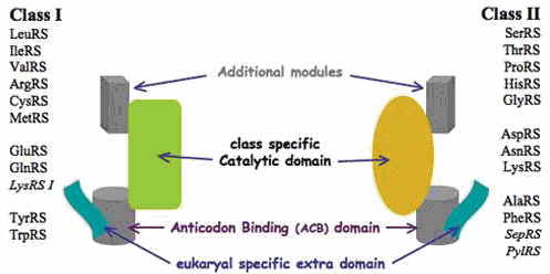
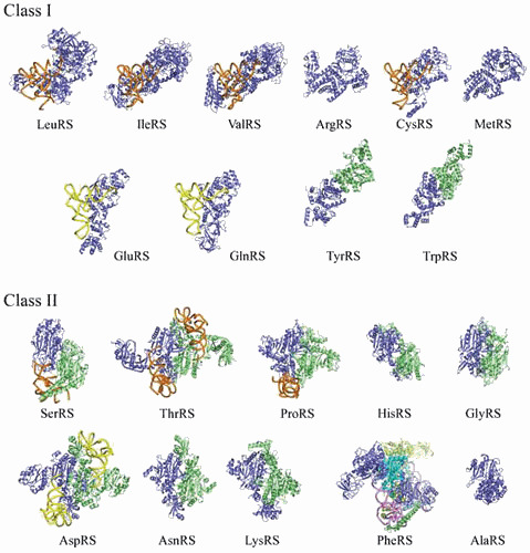
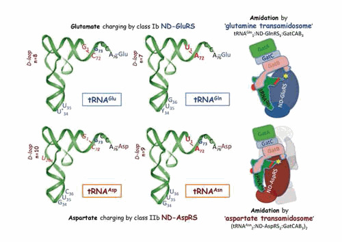
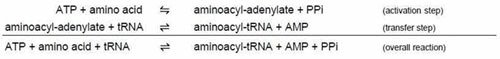
Aminoacyl-tRNA synthetases (aaRSs) are modular enzymes globally conserved in the three kingdoms of life. All catalyze the same two-step reaction, i.e., the attachment of a proteinogenic amino acid on their cognate tRNAs, thereby mediating the correct expression of the genetic code. In addition, some aaRSs acquired other functions beyond this key role in translation. Genomics and X-ray crystallography have revealed great structural diversity in aaRSs (e.g., in oligomery and modularity, in ranking into two distinct groups each subdivided in 3 subgroups, by additional domains appended on the catalytic modules). AaRSs show huge structural plasticity related to function and limited idiosyncrasies that are kingdom or even species specific (e.g., the presence in many Bacteria of non discriminating aaRSs compensating for the absence of one or two specific aaRSs, notably AsnRS and/or GlnRS). Diversity, as well, occurs in the mechanisms of aaRS gene regulation that are not conserved in evolution, notably between distant groups such as Gram-positive and Gram-negative Bacteria. The review focuses on bacterial aaRSs (and their paralogs) and covers their structure, function, regulation, and evolution. Structure/function relationships are emphasized, notably the enzymology of tRNA aminoacylation and the editing mechanisms for correction of activation and charging errors. The huge amount of genomic and structural data that accumulated in last two decades is reviewed, showing how the field moved from essentially reductionist biology towards more global and integrated approaches. Likewise, the alternative functions of aaRSs and those of aaRS paralogs (e.g., during cell wall biogenesis and other metabolic processes in or outside protein synthesis) are reviewed. Since aaRS phylogenies present promiscuous bacterial, archaeal, and eukaryal features, similarities and differences in the properties of aaRSs from the three kingdoms of life are pinpointed throughout the review and distinctive characteristics of bacterium-like synthetases from organelles are outlined.


Figures

Figure 1
Figure 1
Modular architecture of aminoacyl-tRNA synthetases and their partition in two classes with subclasses. Modularity in aaRS structure was first revealed after analysis of the E. coli AlaRS sequence (50) and was confirmed by crystallography of many other aaRSs (51). SepRS (O-phosphoseryl-tRNA synthetase) and PylRS (pyrrolysyl-tRNA synthetase) are noncanonical aaRSs found in Archaea, except a few bacterial PylRSs (see “The ambiguous status of archaeal aaRSs—a short synopsis, below, for details).
Figure 2
Figure 2
A gallery of canonical bacterial aminoacyl-tRNA synthetase structures. AaRSs in each class are displayed with their catalytic domain in the same orientation. Subclass distribution is indicated by tRNA colors, with orange, yellow, and pink backbones for subclasses a, b, and c, respectively (see Table 2 for subclass distribution). For dimeric aaRSs of classes Ic, IIa, and IIb, the second monomer is green. For tetrameric class IIc PheRS, one αβ-heterodimer is shown in blue and the other is shown in green; for AlaRS, the structure corresponds to a monomeric active fragment (see text). Identification of the displayed structures: class I T. thermophilus LeuRS with tRNALeu in posttransfer editing conformation (2bte) (137), S. aureus IleRS with tRNAIle (1qu2) (138), T. thermophilus ValRS with tRNAVal (1gax) (139), T. thermophilus ArgRS (1iq0) (140); E. coli CysRS with tRNACys (1u0b) (141), E. coli MetRS (1qqt) (122), T. thermophilus GluRS with tRNAGlu (2dxi) (142), E. coli GlnRS with tRNAGln (1o0b) (143), E. coli TyrRS (1x8x) (144), and T. thermophilus TrpRS (2el7) (unpublished from RIKEN Structural Genomics Initiative); class II E. coli SerRS with tRNASer (not in PDB) (25), E. coli ThrRS with tRNAThr (1qf6) (145), T. thermophilus ProRS with tRNAPro (1h4s) (146), E. coli HisRS (1kmn) (147), T. thermophilus GlyRS (1ati) (148), E. coli AspRS with tRNAAsp (1c0a) (149), T. thermophilus AsnRS (not in PDB) (150), E. coli LysRS (1e1o) (151), T. thermophilus PheRS with tRNAPhe (2iy5) (152), and A. aeolicus AlaRS (1yfr) (134).
Figure 3
Figure 3
Variability of aminoacyl-tRNA synthetase structures during evolution: the case of AspRS. The figure shows the organization of dimeric AspRS in the three kingdoms of life with bacterial-type (E. coli) (1eqr) (149) AspRS in the middle surrounded by archaeal-type (P. horikoshii) (1b8a) (162) and eukaryal-type (S. cerevisiae) (1eov) (166) AspRS at left and right, respectively. AspRSs are displayed with the anticodon-binding domain in blue, the hinge region in yellow, and the catalytic domain in orange. The second monomer is in light green, and the kingdom-specific domains or extensions are in pink. The exact fold of the N-terminal extension of yeast AspRS is unknown, and the present model is based on structure predictions (89).
Figure 4
Figure 4
Dual tRNA aminoacylation by ND-GluRS and ND-AspRS and tRNA-dependent amino acid amidation, the two steps of the indirect pathway of glutaminyl-tRNAGln and asparaginyl-tRNAAsn formation. (Left) The two tRNA couples (tRNAGlu / tRNAGln and tRNAAsp / tRNAAsn) aminoacylated by ND-GluRS and ND-AspRS, respectively, and (Right) schematized representations of the T. maritima glutamine (3al0) (186) and T. thermophilus aspartate (3kfu) (187) transamidosomes. The main structural and functional features important for tRNA aminoacylation by ND-aaRSs and for tRNA-dependent conversion of the glutamyl and aspartyl residues into glutamine and asparagine are shown, as well as the amidation site (yellow star) in the transamidosomes. The major identity determinants for aminoacylation of the four tRNAs by their cognate aaRSs are shown in blue (6, 188). In Bacteria, position 34 is a modified U in tRNAGlu and a pyrimidine (Y) in tRNAGln. Notice the quite similar identity sets in the tRNAGlu / tRNAGln and tRNAAsp / tRNAAsn couples, in agreement with their dual aminoacylation by the ND-aaRSs. In bold red: U1–A72, the major identity determinants for amidation in tRNAGln and tRNAAsn; in red italics: notably the antideterminant G1–C72 pair that prevents glutamate and aspartate amidation in charged tRNAGlu and tRNAAsp (186, 189). The longer length of the D-loop in tRNAGlu and tRNAAsp (as compared to tRNAGln and tRNAAsn, a feature conserved in Bacteria [19]) is a further antideterminant that prevents amidation. Transamidosomes show an overall conserved organization based on the association of ND-aaRS, tRNA, and heterotrimeric GatCAB. Notice the Yqey domain of GatB that contacts the D-loop of tRNA and thereby plays a key role in transamidation. Notice further the different sizes of the two transamidosomes. While the glutamine transamidosome is formed by five entities (as seen in the figure), the much larger aspartate transamidosome is formed by 14 macromolecular entities (for clarity, only half of the structure is shown, with the second subunit of AspRS and its tRNA ligand shown in light grey). This architectural variation is due to structural differences in ND-GluRSs (class Ib monomers) and ND-AspRSs (class IIb dimers) and the correlated mechanistic differences in the aminoacylation and transamidation steps occurring within the two types of transamidosome (see Fig. 6 in “Aminoacylation of tRNA” and “Indirect pathways of specific tRNA aminoacylation for ribosome-mediated translation,” below, for details).
Figure 5
Figure 5
Shape convergence in biomacromolecular structures and morphology of living organisms, or when macromolecular structures meet zoology. The two panels represent the mimicry of the morphology of a leatherback turtle (Dermochelys coriacea) (left) with the crystal structure of a representative PheRS (e.g., from T. thermophilus [2iy5]) (right). Notice the bilateral symmetry in the shape of both turtle and pseudodimeric (αβ)2 PheRS (with catalytic short α-subunit in green CPK amino acid models displayed on a yellow background and a large β-subunit in cyan). The large front flippers of the turtle show astonishing mimicry with the B1–B5 domains from the β-subunit of the PheRS. Other shape convergences can be found when comparing the structures of dimeric class IIb aaRSs or the dimerization domain of AlaRSC with the symmetric morphology of butterflies (try to find these mimicries, and others, after clicking on the PDB accession codes of aaRS structures given in the text). Whether such shape convergences have biological meaning remains an open question.
Figure 6
Figure 6
Different substrate recognition modes by class I and class II aminoacyl-tRNA synthetases. The differences are illustrated by the structures of E. coli glutamine (left) and aspartate (right) complexes ([1gsg] [179, 244] and [1c0a] [149], respectively): tRNA recognition (top); ATP recognition (bottom). For clarity, only one subunit of the AspRS:tRNAAsp complex is displayed. The tRNAs are shown as yellow ribophosphate backbones with contact residues represented as colored spheres (contacts with CCA are not shown): identity determinants are in green (a few do not contact the aaRS; see text) and other contact residues in orange. The class II adenylate conformation is from the archaeal AspRS from P. kodakaraensis (1b8a) (162). In the E. coli GlnRS:tRNAGln complex, 13 tRNAGln nucleotides (nt) are both determinants and make contact with GlnRS (nt 1, 2, 3, 10, 34, 35, 36, 37, 38, 70, 71, 72, 73) and 10 other nucleotides make additional contact with GlnRS (nt 4, 5, 6, 7, 8, 11, 12, 13, 14, 15). In the E. coli AspRS:tRNAAsp complex, 8 tRNAAsp nucleotides are both determinants and contact residues (nt 2, 10, 34, 35, 36, 38, 71, 73) and 11 other residues make additional contacts with AspRS (nt 11, 12, 25, 28, 32, 33, 67, 68, 69, 70, 72).
None

Similar articles

Cited by

References

    1. de Duve C. 1988. The second genetic code. Nature 333:117–118. [PubMed]10.1038/333117a0 - DOI - PubMed
    1. Schimmel P, Giegé R, Moras D, Yokoyama S. 1993. An operational RNA code for amino acids and possible relationship to genetic code. Proc Natl Acad Sci USA 90:8763–8768. [PubMed]10.1073/pnas.90.19.8763 - DOI - PMC - PubMed
    1. Bullwinkle TJ, Ibba M. 2014. Emergence and evolution. Top Curr Chem 344:43–87. [PubMed]10.1007/128_2013_423 - DOI - PMC - PubMed
    1. Carter CW Jr. 1993. Cognition, mechanism, and evolutionary relationships in aminoacyl-tRNA synthetases. Annu Rev Biochem 62:715–748. [PubMed]10.1146/annurev.bi.62.070193.003435 - DOI - PubMed
    1. Giegé R. 2006. The early history of tRNA recognition by aminoacyl-tRNA synthetases. J Biosci 31:477–488. [PubMed]10.1007/BF02705187 - DOI - PubMed

Publication types

LinkOut - more resources