. 2016 May 25;11(5):e0156074.
doi: 10.1371/journal.pone.0156074. eCollection 2016.

CRISPR-Mediated Slamf1Δ/Δ Slamf5Δ/Δ Slamf6Δ/Δ Triple Gene Disruption Reveals NKT Cell Defects but Not T Follicular Helper Cell Defects

Affiliations

CRISPR-Mediated Slamf1Δ/Δ Slamf5Δ/Δ Slamf6Δ/Δ Triple Gene Disruption Reveals NKT Cell Defects but Not T Follicular Helper Cell Defects

Joyce K Hu et al. PLoS One. .

Abstract

SAP (SH2D1A) is required intrinsically in CD4 T cells to generate germinal center responses and long-term humoral immunity. SAP binds to SLAM family receptors, including SLAM, CD84, and Ly108 to enhance cytokine secretion and sustained T cell:B cell adhesion, which both improve T follicular helper (Tfh) cell aid to germinal center (GC) B cells. To understand the overlapping roles of multiple SLAM family receptors in germinal center responses, Slamf1Δ/Δ Slamf5Δ/Δ Slamf6Δ/Δ triple gene disruption (Slamf1,5,6Δ/Δ) mice were generated using CRISPR-Cas9 gene editing to eliminate expression of SLAM (CD150), CD84, and Ly108, respectively. Gene targeting was highly efficient, with 6 of 6 alleles disrupted in 14 of 23 pups and the majority of alleles disrupted in the remaining pups. NKT cell differentiation in Slamf1,5,6Δ/Δ mice was defective, but not completely absent. The remaining NKT cells exhibited substantially increased 2B4 (SLAMF4) expression. Surprisingly, there were no overt defects in germinal center responses to acute viral infections or protein immunizations in Slamf1,5,6Δ/Δ mice, unlike Sh2d1a-/- mice. Similarly, in the context of a competitive environment, SLAM family receptor expressing GC Tfh cell, GC B cell, and plasma cell responses exhibited no advantages over Slamf1,5,6Δ/Δ cells.


Conflict of interest statement

Competing Interests: The authors have declared that no competing interests exist.

Figures

Fig 1
Fig 1. Generation of Slamf1,5,6 Δ/Δ mice using CRISPR-Cas9 technology.
(A) Surveyor assay performed to determine mutation efficiency in mouse N2A cells that were transfected with gRNA expressing pBT-U6-Cas9-2A-GFP vectors. Gel shows comparison between the original gene product (parent) and the mutated gene products (cut 1, cut 2). One surveyor assay was performed. (B) Genotyping of Slamf1Δ/Δ, Slamf5Δ/Δ, Slamf6Δ/Δ mice. Genomic DNA, guide RNA (gRNA), and sequencing results are aligned, with shaded boxes showing differences in sequence, which include deletions (dashes), insertions, and uncertain base calling that results from heterozygous mutations in each allele. The sequences of each allele of Slamf6 in mouse 11 is shown beneath the chromatogram. Each mutation leads to a stop codon shown in a red box. Genotyping was performed as indicated in Table 2.
Fig 2
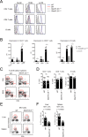
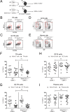
Fig 2. Slamf1,5,6 Δ/Δ mice exhibit deficiencies in NKT cell development but no overt defects in CD4 T cell, CD8 T cell, or B cell development.
(A) Surface expression and (B) MFI of SLAM, CD84, and Ly108 on peripheral CD4 T cells, CD8 T cells, and B cells from WT and SLAMf receptor triple gene disruption mice. (A-B) Two independent experiments are shown, with 2–3 mice per group. (C) Flow cytometry plots and (D) graphs of CD4+ T cell, CD8+ T cell, and B220+ B cell frequencies in spleens of WT and SLAMf receptor triple gene disruption mice. (C-D) Data is representative of two independent experiments, with 4 mice per group. (E) Flow cytometry plots and (F) graphs of B220- CD3+ CD1d Tetramer+ NKT cells in spleens and livers of WT and SLAMf receptor triple gene disruption mice. (E-F) Data are representative of 2 independent experiments, with 4 mice per group.
Fig 3
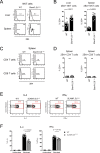
Fig 3. NKT cells from Slamf1,5,6 Δ/Δ mice have higher levels of 2B4 expression and lower functional secretion of cytokines.
(A) Histograms and (B) graphs of 2B4 surface expression on B220- CD3+ CD1d Tetramer+ NKT cells in spleens and livers of WT and Slamf1,5,6 Δ/Δ mice. (C) Histograms and (D) graphs of 2B4 surface expression on splenic CD4 and CD8 T cells in WT and Slamf1,5,6 Δ/Δ mice. (A-D) Data shows two independent experiments, with 4 mice per group. (E) Flow cytometry plots of representative WT and Slamf1,5,6 Δ/Δ NKT cells (gated on B220- CD3int CD1d Tetramer+ cells) after in vivo stimulation with α-GalCer for 45 minutes. Expression of IL-4 and IFN-γ are shown. (F) Frequencies of IL-4 and IFN-γ expression by NKT cells (gated on B220- CD3int CD1d Tetramer+ cells). (E-F) Data represents two independent experiments, with 3 mice per group.
Fig 4
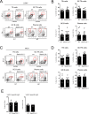
Fig 4. Absence of defects in germinal centers generated in Slamf1,5,6 Δ/Δ mice after infection with LCMV and VACV.
(A) Flow cytometry plots and (B) graphs of CD19- CD4+ CD44+ CXCR5+ BTLA+ Tfh cells, CD19- CD4+ CD44+ CXCR5+ PD1+ GC Tfh cells, CD4- CD19+ Fas+ GL7+ GC B cells, and CD4- CD19+ IgD- CD138+ plasma cells in spleens of WT and Slamf1,5,6 Δ/Δ mice at 8 days post infection with LCMV. (A-B) Data represents three independent experiments, with 4–5 mice per group. (C) Flow cytometry plots and (D) graphs of CD19- CD4+ CD44+ CXCR5+ BTLA+ Tfh cells, CD19- CD4+ CD44+ CXCR5+ PD1+ GC Tfh cells, CD4- CD19+ Fas+ GL7+ GC B cells, and CD4- CD19+ IgD- CD138+ plasma cells in spleens of WT and Slamf1,5,6 Δ/Δ mice at 8 days post infection with VACV. (C-D) Data represents two independent experiments, with 4 mice per group. (E) Endpoint titers and Area Under Curve (AUC) analyses of VACV specific serum IgG at 15 days post infection with VACV. Data represents one experiment, with four mice per group.
Fig 5
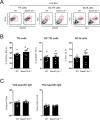
Fig 5. Lack of defects in germinal centers generated in Slamf1,5,6 Δ/Δ mice after immunization with HIV envelope trimer protein.
(A) Flow cytometry plots and (B) graphs of CD19- CD4+ CD44+ CXCR5+ BTLA+ Tfh cells, CD19- CD4+ CD44+ CXCR5+ PD1+ GC Tfh cells, and CD4- CD19+ Fas+ GL7+ GC B cells in draining popliteal lymph nodes of of WT and Slamf1,5,6 Δ/Δ mice at 8 days post immunization with HIV Envelope (YU2 gp140-F) protein. (A-B) Data represents two independent experiments, with 3–4 mice per grouop. (C) Endpoint titers and Area Under Curve (AUC) analyses of HIV Env (YU2-gp140-F) specific serum IgG at 15 days post immunization with YU2-gp140-F. Data represents one experiment, with 4 mice per group.
Fig 6
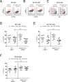
Fig 6. No competitive advantage for Slamf1, Slamf5, and Slamf6-expressing cells after LCMV infection.
(A) Mixed WT CD45.1 and WT CD45.2 control bone marrow (BM) chimeras or mixed WT CD45.1 and SLAMf1,5,6 Δ/Δ CD45.2 BM chimeras were made and infected with LCMV or VACV. (B-I) Bone marrow chimeras infected with LCMV. Flow cytometry plots of CD19- CD4+ CD44+ CXCR5+ BTLA+ Tfh cells (B), CD19- CD4+ CD44+ CXCR5+ Bcl6+ GC Tfh cells (C), CD4- CD19+ IgD- GL7+ GC B cells (D) and CD4- CD19+ IgD- CD138+ plasma cells (E) in spleens of mixed WT:WT control BM chimeras or mixed WT:SLAMf1,5,6 Δ/Δ BM chimeras at 7/8 days post infection with LCMV. (F-I) Frequencies of CD45.2+ WT or CD45.2+ SLAMf1,5,6 Δ/Δ cells in WT:WT and WT:SLAMf1,5,6 Δ/Δ BM chimeras at 7/8 days post LCMV infection. Tfh cells (F), GC Tfh cells (G), GC B cells (H), and plasma cells (I) are shown in relation to their naïve counterparts (naïve T cells or naïve B cells). (B-I) Data represent three independent experiments, with 4–5 mice per group.
Fig 7
Fig 7. No competitive advantage for Slamf1, Slamf5, and Slamf6-expressing cells after VACV infection.
(A-F) Bone marrow chimeras infected with VACV. Flow cytometry plots of CD19- CD4+ CD44+ CXCR5+ BTLA+ Tfh cells (A), CD19- CD4+ CD44+ CXCR5+ Bcl6+ GC Tfh cells (B), and CD4- CD19+ IgD- GL7+ GC B cells (C) in spleens of mixed WT:WT control BM chimeras or mixed WT:SLAMf1,5,6 Δ/Δ BM chimeras at 7 days post infection with VACV. (D-F) Frequencies of CD45.2+ WT or CD45.2+ SLAMf1,5,6 Δ/Δ cells in WT:WT and WT:SLAMf1,5,6 Δ/Δ BM chimeras at 7 days post VACV infection. Tfh cells (D), GC Tfh cells (E), and GC B cells (F) are shown in relation to their naïve counterparts (naïve T cells or naïve B cells). (A-F) Data represent two independent experiments with 5 mice per group.

Similar articles

Cited by

References

    1. Crotty S. T Follicular Helper Cell Differentiation, Function, and Roles in Disease. Immunity. 2014;41: 529–542. 10.1016/j.immuni.2014.10.004 - DOI - PMC - PubMed
    1. Crotty S. Follicular helper CD4 T cells (TFH). Annu Rev Immunol. United States; 2011;29: 621–663. 10.1146/annurev-immunol-031210-101400 - DOI - PubMed
    1. Crotty S. A brief history of T cell help to B cells. Nat Rev Immunol. Nature Publishing Group; 2015;15: 185–189. 10.1038/nri3803 - DOI - PMC - PubMed
    1. Tangye SG, Brink R, Goodnow CC, Phan TG. SnapShot: Interactions between B Cells and T Cells. Cell. 2015;162: 926–926.e1. 10.1016/j.cell.2015.07.055 - DOI - PubMed
    1. Cannons JL, Tangye SG, Schwartzberg PL. SLAM family receptors and SAP adaptors in immunity. Annu Rev Immunol. United States; 2011;29: 665–705. 10.1146/annurev-immunol-030409-101302 - DOI - PubMed

MeSH terms

Substances