. 2016 May 25;11(5):e0156081.
doi: 10.1371/journal.pone.0156081. eCollection 2016.

SDCCAG8 Interacts with RAB Effector Proteins RABEP2 and ERC1 and Is Required for Hedgehog Signaling

Affiliations

SDCCAG8 Interacts with RAB Effector Proteins RABEP2 and ERC1 and Is Required for Hedgehog Signaling

Rannar Airik et al. PLoS One. .

Abstract

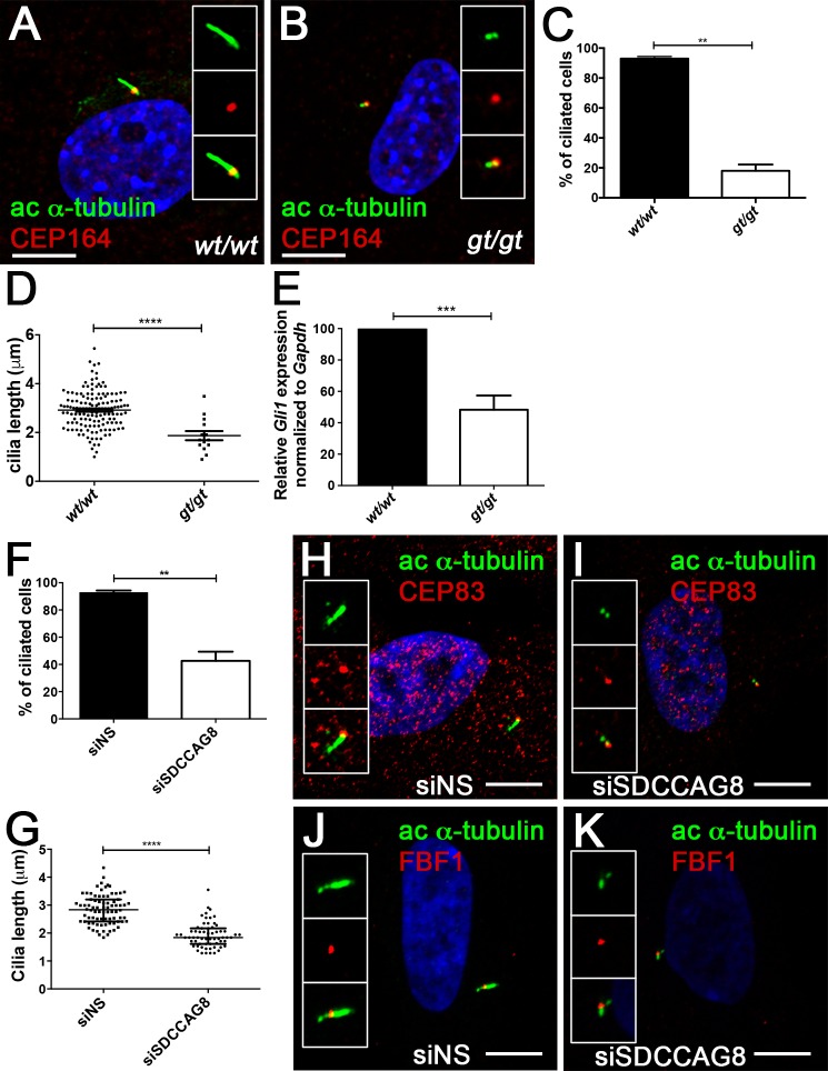
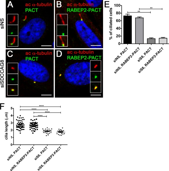
Recessive mutations in the SDCCAG8 gene cause a nephronophthisis-related ciliopathy with Bardet-Biedl syndrome-like features in humans. Our previous characterization of the orthologous Sdccag8gt/gt mouse model recapitulated the retinal-renal disease phenotypes and identified impaired DNA damage response signaling as an underlying disease mechanism in the kidney. However, several other phenotypic and mechanistic features of Sdccag8gt/gt mice remained unexplored. Here we show that Sdccag8gt/gt mice exhibit developmental and structural abnormalities of the skeleton and limbs, suggesting impaired Hedgehog (Hh) signaling. Indeed, cell culture studies demonstrate the requirement of SDCCAG8 for ciliogenesis and Hh signaling. Using an affinity proteomics approach, we demonstrate that SDCCAG8 interacts with proteins of the centriolar satellites (OFD1, AZI1), of the endosomal sorting complex (RABEP2, ERC1), and with non-muscle myosin motor proteins (MYH9, MYH10, MYH14) at the centrosome. Furthermore, we show that RABEP2 localization at the centrosome is regulated by SDCCAG8. siRNA mediated RABEP2 knockdown in hTERT-RPE1 cells leads to defective ciliogenesis, indicating a critical role for RABEP2 in this process. Together, this study identifies several centrosome-associated proteins as novel SDCCAG8 interaction partners, and provides new insights into the function of SDCCAG8 at this structure.


Conflict of interest statement

Competing Interests: The authors have declared that no competing interests exist.

Figures

Fig 1
Fig 1. Sdccag8gt/gt mice exhibit rib cage and pre-axial polydactyly phenotypes.
(AD) Skeletal preparation of E18.5 rib cage and hind limbs, demonstrate misalignment (red arrows) of sternum ossification centers in Sdccag8gt/gt embryos (A), when compared to wild type control (B), and formation of triphalangeal polydactyly (arrow) in hind limbs (C), when compared to wild type control (D). Asterisk indicates a bony pre-axial polydactylous structure in C. Thoracic vertebral segments are identified for T1-T7, R denotes right paw. (EJ) Anatomical overview and skeletal preparations of P30 wild type (E,G,I) and Sdccag8gt/gt (F,H,J) right hind limbs. Fingers are numbered with Roman numerals starting with the thumb (arrows indicate triphalangeal polydactylous pre-axial thumbs in Sdccag8gt/gt hind limbs (F and H). Skeletal preparations of the limbs demonstrate forking of the Sdccag8gt/gt thumb after the first phalange (H). An additional rudimentary finger-like bone is located pre-axial to the defective thumb (arrowhead) (J). wt, Sdccag8 wild type allele; gt, Sdccag8 gene-trap allele. (K) Quantitation of the hind limb polydactyly/triphalangeal thumb phenotype in Sdccag8gt/gt mice reveals a variable expressivity of this feature. The most frequent phenotype is the occurrence of bilateral polydactyly/triphalangeal thumbs in Sdccag8gt/gt mice at 35%. Structurally, less severe phenotypes of right side only or left side only polydactyly/triphalangeal thumbs occur in 27% and 3% of the cases, respectively.
Fig 2
Fig 2. Sdccag8 is required or cilia formation and Hh signaling.
(A,B) Sdccag8gt/gt cells have shorter cilia. Immunofluorescence staining of cultured mouse embryonic fibroblasts derived from wild type (A) and Sdccag8gt/gt (B) mice using antibodies against distal appendage marker CEP164 (red) and cilia marker acetylated tubulin (green), demonstrate shorter cilia in Sdccag8gt/gt cells (B). (C) Quantitation of the percentage of ciliated cells for wild type and Sdccag8gt/gt cells after serum starvation (48 hrs). Significantly less Sdccag8gt/gt cells grow cilia compared to wild type cells (18% vs. 93%, n = 100 for both genotypes, **p = 0.0018). (D) Sdccag8gt/gt MEFs have significantly shorter cilia (1.9 ± 0.2 μm, n = 20) compared to wild type cells (2.9 ± 0.0 μm, n = 142, ****p<0.0001). (E) Sdccag8gt/gt MEFs have attenuated response to Hh signal agonist SAG. qRT-PCR analysis demonstrates reduced levels of Hh pathway target gene Gli1 in SAG treated Sdccag8gt/gt MEFs compared to wild type cells (N = 3). (F) Knockdown of SDCCAG8 causes a reduction in cilia formation in hTERT-RPE1 cells. Only 43% of SDCCAG8 knockdown cells grew cilia compared to 94% wild type cells (**p = 0.0092). (G) Cilia length is significantly reduced in SDCCAG8 knockdown cells (1.9 ± 0.1 μ, n = 63) compared to wild type cells (2.8 ± 0.1 μ., n = 80, ****p<0.0001). Scale bar: (A,B) 7.5 μm. wt, Sdccag8 wild type allele; gt, Sdccag8 gene-trap allele. (H,I,J,K) Immunofluorescence staining of control siNS hTERT-RPE1 cells (H,J) and siSDCCAG8 hTERT-RPE1 cells (I,K) with antibodies against acetylated tubulin (green) and distal appendage markers CEP83 (red) (H,I) or FBF1 (red) (J,K), reveals no abnormalities in the formation of centriolar distal appendages despite the loss of cilia in SDCCAG8 knockdown cells (I,K) compared to control cells transfected with non-specific siRNA (F,H).
Fig 3
Fig 3. SDCCAG8 associates with a characteristic set of proteins at the centrosome.
(A) The Sdccag8 interacting proteins discovered by SILAC as centrosomal components belong to 4 functional groups, i.e. centriolar satellite components, endosomal vesicle components, tRNA synthesis complex proteins, and myosin type II motors involved in ciliogenesis. (B) Overview of the SDCCAG8 constructs used in this study. Numbering corresponds to amino acid positions. Gray boxes designate coiled-coil domains. (C) SDCCAG8 interacts with RABEP2, ERC1, and CEP131. FLAG-tagged full-length and truncation constructs of SDCCAG8 were immunoprecipitated from extracts of HEK293 cells transfected with each construct and analyzed by Western blot. Blots were probed for FLAG, or for endogenous RABEP2, ERC1, and CEP131 respectively. Only full-length FLAG-tagged SDCCAG8, but not truncated SDCCAG8 constructs co-immunoprecipitate RABEP2, ERC1 or CEP131, except for the C-terminal SDCCAG8 fragment that weakly co-immunoprecipitated RABEP2. (D) Western blotting for endogenous proteins in whole cell lysates shows equal loading. (E) CEP131 co-immunoprecipitates FLAG-SDCCAG8. Lysates from cells transfected with FLAG-SDCCAG8 and with non-specific control siRNA or with siCEP131 were subjected to immunoprecipitation using anti-CEP131 antibodies or normal IgG. While anti-CEP131 antibodies co-immunoprecipitated FLAG-SDCCAG8 from control lysates, CEP131 knockdown abolished immunoprecipitation of FLAG-SDCCAG8, demonstrating specificity of the interaction.
Fig 4
Fig 4. Proteins identified by SILAC assay as SDCCAG8 interactors localize to the basal body or centrosome.
(A) Co-staining with the centriolar protein γ-tubulin demonstrates centrosomal localization for the identified SDCCAG8 interacting proteins RABEP2, ERC1, and CEP131 in hTERT-RPE1 cells. Note CEP131 localization also at centriolar satellites. Scale bars: 5 μm. (B) Co-staining with the ciliary axoneme marker poly-glutamylated tubulin (red) demonstrates ciliary and basal body localization of RABEP2, basal body localization of ERC1 and centriolar satellite localization of CEP131 in hTERT-RPE1 cells. Scale bars: 5 μm. (C) Quantification of the co-localization coefficients of RABEP2, ERC1 and CEP131 with γ-tubulin positive centrosomes in cells from (A). In each case n>40 centrosomes; error bars, SEM. (D) Quantification of the co-localization coefficients of RABEP2, ERC1 and CEP131 with polyglutamine-tubulin positive centrosomes in cells from (B). In each case n>40 centrosomes; error bars, SEM.
Fig 5
Fig 5. RABEP2 siRNA knockdown abolishes cilia formation.
(A) Western blot analysis of extracts from control or siRABEP2-depleted hTERT-RPE1 cells, probed with anti-RABEP2 or anti-β-actin, as a loading control. Dharmacon SMARTpool siRNAs were used to knock down endogenous RABEP2 at high efficiency. (B) RABEP2 knockdown abolishes cilia formation in hTERT-RPE1 cells. Control, RABEP2 and SDCCAG8 depleted hTERT-RPE1 cells were serum starved for 48 hours after transfection with siRNA, fixed, and immunostained for acetylated α-tubulin to label primary cilia. Percentage of ciliated cells was determined in each case, control 92.8 ± 5.2%, n = 50; siRABEP2 15 ± 8.6%, n = 50 and siSDCCAG8 31.48 ± 1.694%, n = 50. Error bars, SEM, ****p<0.0001. (C) hTERT-RPE1 cells were transfected with non-specific siRNA (siNS), siRABEP2, or siSDCCAG8. Cells were ciliated by serum starvation and stained with antibodies against SDCCAG8 (green) and acetylated α-tubulin (red). Knockdown of RABEP2 abolishes cilia formation, but does not alter SDCCAG8 localization at the centrioles. Knockdown of SDCCAG8 abolishes cilia formation in hTERT-RPE1 cells. Scale bars: 5 μm. (D) hTERT-RPE1 cells were transfected with non-specific siRNA (siNS), siRABEP2, or siSDCCAG8. Cells were ciliated by serum starvation and stained with antibodies against RABEP2 (red) and polyglutamylated tubulin (green). Knockdown of RABEP2 abolishes cilia formation. Knockdown of SDCCAG8 abolishes cilia formation and depletes RABEP2 localization from the centrioles in hTERT-RPE1 cells. Scale bars: 5 μm. (E) RABEP2 localization at the centrosome is dependent on SDCCAG8. RABEP2- and SDCCAG8-positive pixels were quantitated at acetylated α-tubulin or polyglutamylated-tubulin positive centrosomes in siNS, siRABEP2 and siSDCCAG8 cells (C and D). While control cells (siNS) have 69.40 ± 1.194, n = 30, RABEP2-positive pixels at the centrosome, RABEP2-positive pixels are significantly reduced in siRABEP2 cells (13.00 ± 1.678, n = 30, ****p<0.0001) and in siSDCCAG8 cells (11.84 ± 1.231, n = 30, ****p<0.0001). Control cells (siNS) have 75.25 ± 2.145, n = 30 SDCCAG8-positive pixels at the centrosome, which is not changed in siRABEP2 cells (73.83 ± 2.138, n = 30), but is significantly reduced in siSDCCAG8 cells (10.76 ± 0.9907, n = 30, ****p<0.0001). Error bars, SEM; siNS, non-specific siRNA; siR2, RABEP2 siRNA; siS8, SDCCAG8 siRNA.
Fig 6
Fig 6. RABEP2-PACT does not rescue siSDCCAG8 ciliogenesis defect.
(A,B) hTERT-RPE1 cells were transfected with non-specific siRNA (siNS) and 24 hours later transfected with EGFP-PACT (A) or EGFP-RABEP2-PACT (B) constructs. Either of the tagged proteins localized to the centrosome and were detected with anti-GFP antibody (green). Ciliogenesis (acetylated-α-tubulin, red) was not affected in either of the situations. (C,D) hTERT-RPE1 cells were transfected with siSDCCAG8 and 24 hours later transfected with EGFP-PACT (C) or EGFP-RABEP2-PACT (D) constructs. Although, the tagged proteins localized to the centrosome (green), they failed to rescue the ciliogenesis defect (acetylated α-tubulin, red) in SDCCAG8 knockdown cells. Scale bars: 4 μm. (E) Quantitation of the percentage of ciliated cells in (A, B, C and D), demonstrates that over expression of the EGFP-PACT or EGFP-RABEP2-PACT construct does not affect normal ciliation in siNS hTERT-RPE1 cells. Moreover, EGFP-RABEP2-PACT construct fails to rescue the ciliation defect in SDCCAG8 depleted cells (**p = 0.0068). Error bars, SEM. (F) We measured the length of cilia in EGFP-PACT and EGFP-RABEP2-PACT overexpression cells to examine whether it was affected. There was no significant difference in the length of cilia between siNS cells expressing EGFP-PACT and EGFP-RABEP2-PACT (73.08 ± 4.417 μm, n = 3, vs. 67.80 ± 2.446 μm, n = 3, p = 0.41). Similarly, the cilia length was uncorrected by the overexpression of EGFP-PACT and EGFP-RABEP2-PACT in siSDCCAG8 cells (13.36 ± 2.194 μm, n = 3 vs. 14.58 ± 2.083 μm, n = 3, p = 0.72). Error bars, SEM.

References

    1. Otto EA, Hurd TW, Airik R, Chaki M, Zhou W, Stoetzel C, et al. Candidate exome capture identifies mutation of SDCCAG8 as the cause of a retinal-renal ciliopathy. Nat Genet. 2010;42:840–50. Epub 2010/09/14. doi: ng.662 [pii] 10.1038/ng.662 . - DOI - PMC - PubMed
    1. Schaefer E, Durand M, Stoetzel C, Doray B, Viville B, Helle S, et al. Molecular diagnosis reveals genetic heterogeneity for the overlapping MKKS and BBS phenotypes. Eur J Med Genet. 2011;54(2):157–60. Epub 2010/11/04. doi: S1769-7212(10)00117-5 [pii] 10.1016/j.ejmg.2010.10.004 . - DOI - PubMed
    1. Airik R, Slaats GG, Guo Z, Weiss AC, Khan N, Ghosh A, et al. Renal-retinal ciliopathy gene Sdccag8 regulates DNA damage response signaling. Journal of the American Society of Nephrology: JASN. 2014;25(11):2573–83. 10.1681/ASN.2013050565 - DOI - PMC - PubMed
    1. Insolera R, Shao W, Airik R, Hildebrandt F, Shi SH. SDCCAG8 regulates pericentriolar material recruitment and neuronal migration in the developing cortex. Neuron. 2014;83(4):805–22. 10.1016/j.neuron.2014.06.029 - DOI - PMC - PubMed
    1. Huangfu D, Liu A, Rakeman AS, Murcia NS, Niswander L, Anderson KV. Hedgehog signalling in the mouse requires intraflagellar transport proteins. Nature. 2003;426(6962):83–7. . - PubMed

Publication types

MeSH terms