. 2016 May 25;11(5):e0156303.
doi: 10.1371/journal.pone.0156303. eCollection 2016.

Sphingosine-1-Phosphate Receptor 2 Regulates Proinflammatory Cytokine Production and Osteoclastogenesis

Affiliations

Sphingosine-1-Phosphate Receptor 2 Regulates Proinflammatory Cytokine Production and Osteoclastogenesis

Hong Yu. PLoS One. .

Abstract

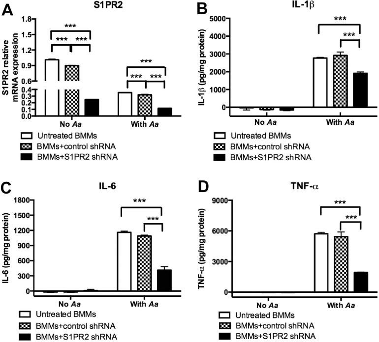
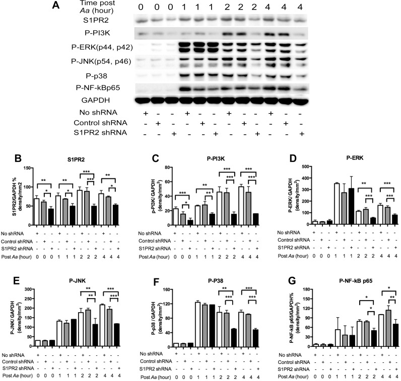
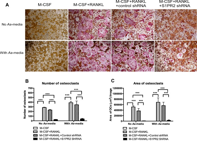
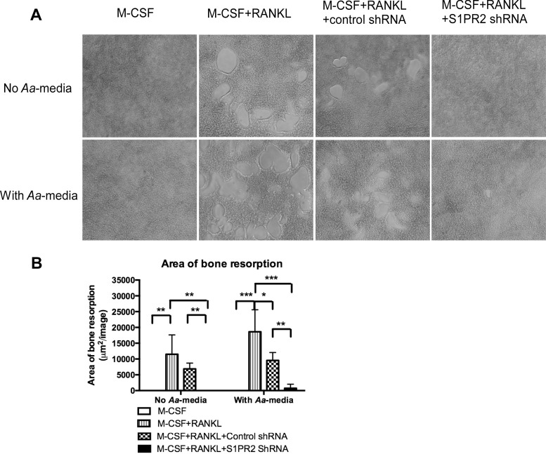
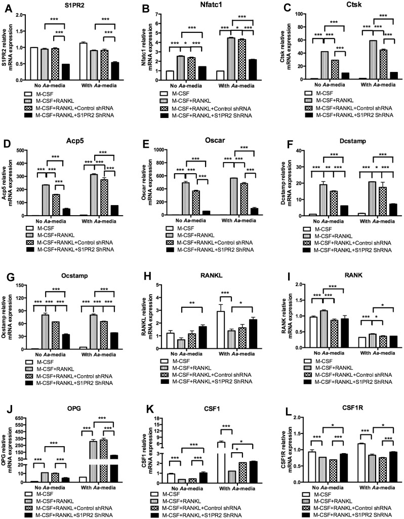
Sphingosine-1-phosphate receptor 2 (S1PR2) couples with the Gi, Gq, and G12/13 group of proteins, which modulate an array of cellular signaling pathways and affect immune responses to multiple stimuli. In this study, we demonstrated that knockdown of S1PR2 by a specific S1PR2 shRNA lentiviral vector significantly inhibited IL-1β, IL-6, and TNF-α protein levels induced by oral pathogen Aggregatibacter actinomycetemcomitans (A. actinomycetemcomitans) in murine bone marrow-derived monocytes and macrophages (BMMs) compared with controls. In addition, knockdown of S1PR2 by the S1PR2 shRNA lentiviral vector suppressed p-PI3K, p-ERK, p-JNK, p-p38, and p-NF-κBp65 protein expressions induced by A. actinomycetemcomitans. Furthermore, bone marrow cells treated with the S1PR2 shRNA lentiviral vector inhibited osteoclastogenesis induced by RANKL compared with controls. The S1PR2 shRNA suppressed the mRNA levels of six osteoclastogenic factors including nuclear factor of activated T-cells cytoplasmic calcineurin-dependent 1 (NFATc1), cathepsin K (Ctsk), acid phosphatase 5 (Acp5), osteoclast-associated receptor (Oscar), dendritic cells specific transmembrane protein (Dcstamp), and osteoclast stimulatory transmembrane protein (Ocstamp) in bone marrow cells. We conclude that S1PR2 plays an essential role in modulating proinflammatory cytokine production and osteoclastogenesis. Blocking S1PR2 signaling might be a novel therapeutic strategy to treat inflammatory bone loss diseases.


Conflict of interest statement

Competing Interests: The author declares no potential conflicts of interest with respect to the authorship and/or publication of this article.

Figures

Fig 1
Fig 1. Knockdown of S1PR2 significantly decreased IL-1b, IL-6, and TNF-a protein expressions induced by A. actinomycetemcomitans (Aa) in BMMs.
Murine BMMs were uninfected, infected with a S1PR2 shRNA lentivirus, or infected with a control shRNA lentivirus (moi 50) for 72 h. Then the cells were either unstimulated or stimulated with Aa (1.5 CFU/cell) for 4 h (S1PR2 mRNA assay) or 8 h (cytokine protein assays). (A) S1PR2 mRNA expression was determined by real time PCR, (B) IL-1β, (C) IL-6, and (D) TNF-α protein levels were quantified by ELISA and normalized by protein levels in cell lysates. The data are representatives from three separate experiments (n = 3, ***P<0.001).
Fig 2
Fig 2. Knockdown of S1PR2 attenuated S1PR2, p-PI3K, p-ERK, p-JNK, p-p38, and p-NF-kBp65 protein expressions in BMMs.
Murine BMMs were uninfected, infected with a S1PR2 shRNA lentivirus, or infected with a control shRNA lentivirus (moi 50) for 72 h. Then the cells were either unstimulated or stimulated with Aa (1.5 CFU/cell) for 1 to 4 h. (A) S1PR2, p-PI3K, p-ERK, p-JNK, p-p38, and p-NF-κBp65 protein expressions were evaluated by Western Blot. (B) S1PR2 protein density, (C) p-PI3K protein density, (D) p-ERK protein density, (E) p-JNK protein density, (F) p-p38 protein density, and (G) p-NF-κBp65 protein density were analyzed and normalized by GAPDH protein expression. The data are representatives from three separate experiments (n = 3, *P<0.05, **P<0.01, ***P<0.001).
Fig 3
Fig 3. Knockdown of S1PR2 suppressed osteoclastogenesis in BM cells induced by RANKL.
BM cells were uninfected, infected with a S1PR2 shRNA lentivirus, or infected with a control shRNA lentivirus (moi 20), and co-cultured with M-CSF and RANKL as described in Methods. A control group of cells were cultured only with M-CSF. BM cells were either untreated or treated with A. actinomycetemcomitans-stimulated media (Aa-media) for 24 h. (A) Representative images show TRAP-stained cells with and without Aa-media stimulation. Pictures were taken at 100x magnification. (B) Number of TRAP+ multinucleated (more than 3 nuclei) osteoclasts/well (96-well) and (C) Total areas of osteoclasts/image were quantified. The data are representatives from three separate experiments (n = 3, ***P<0.001).
Fig 4
Fig 4. Knockdown of S1PR2 inhibited bone resorption in BM cells induced by RANKL.
BM cells were uninfected, infected with a S1PR2 shRNA lentivirus, or infected with a control shRNA lentivirus (moi 20), and co-cultured with M-CSF and RANKL, with or without A. actinomycetemcomitans-stimulated media (Aa-media) treatment as described in Methods. Control groups of cells were cultured only with M-CSF with or without Aa-media treatment. (A) Representative images show bone resorption pits. Pictures were taken at 100x magnification. (B) Total areas of bone resorption pits /image were quantified. The data are representatives from three separate experiments (n = 3, *P<0.05, ** P<0.01, *** P<0.001).
Fig 5
Fig 5. Knockdown of S1PR2 significantly attenuated Nfatc1, Ctsk, Acp5, Oscar, Dcstamp, and Ocstamp mRNA expressions in BM cells with or without treatment with A. actinomycetemcomitans-stimulated media (Aa-media).
BM cells were uninfected, infected with a S1PR2 shRNA lentivirus, or infected with a control shRNA lentivirus (moi 20), and co-cultured with M-CSF and RANKL as described in Methods. A control group of cells were cultured only with M-CSF. BM cells were either unstimulated or stimulated with Aa- media for 4 h. (A) S1PR2 mRNA, (B) Nfatc1 mRNA, (C) Ctsk mRNA, (D) Acp5 mRNA, (E) Oscar mRNA, (F) Dcstamp mRNA, (G) Ocstamp mRNA, (H) RANKL mRNA, (I) RANK mRNA, (J) OPG mRNA, (K) CSF1 mRNA, and (L) CSF1R mRNA levels were quantified by real time PCR and normalized by GAPDH expression. The data are representatives from three separate experiments (n = 3, *P<0.05, ***P<0.001).

References

    1. Spiegel S, Milstien S. The outs and the ins of sphingosine-1-phosphate in immunity. Nat Rev Immunol. 2011;11(6):403–15. 10.1038/nri2974 - DOI - PMC - PubMed
    1. Xia P, Wadham C. Sphingosine 1-phosphate, a key mediator of the cytokine network: juxtacrine signaling. Cytokine & growth factor reviews. 2011;22(1):45–53. - PubMed
    1. Yu H, Valerio M, Bielawski J. Fenretinide inhibited de novo ceramide synthesis and proinflammatory cytokines induced by Aggregatibacter actinomycetemcomitans. Journal of lipid research. 2013;54(1):189–201. 10.1194/jlr.M031427 - DOI - PMC - PubMed
    1. Yu H, Sun C, Argraves KM. Periodontal inflammation and alveolar bone loss induced by Aggregatibacter actinomycetemcomitans is attenuated in sphingosine kinase 1-deficient mice. Journal of periodontal research. 2016;51(1):38–49. 10.1111/jre.12276 - DOI - PMC - PubMed
    1. Vaananen HK, Laitala-Leinonen T. Osteoclast lineage and function. Arch Biochem Biophys. 2008;473(2):132–8. 10.1016/j.abb.2008.03.037 - DOI - PubMed

MeSH terms