. 2016 May 24;110(10):2169-75.
doi: 10.1016/j.bpj.2016.04.031.

Stable Domain Assembly of a Monomolecular DNA Quadruplex: Implications for DNA-Based Nanoswitches

Affiliations

Stable Domain Assembly of a Monomolecular DNA Quadruplex: Implications for DNA-Based Nanoswitches

Besik Kankia et al. Biophys J. .

Abstract

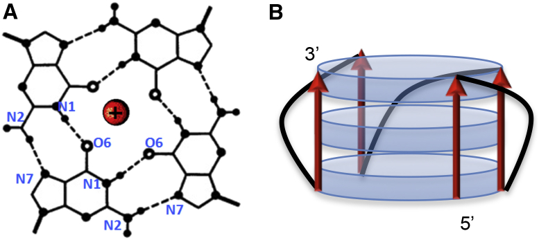
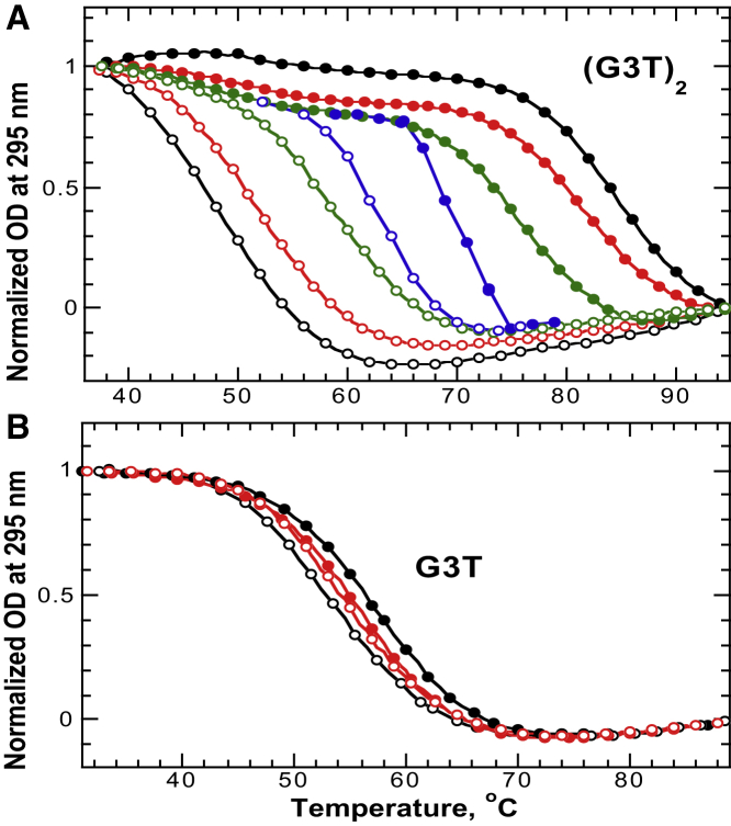
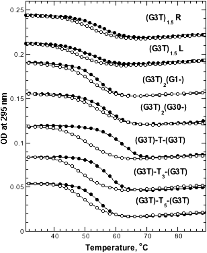
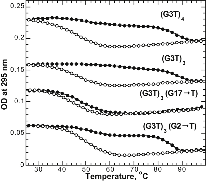
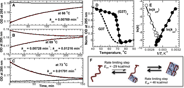
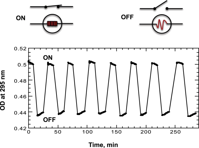
In the presence of K(+) ions, the 5'-GGGTGGGTGGGTGGG-3' (G3T) sequence folds into a monomolecular quadruplex with unusually high thermal stability and unique optical properties. In this study we report that although single G3T molecules unfold and fold rapidly with overlapping melting and refolding curves, G3T multimers (G3T units covalently attached to each other) demonstrate highly reproducible hysteretic behavior. We demonstrate that this behavior necessitates full-length tandem G3T monomers directly conjugated to each other. Any modification of the tandem sequences eliminates the hysteresis. The experimentally measured kinetic parameters and equilibrium transition profiles suggest a highly specific two-state transition in which the folding and unfolding of the first G3T monomer is rate-limiting for both annealing and melting processes. The highly reproducible hysteretic behavior of G3T multimers has the potential to be used in the design of heat-stimulated DNA switches or transistors.


Figures

Figure 1
Figure 1
(A) G-quartet with cation (red) in the center; and (B) schematic representation of G3T quadruplex with all parallel G-tracts (red) and chain-reversal T-loops (black). Blue discs represent G-quartets. To see this figure in color, go online.
Figure 2
Figure 2
UV unfolding and refolding experiments of (A) (G3T)2 and (B) G3T in 0.1 mM KCl. Color code for the temperature gradient: 1°C/min (black), 0.5°C/min (red), 0.1°C/min (green), and 0.02°C/min (blue). Solid and open circles correspond to unfolding and refolding curves, respectively. To see this figure in color, go online.
Figure 3
Figure 3
UV unfolding and refolding experiments of (G3T)2 variants measured using a 1°C/min temperature gradient in 0.1 mM KCl. Upper four curves correspond to truncations and lower three curves correspond to T-insertions. Curves are offset for clarity.
Figure 4
Figure 4
UV unfolding and refolding experiments of (G3T)3, (G3T)4, and variants measured using a 1°C/min temperature gradient in 0.1 mM KCl. Curves are offset for clarity.
Figure 5
Figure 5
Isothermal folding and unfolding of (G3T)2 in 0.1 mM KCl. (AC) Representative kinetics of simultaneous folding and unfolding at different temperatures are shown. At 66°C we observe only folding; at 73°C, only unfolding; and at 69°C, both processes are visible. The red lines correspond to a single-exponential fit of the data. (D) The reversible melting curve of (G3T)2 constructed from isothermal experiments (solid circles) is shown. The reversible melting of G3T from Fig. 2B (dashed line) is shown for clarity. (E) Arrhenius plot to determine activation energies for folding (solid circles) and unfolding (open circles) of (G3T)2 is shown. (F) Suggested model describing the two-state phase transition is shown. To see this figure in color, go online.
Figure 6
Figure 6
Heat-induced reversible folding and unfolding of a (G3T)2 quadruplex at constant temperature, 70°C, monitored by light absorption. Folding and unfolding was achieved by removing the optical cell from the UV spectrometer and incubating the cuvette at 45°C for 3 min to achieve the initial ON state. The cell was returned to the cell holder and following a 2 min equilibration to reach the reaction temperature of 70°C, the UV absorption was recorded for 10–15 min. The OFF state was then achieved by removing the cell and incubating the sample at 95°C for 3 min. The measurement was continued after returning the cell into the cell holder and equilibrating for 2 min to reach the reaction temperature of 70°C. This procedure was repeated a total of eight times. To see this figure in color, go online.

References

    1. Gellert M., Lipsett M.N., Davies D.R. Helix formation by guanylic acid. Proc. Natl. Acad. Sci. USA. 1962;48:2013–2018. - PMC - PubMed
    1. Maizels N. Dynamic roles for G4 DNA in the biology of eukaryotic cells. Nat. Struct. Mol. Biol. 2006;13:1055–1059. - PubMed
    1. Maizels N. G4-associated human diseases. EMBO Rep. 2015;16:910–922. - PMC - PubMed
    1. Rhodes D., Lipps H.J. G-quadruplexes and their regulatory roles in biology. Nucleic Acids Res. 2015;43:8627–8637. - PMC - PubMed
    1. Simone R., Fratta P., Isaacs A.M. G-quadruplexes: emerging roles in neurodegenerative diseases and the non-coding transcriptome. FEBS Lett. 2015;589:1653–1668. - PubMed

Publication types

Substances

LinkOut - more resources