Clinical Trial
. 2016 May 25;11(5):e0156044.
doi: 10.1371/journal.pone.0156044. eCollection 2016.

Overexpression of L-Type Amino Acid Transporter 1 (LAT1) and 2 (LAT2): Novel Markers of Neuroendocrine Tumors

Affiliations
Clinical Trial

Overexpression of L-Type Amino Acid Transporter 1 (LAT1) and 2 (LAT2): Novel Markers of Neuroendocrine Tumors

Susi Barollo et al. PLoS One. .

Abstract

Background: 6-18F-fluoro-L-3,4-dihydroxyphenylalanine (18F-FDOPA) PET is a useful tool in the clinical management of pheochromocytoma (PHEO) and medullary thyroid carcinoma (MTC). 18F-FDOPA is a large neutral amino acid biochemically resembling endogenous L-DOPA and taken up by the L-type amino acid transporters (LAT1 and LAT2). This study was conducted to examine the expression of the LAT system in PHEO and MTC.

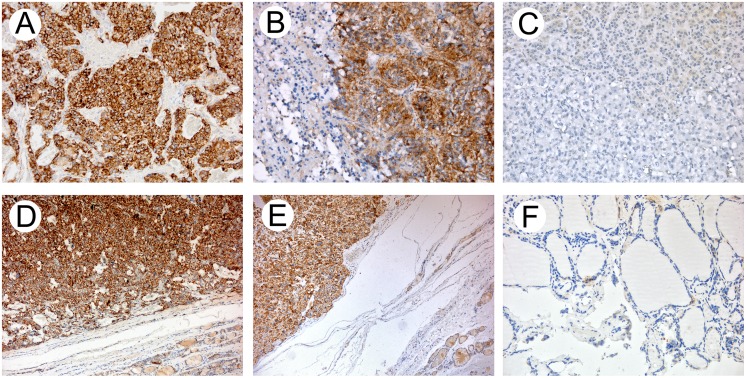
Methods: Real-time PCR and Western blot analyses were used to assess LAT1 and LAT2 gene and protein expression in 32 PHEO, 38 MTC, 16 normal adrenal medulla and 15 normal thyroid tissue samples. Immunohistochemistry method was applied to identify the proteins' subcellular localization.

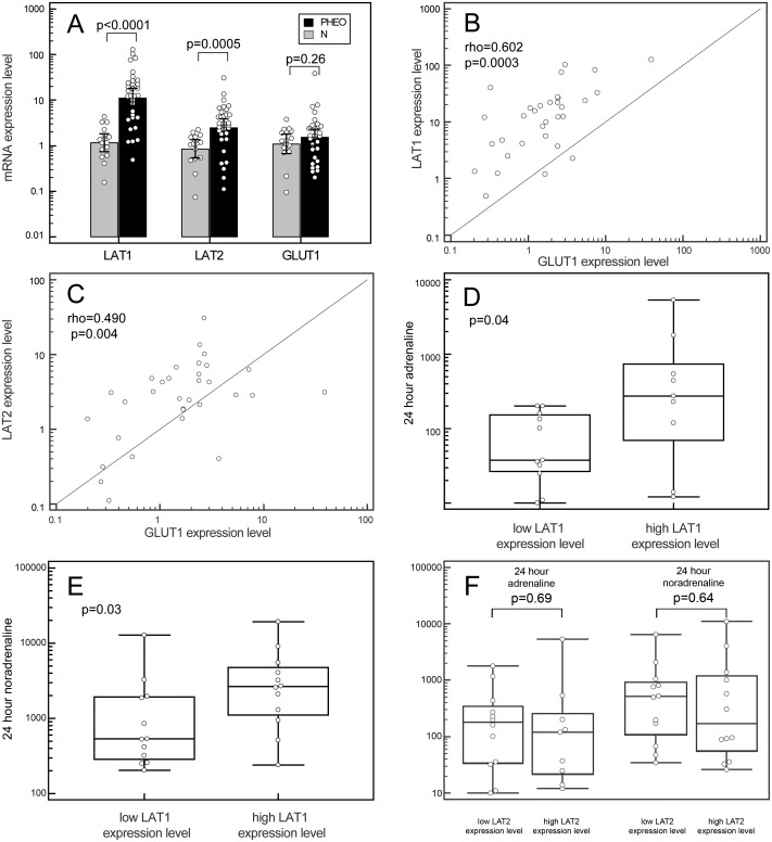
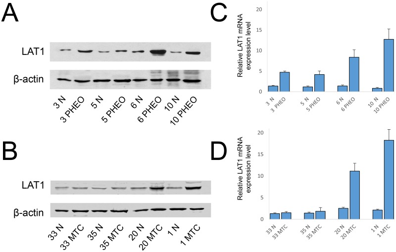
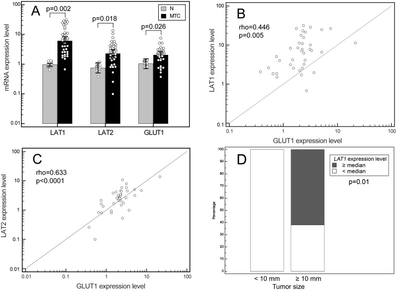
Results: LAT1 and LAT2 were overexpressed in both PHEO and MTC by comparison with normal tissues. LAT1 presented a stronger induction than LAT2, and their greater expression was more evident in PHEO (15.1- and 4.1-fold increases, respectively) than in MTC (9.9- and 4.1-fold increases, respectively). Furthermore we found a good correlation between LAT1/2 and GLUT1 expression levels. A positive correlation was also found between urinary noradrenaline and adrenaline levels and LAT1 gene expression in PHEO. The increased expression of LAT1 is also confirmed at the protein level, in both PHEO and MTC, with a strong cytoplasmic localization.

Conclusions: The present study is the first to provide experimental evidence of the overexpression in some NET cancers (such as PHEO or MTC) of L-type amino acid transporters, and the LAT1 isoform in particular, giving the molecular basis to explain the increase of the DOPA uptake seen in such tumor cells.


Conflict of interest statement

Competing Interests: The authors have declared that no competing interests exist.

Figures

Fig 1
Fig 1. LAT1, LAT2 and GLUT1 genes expression in PHEO.
(A) Box plots of relative qPCR gene expression measurements of LAT1, LAT2 and GLUT1 in PHEO specimens and the relative paired normal tissues. Each value was referred to a pool of normal thyroid tissues that was set to 1. Each dot represents a single sample. (B) and (C) Associations between the LAT1 and GLUT1 (B) and the LAT2 and GLUT1 (C) gene expressions in PHEO specimens, by Spearman’s rank correlation. (D) and (E) Box plots representing the correlation between the LAT1 expression levels in PHEO, dichotomized according to the median value, with the urinary levels of adrenaline (D) and noradrenaline (E). (F) Box plots representing the correlation between LAT2 expression levels in PHEO, dichotomized according to the median value, with the urinary levels of adrenaline (left) and noradrenaline (right). Boxes indicate the range from lower to upper quartile values, with the line inside the box representing the median. The vertical lines mark the highest and lowest value observed within a distance of 1.5 times the inter-quartile range from the bottom and the top of the boxes, respectively.
Fig 2
Fig 2. Western blot analysis and corresponding LAT1 mRNA expression level.
Representative Western blot analysis of normal (N)/tumoral match-pair samples for the expression of LAT1 in PHEO (A) and MTC (B) samples. Samples were corrected for protein loading by β-actin. Corresponding bar graphs for relative mRNA expression level of LAT1 in (C) PHEO and MTC (D).
Fig 3
Fig 3. LAT1, LAT2 and GLUT1 genes expression in MTC.
(A) Box plots of relative qPCR gene expression measurements of LAT1, LAT2 and GLUT1 in MTC specimens and the relative paired normal tissues. Each value was referred to a pool of normal thyroid tissues that was set to 1. Each dot represents a single sample. (B) and (C) Associations between the LAT1 and GLUT1 (B) and the LAT2 and GLUT1 (C) gene expressions in MTC specimens, by Spearman’s rank correlation. (D) Frequency chart representing the Fisher’s exact test correlation between the LAT1 expression levels in MTC, dichotomized according to the median value, with the tumor size dichotomized for the size of 10 millimeters.
Fig 4
Fig 4. LAT1 immunohistochemistry.
(A-B) Pheochromocytoma, strong cytoplasmic staining of LAT1 with membranous enhancement sparing of the neoplastic nuclei and normal adrenal tissue (C). (D-E) Medullary thyroid carcinoma, strong cytoplasmic staining of LAT1 with membranous enhancement sparing of the neoplastic nuclei and normal thyroid tissue (F). (A-C and F) 20× original magnification; (D-E) 10× original magnification.

References

    1. Taieb D, Timmers HJ, Hindie E, Guillet BA, Neumann HP, Walz MK, et al. (2012) EANM 2012 guidelines for radionuclide imaging of phaeochromocytoma and paraganglioma. Eur J Nucl Med Mol Imaging 39: 1977–1995. 10.1007/s00259-012-2215-8 - DOI - PMC - PubMed
    1. Becherer A, Szabo M, Karanikas G, Wunderbaldinger P, Angelberger P, Raderer M, et al. (2004) Imaging of advanced neuroendocrine tumors with (18)F-FDOPA PET. J Nucl Med 45: 1161–1167. - PubMed
    1. Hoegerle S, Altehoefer C, Ghanem N, Koehler G, Waller CF, Scheruebl H, et al. (2001) Whole-body 18F dopa PET for detection of gastrointestinal carcinoid tumors. Radiology 220: 373–380. - PubMed
    1. Pearse AG (1969) The cytochemistry and ultrastructure of polypeptide hormone-producing cells of the APUD series and the embryologic, physiologic and pathologic implications of the concept. J Histochem Cytochem 17: 303–313. - PubMed
    1. Jager PL, Chirakal R, Marriott CJ, Brouwers AH, Koopmans KP, Gulenchyn KY (2008) 6-L-18F-fluorodihydroxyphenylalanine PET in neuroendocrine tumors: basic aspects and emerging clinical applications. J Nucl Med 49: 573–586. 10.2967/jnumed.107.045708 - DOI - PubMed

Publication types

MeSH terms

Substances

Supplementary concepts