. 2016 Jun 21;7(25):38451-38466.
doi: 10.18632/oncotarget.9498.

Increased translocation of antigens to endosomes and TLR4 mediated endosomal recruitment of TAP contribute to nicotine augmented cross-presentation

Affiliations

Increased translocation of antigens to endosomes and TLR4 mediated endosomal recruitment of TAP contribute to nicotine augmented cross-presentation

Yan Yan Wang et al. Oncotarget. .

Abstract

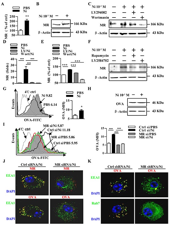
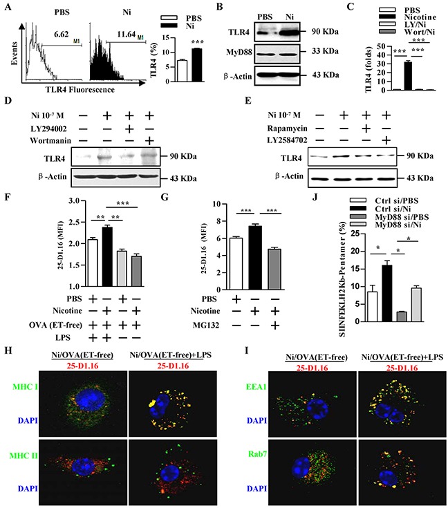
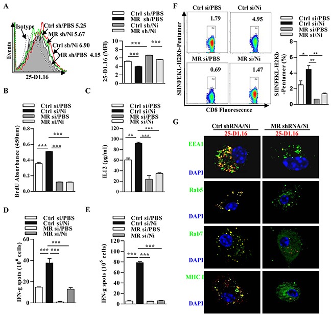
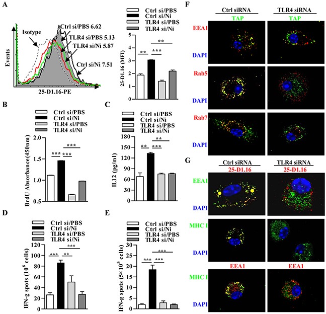
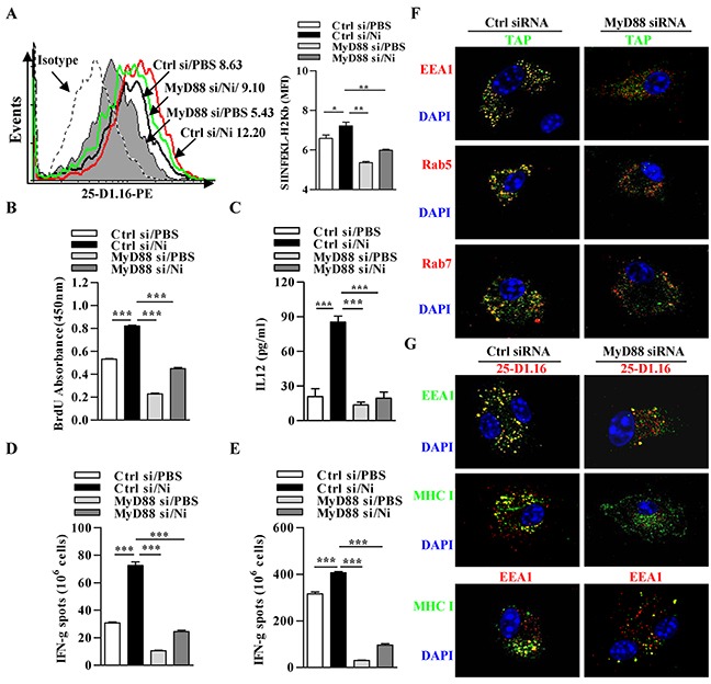
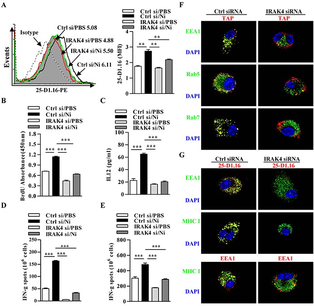
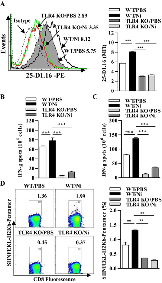
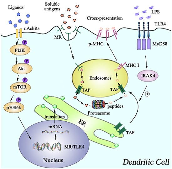
Cross-presentation by dendritic cells (DCs) requires surface molecules such as lectin, CD40, langerin, heat shock protein, mannose receptor, mediated endocytosis, the endosomal translocation of internalized antigen, and the relocation of transporter associated with antigen processing (TAP). Although the activation of α7 nicotinic acetylcholine receptor (α7 nAchR) up-regulate surface molecule expression, augment endocytosis, and enhance cross-presentation, the molecular mechanism of α7 nAchR activation-increased cross-presentation is still poorly understood. In this study, we investigated the role of mannose receptor in nicotine-increased cross-presentation and the mechanism that endotoxins orchestrating the recruitment of TAP toward endosomes. We demonstrated that nicotine increase the expressiones of mannose receptor and Toll-like receptor 4 (TLR4) via PI3K-Akt-mTOR-p70S6 pathway. Both endosomal translocation of mannose receptor-internalized antigens and TLR4 sig- naling are necessary for nicotine-augmented cross-presentation and cross-priming. Importantly, the recruitment of TAP toward endosomes via TLR4-MyD88-IRAK4 signaling contributes to nicotine-increased cross-presentation and cross-activation of T cells. Thus, these data suggest that increased recruitment of TAP to Ag-containing vesicles contributes to the superior cross-presentation efficacy of α7 nAchR activated DCs.

Keywords: cross-presentation; dendritic cells; mannose receptor; α7 nicotinic acetylcholine receptor; Toll-like receptor 4.


Conflict of interest statement

The authors have no conflict of interest to declare.

Figures

Figure 1
Figure 1. Nicotine-increased mannose receptor expression via PI3K-Akt-mTOR-p70S6 pathway contributes to receptor-mediated endocytosis and the endosomal translocation of antigens
A–F. Murine DCs were pretreated with PBS or kinase inhibitors (10 μmol/l) LY294002, wortmannin, Rapamycin, LY2584702 2 h prior to nicotine (10−7 mol/l) 12~15 h stimulation. MR expression was determined via flow cytometry (A, E), western blot analyses (B, C, F) and Q-PCR (D). β-actin was used as an internal control. G–K. After pulsing with FITC-labeled (G, I) or unlabeled (H, J, K) ‘ordinary’ OVA (50 μg/ml), MR deficient and control DCs were conferred flow cytometric assay (G, I), western blot analyses (H), or Immunofluorescence (J-K) to monitor endocytosis (G-I) or antigenic endosomal translocation (J, K). For flow cytometry (G, I), numbers in histogram indicate MFI of analyzed population (left). Statistical analysis of MFI (right) is shown. The data are presented as the mean±SEM, *p<0.05, **p<0.01, ***p<0.001, student t test or one-way ANOVA with Newman-Keulspost test. One representative from 3 independent experiments is shown. Original magnification, ×600. Ni: nicotine; MR: mannose receptor; LY: LY294002; Wort: wortmannin; OVA: ovalbumin; si: siRNA.
Figure 2
Figure 2. Nicotine-increased TLR4 expression via PI3K-Akt-mTOR-p70S6 pathway augments cross-presentation
A–E. Murine DCs were pretreated with PBS or kinase inhibitors (10 μmol/l) LY294002, wortmannin, Rapamycin, LY2584702 2 h prior to nicotine (10−7 mol/l) 12~15 h stimulation. TLR4 expression was determined via flow cytometry (A), western blot analyses (B, D, E) and Q-PCR (C). β-actin was used as an internal control. F. Flow cytometric analyses of DCs previously exposed to ‘ordinary’ OVA or endotoxin-free OVA (OVA(ET-free)) with or without LPS exposure. G. Flow cytometric analyses of DCs conferred with PBS or proteasome inhibitor MG132 (20 μmol/l) 2 h prior to ‘ordinary’ OVA pulse. H, I. Immunofluorescence observation of nicotine-increased cross-presentation. Cross-presented OVA is stained with 25-D1.16 (red); MHC class I and II molecules are stained green (H); EEA1, Rab7 (all green); nuclei are counterstained with DAPI (blue). Original magnification, ×600. J. Flow cytometric analyses of OVA-specific CD8+ T cell priming in splenocytes of the recipients by SIINFEKL-H2Kb-pentamers staining. The data are presented as the mean±SEM, *p<0.05, **p<0.01, ***p<0.001, student t test or one-way ANOVA with Newman-Keulspost test. One representative from 3 independent experiments is shown. Ni: nicotine; TLR4: Toll like receptor; LY: LY294002; Wort: wortmannin.
Figure 3
Figure 3. The up-regulation of mannose receptor is required for nicotine-increased cross-presentation
MR deficient and control DCs were stimulated with nicotine and further incubated with endotoxin-free OVA with short term exposure of LPS. A. Flow cytometric determination of cross-presented OVA in DCs. Numbers in histogram indicates MFI of analyzed population. B. BrdU cell proliferation assay of splenocytes co-cultured with OVA-pulsed DCs. C. ELISA of IL-12 in supernatants of splenocytes co-cultured with OVA-pulsed DCs. IFN-γ Elispot assay of OVA-specific CD8+ T cells in the splenocytes D. and lymph nodes E. of the recipients which conferred intraperitoneal DCs transfer. F. Flow cytometric analyses of OVA-specific CD8+ T cell priming in splenocytes of the recipients by SIINFEKL-H2Kb-pentamers staining. Numbers in dot plot indicate positive percentages of analyzed population. G. Immunofluorescence observation of nicotine-increased cross-presentation. Cross-presented OVA is stained with 25-D1.16 (red); MHC class I, Rab5, EEA1, Rab7 (all green); nuclei are counterstained with DAPI (blue). Original magnification, ×600. The data are presented as the mean±SEM, **p<0.01, ***p<0.001, one-way ANOVA with Newman-Keulspost test. One representative from 3 independent experiments is shown. Ni: nicotine; MR: mannose receptor; si: siRNA; sh: shRNA.
Figure 4
Figure 4. Nicotine-increased cross-presentation requires the endosomal recruitment of TAP via TLR4 signaling
Nicotine-treated TLR4 deficient and control DCs were incubated with endotoxin-free OVA with short term exposure of LPS. A. Flow cytometric determination of cross-presented OVA in DCs. Numbers in histogram indicates MFI of analyzed population. B. BrdU cell proliferation assay of splenocytes co-cultured with OVA-pulsed DCs. C. ELISA of IL-12 in supernatants of splenocytes co-cultured with OVA-pulsed DCs. IFN-γ Elispot assay of OVA-specific CD8+ T cells in the splenocytes D. and lymph nodes E. of the recipients which conferred intraperitoneal DCs transfer. F. Immunofluorescence observation of the recruitment of TAP toward endosomes. TAP (green); Rab5, EEA1, Rab7 (all red); nuclei are counterstained with DAPI (blue). Original magnification, ×600. G. Immunofluorescence observation of TLR4 deficiency on nicotine-increased cross-presentation. Cross-presented OVA is stained with 25-D1.16 (red); MHC class I (green); EEA1 in 25-D1.16 co-localization is green and in MHC class I co-localization is red; nuclei are counterstained with DAPI (blue). Original magnification, ×600. The data are presented as the mean±SEM, **p<0.01, ***p<0.001, one-way ANOVA with Newman-Keulspost test. One representative from 3 independent experiments is shown. Ni: nicotine; TLR4: Toll like receptor; si: siRNA.
Figure 5
Figure 5. Nicotine-increased cross-presentation requires the endosomal recruitment of TAP via MyD88 signaling
Nicotine-treated MyD88 deficient and control DCs were incubated with endotoxin-free OVA with short term exposure of LPS. A. Flow cytometric determination of cross-presented OVA in DCs. Numbers in histogram indicates MFI of analyzed population. B. BrdU cell proliferation assay of splenocytes co-cultured with OVA-pulsed DCs. C. ELISA of IL-12 in supernatants of splenocytes co-cultured with OVA-pulsed DCs. IFN-γ Elispot assay of OVA-specific CD8+ T cells in the splenocytes D. and lymph nodes E. of the recipients which conferred intraperitoneal DCs transfer. F. Immunofluorescence observation of the recruitment of TAP toward endosomes. TAP (green); Rab5, EEA1, Rab7 (all red); nuclei are counterstained with DAPI (blue). Original magnification, ×600. G. Immunofluorescence observation of MyD88 deficiency on nicotine-increased cross-presentation. Cross-presented OVA is stained with 25-D1.16 (red); MHC class I (green); EEA1 in 25-D1.16 co-localization is green and in MHC class I co-localization is red; nuclei are counterstained with DAPI (blue). Original magnification, ×600. The data are presented as the mean±SEM, *p<0.05, **p<0.01, ***p<0.001, one-way ANOVA with Newman-Keulspost test. One representative from 3 independent experiments is shown. Ni: nicotine; si: siRNA.
Figure 6
Figure 6. Nicotine-increased cross-presentation requires the endosomal recruitment of TAP via IRAK4 signaling
Nicotine-treated IRAK4 deficient and control DCs were incubated with endotoxin-free OVA with short term exposure of LPS. A. Flow cytometric determination of cross-presented OVA in DCs. Numbers in histogram indicates MFI of analyzed population. B. BrdU cell proliferation assay of splenocytes co-cultured with OVA-pulsed DCs. C. ELISA of IL-12 in supernatants of splenocytes co-cultured with OVA-pulsed DCs. IFN-γ Elispot assay of OVA-specific CD8+ T cells in the splenocytes D. and lymph nodes E. of the recipients which conferred intraperitoneal DCs transfer. F. Immunofluorescence observation of the recruitment of TAP toward endodomes. TAP (green); Rab5, EEA1, Rab7 (all red); nuclei are counterstained with DAPI (blue). Original magnification, ×600. G. Immunofluorescence observation of IRAK4 deficiency on nicotine-increased cross-presentation. Cross-presented OVA is stained with 25-D1.16 (red); MHC class I (green); EEA1 in 25-D1.16 co-localization is green and in MHC class I co-localization is red; nuclei are counterstained with DAPI (blue). Original magnification, ×600. The data are presented as the mean±SEM, *p<0.05, **p<0.01, ***p<0.001, one-way ANOVA with Newman-Keulspost test. One representative from 3 independent experiments is shown. Ni: nicotine; si: siRNA.
Figure 7
Figure 7. TLR4 signaling-promoted endosomal recruitment of TAP facilitates nicotine-increased cross-presentation
Wild-type and TLR4 deficient DCs were stimulated with nicotine and further incubated with endotoxin-free OVA with short term exposure of LPS. A. Flow cytometric determination of cross-presented OVA in Wild-type and TLR4 deficient DCs. Numbers in histogram indicate MFI of analyzed population. B-C. IFN-γ Elispot assay of OVA-specific CD8+ T cells in the splenocytes (B) and lymph nodes (C) of the recipients which conferred intraperitoneal DCs transfer. D. Flow cytometric analyses of SIINFEKL-H2Kb pentramers positive cells in the splenocytes of DCs-transferred recipients. Numbers in dot plot indicate positive percentages of analyzed population. The data are presented as the mean±SEM, **p<0.01, ***p<0.001, one-way ANOVA with Newman-Keulspost test. One representative from 3 independent experiments is shown. Ni: nicotine; TLR4 KO: Toll like receptor 4 deficient; WT: wild type.
Figure 8
Figure 8. Model of mannose receptor and TLR4 signaling in nicotine-increased cross-presentation
The up-regulation of MR and TLR4, which was achieved via PI3K-Akt-mTOR-p70S6 pathway, lead to the endosomal translocation of internalized antigens, and the endosomal recruitment of TAP via TLR4-MyD88-IRAK4 signal, respectively. Increased translocations of internalized antigens toward endosomes, together with the endosomal recruitment of TAP, facilitate α7 nAChR activation-increased cross-presentation and subsequent cross-priming.

Similar articles

Cited by

References

    1. Mellman I, Steinman RM. Dendritic cells: specialized and regulated antigen processing machines. Cell. 2001;106:255–8. - PubMed
    1. Seya T, Shime H, Takeda Y, Tatematsu M, Takashima K, Matsumoto M. Adjuvant for vaccine immunotherapy of cancer - focusing on Toll-like receptor 2 and 3 agonists for safely enhancing antitumor immunity. Cancer Sci. 2015;106:1659–68. - PMC - PubMed
    1. Villadangos JA, Heath WR, Carbone FR. Outside looking in: the inner workings of the cross-presentation pathway within dendritic cells. Trends Immunol. 2007;28:45–7. - PubMed
    1. Wessler IK, Kirkpatrick CJ. The non-neuronal cholinergic system: an emerging drug target in the airways. Pulm Pharmacol Ther. 2001;14:423–34. - PubMed
    1. Printz C. Gap narrows in African American smoking-related cancers, increases in breast and colorectal cancers. Cancer. 2011;117:2357. - PubMed

MeSH terms

Substances