. 2016 Aug 16;7(33):52940-52956.
doi: 10.18632/oncotarget.9507.

FOXP2-positive diffuse large B-cell lymphomas exhibit a poor response to R-CHOP therapy and distinct biological signatures

Affiliations

FOXP2-positive diffuse large B-cell lymphomas exhibit a poor response to R-CHOP therapy and distinct biological signatures

Kah Keng Wong et al. Oncotarget. .

Abstract

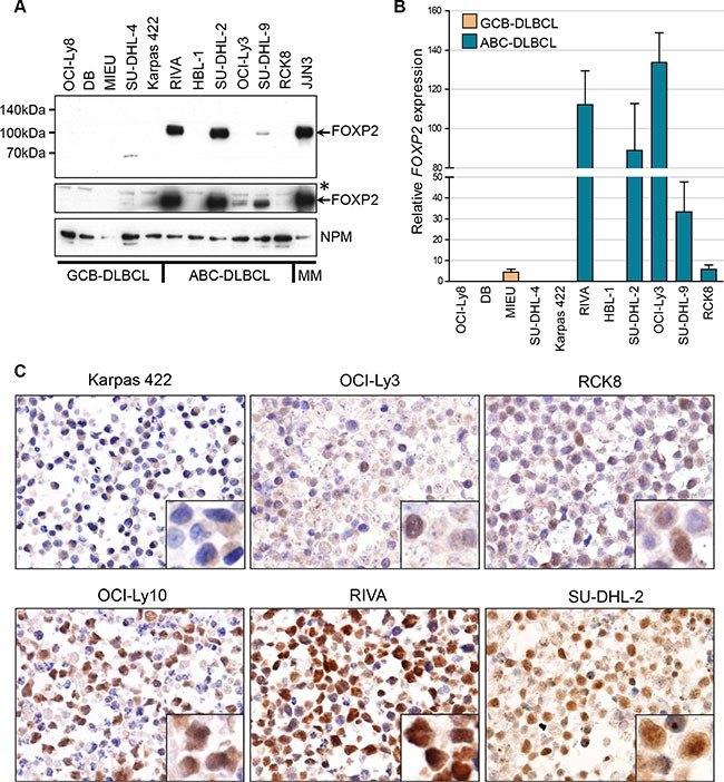
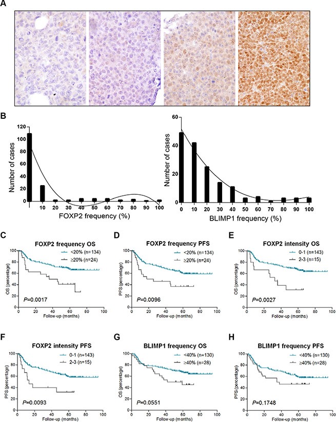
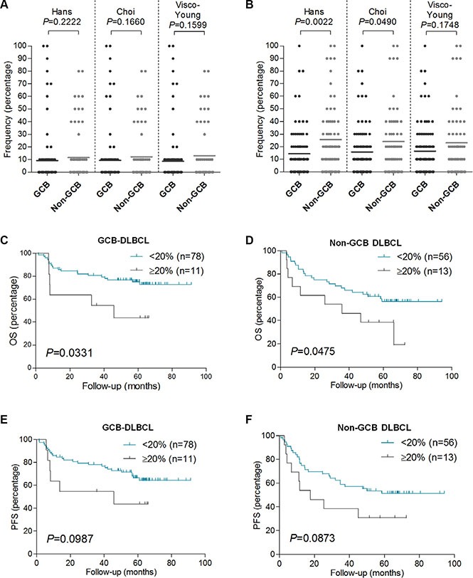
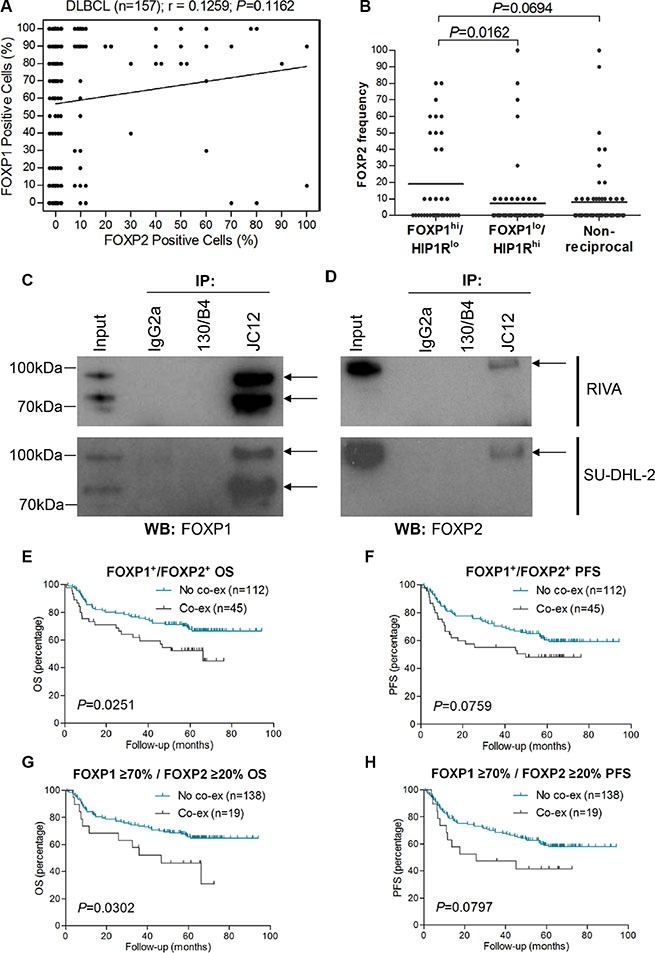
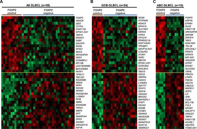
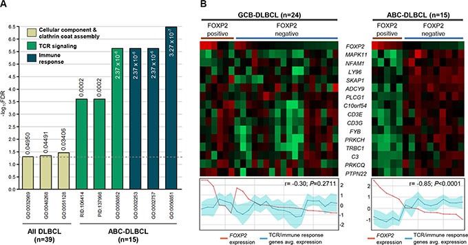
FOXP2 shares partially overlapping normal tissue expression and functionality with FOXP1; an established diffuse large B-cell lymphoma (DLBCL) oncogene and marker of poor prognosis. FOXP2 is expressed in the plasma cell malignancy multiple myeloma but has not been studied in DLBCL, where a poor prognosis activated B-cell (ABC)-like subtype display partially blocked plasma cell differentiation. FOXP2 protein expression was detected in ABC-DLBCL cell lines, and in primary DLBCL samples tumoral FOXP2 protein expression was detected in both germinal center B-cell-like (GCB) and non-GCB DLBCL. In biopsies from DLBCL patients treated with immunochemotherapy (R-CHOP), ≥ 20% nuclear tumoral FOXP2-positivity (n = 24/158) correlated with significantly inferior overall survival (OS: P = 0.0017) and progression-free survival (PFS: P = 0.0096). This remained significant in multivariate analysis against either the international prognostic index score or the non-GCB DLBCL phenotype (P < 0.05 for both OS and PFS). Expression of BLIMP1, a marker of plasmacytic differentiation that is commonly inactivated in ABC-DLBCL, did not correlate with patient outcome or FOXP2 expression in this series. Increased frequency of FOXP2 expression significantly correlated with FOXP1-positivity (P = 0.0187), and FOXP1 co-immunoprecipitated FOXP2 from ABC-DLBCL cells indicating that these proteins can co-localize in a multi-protein complex. FOXP2-positive DLBCL had reduced expression of HIP1R (P = 0.0348), which is directly repressed by FOXP1, and exhibited distinct patterns of gene expression. Specifically in ABC-DLBCL these were associated with lower expression of immune response and T-cell receptor signaling pathways. Further studies are warranted to investigate the potential functional cooperativity between FOXP1 and FOXP2 in repressing immune responses during the pathogenesis of high-risk DLBCL.

Keywords: FOXP2; diffuse large B-cell lymphoma; survival.


Conflict of interest statement

The authors declare no conflicts of interest.

Figures

Figure 1
Figure 1. FOXP2 is expressed in ABC-DLBCL cell lines
(A) Western blotting of nuclear lysates from GCB-DLBCL, ABC-DLBCL and myeloma cell lines with anti-FOXP2 (clone 73A/8) and anti-NPM (clone NA24) antibodies. An overexposed image (middle panel) shows very weak FOXP2 expression in OCI-Ly3 and just detectable expression in RCK8. *Indicates an upper band that is thought to be non-specific. MIEU remained negative in other experiments with more abundant sample loading. (B) qRT-PCR for FOXP2 transcripts in GCB- and ABC-DLBCL cell lines. FOXP2 expression was normalized according to the expression of 18S rRNA housekeeping gene and expressed relative to JJN3 (set to 100%). (C) Immunohistochemical labeling of FOXP2 nuclear expression in DLBCL cell lines: showing negative (Karpas 422), weak (OCI-Ly3, RCK8) and strongly positive (OCI-Ly10, RIVA, SU-DHL-2) examples. Higher power insets are shown in the bottom right corners of each panel.
Figure 2
Figure 2. FOXP2 expression confers inferior survival in R-CHOP-treated DLBCL cases
(A) Immunohistochemical labeling of FOXP2 expression in primary DLBCL cases with variable intensity and frequency of expression. Left to right these were scored as negative, intensity 1/frequency 55%, intensity 2/frequency 80%, intensity 3/frequency 100%. (B) Frequency distribution of FOXP2 (left panel) and BLIMP1 (right panel) at each 10% cut-off level in DLBCL cases (n = 158). (CH) Overall survival (OS) and progression free survival (PFS) of DLBCL cases (n = 158) according to FOXP2 frequency at 20% cut-off (C–D) or intensity (E–F), and BLIMP1 frequency at 40% cut-off (G–H).
Figure 3
Figure 3. FOXP2 expression is not associated with non-GCB DLBCL primary cases, and it confers worse OS in GCB or non-GCB DLBCL subtypes
(AB) Relationship of FOXP2 (A) or BLIMP1 (B) protein expression frequency with GCB or non-GCB DLBCL subtype according to Hans (n = 158), Choi (n = 155) or Visco-Young (n = 157) immunohistochemical algorithms. (CF) OS (C–D) and PFS (E–F) in GCB-DLBCL (n = 89) or non-GCB DLBCL (n = 69) according to FOXP2 20% cut-off. The DLBCL cases were stratified according to Hans algorithm.
Figure 4
Figure 4. FOXP2 is co-immunoprecipitated by FOXP1 and their co-expression confers worse survival in R-CHOP-treated DLBCL cases
(A) Correlation of FOXP1 and FOXP2 expression frequency in DLBCL (n = 157). (B) Relationship of FOXP2 frequency with FOXP1hi/HIP1Rlo (n = 36), FOXP1lo/HIP1Rhi (n = 58) or non-reciprocal (n = 62) DLBCL cases. (CD) Co-immunoprecipitation of endogenous FOXP1 in RIVA and SU-DHL-2 cell lysates with anti-FOXP1 JC12 antibody or with non-specific IgG2a isotype and 130/B4 (“in house” anti-NFIL3) control antibodies. Input indicates non-immunoprecipitated cell lysates. JC12 blotting; top arrow indicates FOXP1FL, bottom arrow indicates FOXP1s (C). Co-immunoprecipitated FOXP2 was detected by Western blotting the JC12 immunoprecipitated proteins with the FOXP2-73A/8 antibody (D).
Figure 5
Figure 5. Genes most highly-correlated with FOXP2 protein expression in primary DLBCL cases
(AC) Top 20 genes positively- or inversely-correlated with FOXP2 protein positive expression in all DLBCL cases (n = 39; A), GCB-DLBCL (n = 24; B) or ABC-DLBCL (n = 15; C) subtype. The cases were stratified according to FOXP2 protein positive (i.e. absolute positivity without regarding frequency cut-off) or negative (i.e. 0% frequency). FOXP2 protein was positive in six (of 24) GCB-DLBCL and five (of 15) ABC–DLBCL cases.
Figure 6
Figure 6. TCR signaling and immune response signatures were significantly reduced in FOXP2-positive ABC-DLBCL cases
(A) Gene ontology and pathway signatures suppressed in FOXP2-positive DLBCL cases compared with their FOXP2-negative counterparts. All DLBCL cases (n = 39) or ABC-DLBCL (n = 15) molecular subtype were analyzed separately. The bar graph was constructed on a negative log10 scale with higher -log10 value denoting more significant FDR value. The linear FDR values were shown next to or within each bar, and the horizontal dotted line denotes linear FDR threshold of 0.05. GO: Gene Ontology; PID: Pathway Interaction Database; RID: Reactome ID. (B) Heatmap visualization of genes involved in TCR and immune response signatures compared with FOXP2 microarray transcript expression (probe: 1555516_at) in FOXP2 protein positive or negative GCB-DLBCL (n = 24) or ABC-DLBCL (n = 15) cases. Line graphs comparing FOXP2 transcript expression with the averaged expression values of TCR/immune response genes (the region shaded in turquoise represents the standard deviation of the averaged values) were plotted corresponding to each case present on the heatmap.

Similar articles

Cited by

References

    1. Engelhard M, Brittinger G, Huhn D, Gerhartz HH, Meusers P, Siegert W, Thiel E, Wilmanns W, Aydemir U, Bierwolf S, Griesser H, Tiemann M, Lennert K. Subclassification of diffuse large B-cell lymphomas according to the Kiel classification: distinction of centroblastic and immunoblastic lymphomas is a significant prognostic risk factor. Blood. 1997;89:2291–2297. - PubMed
    1. Diebold J, Anderson JR, Armitage JO, Connors JM, Maclennan KA, Muller-Hermelink HK, Nathwani BN, Ullrich F, Weisenburger DD. Diffuse large B-cell lymphoma: a clinicopathologic analysis of 444 cases classified according to the updated Kiel classification. Leuk Lymphoma. 2002;43:97–104. - PubMed
    1. De Paepe P, Achten R, Verhoef G, Wlodarska I, Stul M, Vanhentenrijk V, Praet M, De Wolf-Peeters C. Large cleaved and immunoblastic lymphoma may represent two distinct clinicopathologic entities within the group of diffuse large B-cell lymphomas. J Clin Oncol. 2005;23:7060–7068. - PubMed
    1. Ott G, Ziepert M, Klapper W, Horn H, Szczepanowski M, Bernd HW, Thorns C, Feller AC, Lenze D, Hummel M, Stein H, Muller-Hermelink HK, Frank M, et al. Immunoblastic morphology but not the immunohistochemical GCB/nonGCB classifier predicts outcome in diffuse large B-cell lymphoma in the RICOVER-60 trial of the DSHNHL. Blood. 2010;116:4916–4925. - PubMed
    1. Alizadeh AA, Eisen MB, Davis RE, Ma C, Lossos IS, Rosenwald A, Boldrick JC, Sabet H, Tran T, Yu X, Powell JI, Yang L, Marti GE, et al. Distinct types of diffuse large B-cell lymphoma identified by gene expression profiling. Nature. 2000;403:503–511. - PubMed

MeSH terms