. 2017 Feb 28;8(9):14325-14342.
doi: 10.18632/oncotarget.9527.

Antivascular and antitumor properties of the tubulin-binding chalcone TUB091

Affiliations

Antivascular and antitumor properties of the tubulin-binding chalcone TUB091

María-Dolores Canela et al. Oncotarget. .

Abstract

We investigated the microtubule-destabilizing, vascular-targeting, anti-tumor and anti-metastatic activities of a new series of chalcones, whose prototype compound is (E)-3-(3''-amino-4''-methoxyphenyl)-1-(5'-methoxy-3',4'-methylendioxyphenyl)-2-methylprop-2-en-1-one (TUB091). X-ray crystallography showed that these chalcones bind to the colchicine site of tubulin and therefore prevent the curved-to-straight structural transition of tubulin, which is required for microtubule formation. Accordingly, TUB091 inhibited cancer and endothelial cell growth, induced G2/M phase arrest and apoptosis at 1-10 nM. In addition, TUB091 displayed vascular disrupting effects in vitro and in the chicken chorioallantoic membrane (CAM) assay at low nanomolar concentrations. A water-soluble L-Lys-L-Pro derivative of TUB091 (i.e. TUB099) showed potent antitumor activity in melanoma and breast cancer xenograft models by causing rapid intratumoral vascular shutdown and massive tumor necrosis. TUB099 also displayed anti-metastatic activity similar to that of combretastatin A4-phosphate. Our data indicate that this novel class of chalcones represents interesting lead molecules for the design of vascular disrupting agents (VDAs). Moreover, we provide evidence that our prodrug approach may be valuable for the development of anti-cancer drugs.

Keywords: cancer; drug research; tubulin; vascular-disrupting.


Conflict of interest statement

CONFLICTS OF INTEREST

Maria-Dolores Canela is currently working at Teva Pharmaceuticals as Medical Advisor.

Figures

Figure 1
Figure 1. Structural formulae of reference compounds and newly synthesized chalcones
Figure 2
Figure 2. Tubulin-binding activity of chalcones
(A) Growth curve of MDA-MB-231 cells in the presence of different concentrations of TUB091. The graph presents the normalized cell index (based on real-time impedance measurements) from 18 to 46 h after cell seeding. Compound was added 20 h after cell seeding (vertical line). Fast reduction in cell index with recovery of surviving cells after 14 h treatment (as seen at 5 nM) is specific for tubulin-binding compounds. (B) MDA-MB-231 cells were treated for 15 h with different concentrations of TUB091, fixed and stained with anti-β-tubulin antibody (green) to visualize the microtubules and Hoechst for DNA (blue). Cell cycle phase and spindle organization were investigated in individual cells (minimum 100 per condition) and quantified based on spindle morphology.
Figure 3
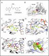
Figure 3. X-ray analysis of the chalcone-tubulin complex
(A) Chemical structure of TUB092. (B) Overall view of the complex formed between αβ-tubulin and TUB092. α- and β-tubulin are in dark and light grey, respectively. (C, D). Close-up views of the interaction network observed between TUB092 (green) and tubulin (gray). Interacting residues of tubulin are shown in stick representation and are labeled. Oxygen and nitrogen atoms are colored red and blue, respectively; carbon atoms are in green (TUB092) or gray (tubulin). Hydrogen bonds are depicted as black broken lines. Secondary structure elements of tubulin are labeled in blue. (D) 90° rotation of C. (E-F) Comparison of TUB092 and colchicine tubulin-binding modes. (E) Superimposition of the tubulin–TUB092 (dark gray ribbons) and tubulin–colchicine (PDB ID 4O2B, light gray ribbons) structures. TUB092 and colchicine are in green and violet-purple stick representation, respectively. Water molecules are displayed as red (TUB092 structure) and grey (colchicine structure) spheres, respectively. (F) Superimposition of the “curved” (tubulin–TUB092; light gray ribbons) and “straight” (PDB ID 1JFF; light blue ribbons) tubulin conformational states. TUB092 is shown in green sphere representation. Arrows highlight regions of steric clashes between straight tubulin and TUB092.
Figure 4
Figure 4. TUB091 induces G2/M phase arrest and apoptosis in human breast cancer MDA-MB-231 cells
(A) Flow cytometric analysis of cell cycle distribution of MDA-MB-231 cells that were treated for 24 h with different concentrations of TUB091. The experiment was repeated 3 times with similar results. Results from 1 experiment are shown. (B) Caspase-3 activity in MDA-MB-231 cells. Different concentrations of compound and 2 μM of the caspase-3 substrate DEVD-NucView488 were added to MDA-MB-231 cells. At indicated time-points, the cells were incubated with 2 μg/ml Hoechst 33342 to stain the nucleus, and imaged. Data are the result of 3 experiments performed in duplicate and are expressed as mean ± SD. *p < 0.05 compared with control.
Figure 5
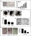
Figure 5. Effects of TUB091 on endothelial cell functions in vitro
(A) Endothelial cell proliferation. HUVEC and HMEC-1 cells were seeded at 20,000 cells/cm2. After 24 h, compounds were added. The cells were allowed to grow for an additional 3 days, trypsinized and counted. Mean of 3 independent experiments with similar results is shown. (B) Flow cytometric analysis of cell cycle distribution of HMEC-1 cells that were treated for 24 h with different concentrations of TUB091. The experiment was repeated 3 times with similar results. Results from 1 experiment are shown. (C) Caspase-3 activity in HUVECs. Different concentrations of TUB091 or CA-4P and 2 μM of the caspase-3 substrate DEVD-NucView488 were added to HUVECs. At 24 h, the cells were incubated with 2 μg/ml Hoechst 33342 to stain the nucleus, and imaged. (D) Endothelial cell migration. Wounds were created in confluent HMEC-1 monolayers. Then, cells were incubated in fresh medium in the presence of the test compounds and 1 μg/ml of mitomycine C to inhibit cell proliferation. After 18 h, the wounds were photographed and wound repair was quantified by computerized analysis. (C, D) Data are the result of 3 experiments performed in duplicate and are expressed as mean ± SD. *p < 0.05 compared with control.
Figure 6
Figure 6. Anti-angiogenic and VDA activity of TUB091
(A) Disruption of the vascular network. HMEC-1 cells were cultured on matrigel for 3 h to allow the formation of tube-like structures. Then different concentrations of compounds were added. After 90 min, tube formation was quantified. Data are the result of 3 experiments performed in duplicate and are expressed as mean ± SD. *p < 0.05 compared with control. (BC) Gelatin sponge CAM assay. Sponges containing 3 nmol of CA-4P or TUB091 were added onto the CAM. (B) Newly formed microvessels directed at the sponges were counted 4 days later (lower panel). Hundred eggs were used per condition. Data are expressed as mean ± SD. *p < 0.05 compared with control. Macroscopic pictures of the CAM are shown (upper panel) (C) Histological examination demonstrates a complete disappearance of the intermediate mesenchyme without any cellular and vascular components in the TUB091-treated CAMs, as compared to control. CH: chorion; M: mesenchyme, A: allantois. (D) Plastic discs containing the dried compound were added onto the CAM. Graph shows the percentage of CAMs with complete inhibition of blood vessel formation 2 days later. Data are the result of 2 independent experiments using 8 eggs per condition. *p < 0.05 compared with control.
Figure 7
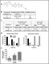
Figure 7. Biological evaluation of TUB091 prodrugs
(A) Structure of TUB099, the L-lysyl-L-prolyl derivative of TUB091. (B) Aqueous solubility of TUB091 and its prodrugs in PBS. (C) Growth-inhibitory activity is presented as IC50, i.e. concentration that reduces cell growth by 50%. HMEC-1: human microvascular endothelial cell line-1; BAEC: bovine aortic endothelial cells; Cem: human lymphocytic leukemia cells; HeLa, human cervical carcinoma cells. Data are mean ± SD. (D) Release of parent compound (TUB091) in human serum and murine liver extract. TUB099 (100 μM) was incubated for 60 and 180 min at 37°C in human serum or mouse liver extract. Aliquots were quantified by HPLC with detection at 305 nm. (E) Vascular-disrupting activity of TUB099. HMEC-1 cells were cultured on matrigel for 3 h to allow the formation of tube-like structures. Then different concentrations of TUB099 were added. After 90 min, tube formation was quantified. Data are the result of 3 experiments performed in duplicate and are expressed as mean ± SD. *p < 0.05 compared with control.
Figure 8
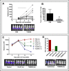
Figure 8. Intratumoral (i.t.) TUB099 impairs primary tumor growth and metastasis
(A, B) Effect of TUB099 on subcutaneous growth of melanoma. Thirty thousand B16.F10.luc2 cells were injected subcutaneously in SCID mice. Intratumoral (i.t.) treatment with TUB099 (10 mg/kg; full line) or vehicle (dashed line) was started 3 days after cell injection and continued for 5 consecutive days (horizontal line in A). The mice were imaged (A) at regular time intervals. Each line in A represents one single mouse. Representative pictures of bioluminescence in the tumors of control and TUB099-treated mice at day 10 are shown. (B) Tumor weight at day 17 after cell inoculation, i.e. 10 days after treatment was terminated. Data are mean ± SEM, n = 5. *p < 0.05. (C, D) Effect of TUB099 on primary tumor growth and metastasis of MDA-MB-231/4mRL.luc2 human breast cancer cells. 4mRL.luc2 cells (106) were injected in the mammary fat pad of SCID mice. PBS, TUB099 or CA-4P was administered i.t. at day 12 only (1×) or from day 12 till day 15 (4×). (C) The mice were imaged at regular time intervals. Data are mean ± SEM of luciferase measurements, n = 5. At day 16, a significant reduction in luminescent signal is visible in the CA-4P- but particularly in the TUB099-treated tumors. (D) Lung and/or lymph node metastasis 19 days after removal of the primary tumor (i.e. 35 days after cell inoculation). Experiments were performed twice with similar results. Results of one experiment are shown.
Figure 9
Figure 9. Vascular disrupting activity of systemic (i.p.) TUB099 treatment
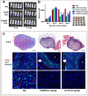
(AC) MDA-MB-231/4mRL.luc2 cells (106) were injected in the mammary fat pad of SCID mice. PBS, TUB099 or CA-4P (15 or 30 mg/kg) was administered once, intraperitoneally, at day 14. (A) Luminescence in the tumor before (left) and 2 h after administration (right) of compound. (B) The mice were imaged at regular time intervals. Data are mean ± SEM, n = 5.No difference in primary tumor size was observed at day 16, when the tumors were resected and processed for histological evaluation (inset). (C) Total tumor sections were stained with H&E or were double immunostained with anti-CD31 (red) and anti-Ki67 (green) antibodies, followed by nuclear counterstaining with Hoechst (blue). Asterisks indicate necrotic tumor center. Higher magnification of tumor center (bottom panel). Arrow indicates viable tumor rim. Scale bar = 1 mm. Two independent experiments yielded comparable data.

Similar articles

Cited by

References

    1. Torre LA, Bray F, Siegel RL, Ferlay J, Lortet-Tieulent J, Jemal A. Global cancer statistics, 2012. CA Cancer J Clin. 2015;65:87–108. - PubMed
    1. Holohan C, Van Schaeybroeck S, Longley DB, Johnston PG. Cancer drug resistance: an evolving paradigm. Nat Rev Cancer. 2013;13:714–726. - PubMed
    1. Tozer GM, Kanthou C, Baguley BC. Disrupting tumour blood vessels. Nat Rev Cancer. 2005;5:423–435. - PubMed
    1. McKeage MJ, Baguley BC. Disrupting established tumor blood vessels: an emerging therapeutic strategy for cancer. Cancer. 2010;116:1859–1871. - PubMed
    1. Greene LM, Meegan MJ, Zisterer DM. Combretastatins: More Than Just Vascular Targeting Agents? J Pharmacol Exp Ther. 2015;355:212–227. - PubMed

MeSH terms