. 2016 May 26;533(7604):499-503.
doi: 10.1038/nature18015. Epub 2016 May 11.

Synchronized mitochondrial and cytosolic translation programs

Affiliations

Synchronized mitochondrial and cytosolic translation programs

Mary T Couvillion et al. Nature. .

Abstract

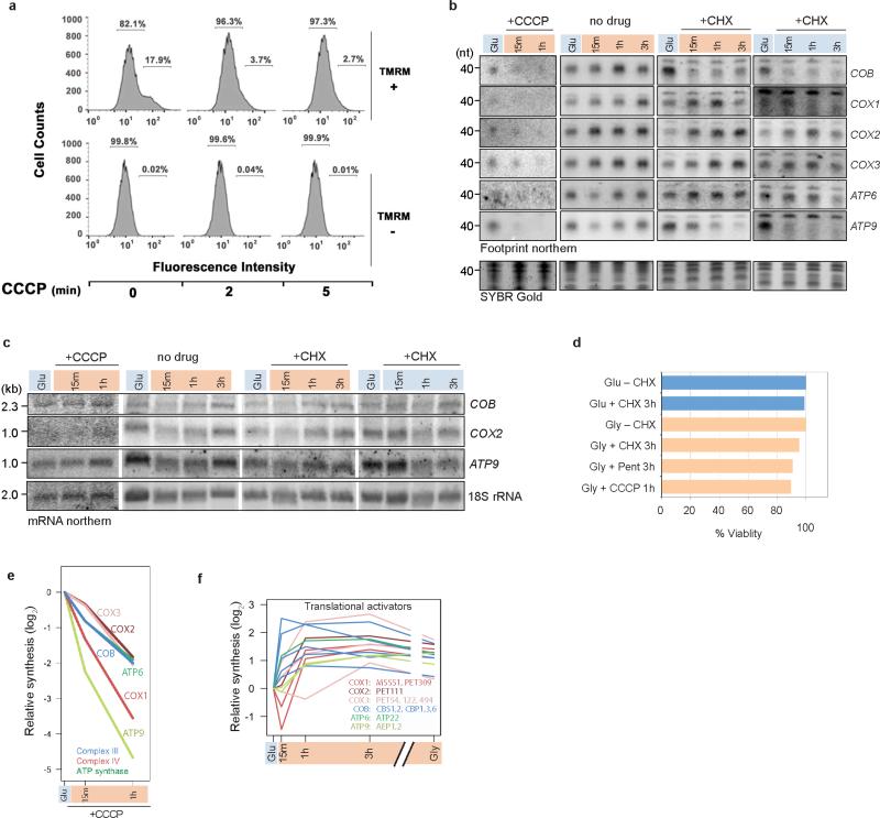
Oxidative phosphorylation (OXPHOS) is a vital process for energy generation, and is carried out by complexes within the mitochondria. OXPHOS complexes pose a unique challenge for cells because their subunits are encoded on both the nuclear and the mitochondrial genomes. Genomic approaches designed to study nuclear/cytosolic and bacterial gene expression have not been broadly applied to mitochondria, so the co-regulation of OXPHOS genes remains largely unexplored. Here we monitor mitochondrial and nuclear gene expression in Saccharomyces cerevisiae during mitochondrial biogenesis, when OXPHOS complexes are synthesized. We show that nuclear- and mitochondrial-encoded OXPHOS transcript levels do not increase concordantly. Instead, mitochondrial and cytosolic translation are rapidly, dynamically and synchronously regulated. Furthermore, cytosolic translation processes control mitochondrial translation unidirectionally. Thus, the nuclear genome coordinates mitochondrial and cytosolic translation to orchestrate the timely synthesis of OXPHOS complexes, representing an unappreciated regulatory layer shaping the mitochondrial proteome. Our whole-cell genomic profiling approach establishes a foundation for studies of global gene regulation in mitochondria.


Figures

Extended Data Figure 1
Extended Data Figure 1. OXPHOS proteins are induced during mitochondrial biogenesis
Western blot analysis of mitochondrial (Cob, Cox1, Cox2) and nuclear (Cox4) OXPHOS proteins compared to FLAG-tagged mitoribosome small subunit protein MrpS17 and GAPDH. For gel source data, see Supplementary Fig. 1.
Extended Data Figure 2
Extended Data Figure 2. Dynamics of non-OXPHOS RNAs through mitochondrial biogenesis
a, b, c, RNA levels (reads per kb) normalized to spike-in controls and plotted as -fold change compared to levels in log phase glucose growth for (a) all nuclear-encoded structural components of the complexes shown, (b) intron-encoded maturases, and (c) nuclear and mitochondrial-encoded mitoribosome subunits. To calculate values for maturase transcripts, only reads not overlapping the main ORF (COX1 or COB) were considered. Group II intron splicing intermediates stably accumulate and may not represent translation-competent transcripts.
Extended Data Figure 3
Extended Data Figure 3. Optimization of affinity purification for intact mitoribosomes
a, Spot tests verifying tagged mitoribosome subunits are functional as they support respiratory growth on glycerol (YPG). ρ0 is a strain without mitochondrial DNA. b, Frequency of petite colonies in our corrected S288c strain (see Methods) after growth for 5 days on 0.1% glucose + 3% glycerol. BY4742 is S288c background with designer auxotrophies. Σ1278b is a strain with wild-type HAP1, and a high-fidelity allele of MIP1, MIP1[Σ], along with other differences compared to S288c. Error bars show variation due to counting, with 175–750 colonies counted for each sample. c, Lysis and IP buffer conditions affect mitoribosome subunit association and thus footprint retention. Left panel: silver staining after IP of the large subunit (LSU) with Mrp20-F and of the small subunit (SSU) with MrpS17-F in Condition 1 (20 mM Tris, pH 8.0, 200 mM KCl, 5 mM MgCl2, 0.5% lauryl maltoside), and in Condition 2 (10 mM Tris, pH 8.0, 50 mM NH4Cl, 10 mM MgCl2, 0.5% lauryl maltoside). Arrowheads indicate bands that appear in both IPs in Condition 2 that can be assigned to the LSU or SSU by comparison to Condition 1. Asterisks mark the expected mobility of the tagged proteins. Right panel: Northern blotting of the co-purifying RNA in each condition. For gel source data, see Supplementary Fig. 1. d, Western blot showing fractions from IP using optimized buffer conditions. FLAG IP targeting the mitoribosome SSU co-purifies an HA-tagged LSU protein. For gel source data, see Supplementary Fig. 1.
Extended Data Figure 4
Extended Data Figure 4. Mitoribosome profiling is robust, reproducible, and does not require translation inhibitors
a, Mapping statistics for representative mitoribosome and cytoribosome profiling libraries from log phase glycerol-grown cells. b, Fraction of reads mapping to each frame of mitochondrial ORFs (left panel) and nuclear ORFs (right panel) in mitoribosome profiling and cytoribosome profiling data, respectively. RNA-seq reads in the left panel were treated identically to footprint reads, including size selection for library generation. c, Reproducibility between biological replicates. Each dot corresponds to the number of reads mapped to a particular position on mRNA (rpm, left panel), or summed number of reads mapped across each mRNA then normalized to length (RPKM, right panel). d, Reproducibility with and without translation inhibitors thiamphenicol (50 μg/mL) and GMPPNP (1 mM).
Extended Data Figure 5
Extended Data Figure 5. Mitochondrial and cytosolic protein synthesis on OXPHOS mRNAs is rapidly regulated
a, b, -Fold change in relative protein synthesis (footprint RPKM values) compared to log phase glucose growth for the OXPHOS subunits synthesized in (a) the mitochondria (values are averages of two experiments) and (b) the cytosol. Asterisks on heat maps indicate the subunits shown in the line plots.
Extended Data Figure 6
Extended Data Figure 6. Mitoribosome TE -fold changes are reproducible
–Fold change data identical to that shown in Fig. 3a, but including range bars for two experiments performed from independent cultures on different days (left panel), and -fold change TE data plotted as a scatter with the Pearson correlation coefficient (right panel). Dotted lines mark 2-fold difference. RNA-seq data used in calculating TE is from a single experiment.
Extended Data Figure 7
Extended Data Figure 7. Global translation is transiently inhibited upon shift to glycerol
Polysome profiles from samples used for cytoribosome profiling, but without RNase I treatment. Gradients were loaded with lysate from equal cell numbers, allowing overall ribosome abundances to be compared between samples. Doubling time during log phase in glucose is ~1.2 h, and in glycerol is ~3.7 h.
Extended Data Figure 8
Extended Data Figure 8. Cytosolic translation controls mitochondrial translation response
a, FACS analysis of yeast cultures treated with CCCP. Wild-type cultures were grown in glucose to mid-log phase and treated with 40 μM of CCCP for the indicated times. Mitochondrial membrane potential (ΔΨm) was assessed using 1 μM tetramethylrhodamine (TMRM) that only a fraction of the cell population takes up (17.9% in this experiment). TMRM accumulates inside negatively charged mitochondria producing increased fluorescence intensity (102). Loss of membrane potential dissipates probe, measured as loss of high-intensity fluorescence. b, Representative northern blots for data in Fig. 4b,c and Extended Data Fig. 8e. For quantification, northern signals were normalized by relative mitoribosome recovery measured by MrpS17-F signal in western blots. For gel source data, see Supplementary Fig. 1. c, Northern blotting of total RNA for the indicated transcripts. For gel source data, see Supplementary Fig. 1. d, Quantification of viability assay. Cells were grown in YPD (Glu) or YPG (Gly) with or without drug for the time indicated. Cells were washed out of drug and plated on YPD for calculation of colony-forming units. CHX (100 μg mL−1). Pent: pentamidine (10 μM). CCCP (40 μM). e, Mitochondrial translation response, measured by northern blotting for footprints (see b), to inhibition of mitochondrial import with CCCP. f, -Fold change in synthesis measured by cytoribosome profiling of the nuclear-encoded mitochondrial mRNA-specific translational activators (color-coded by mRNA target). For each mitochondrial mRNA, the names of the known translation activators is listed.
Extended Data Figure 9
Extended Data Figure 9. Cytosolic OXPHOS translation response is independent of mitochondrial gene expression
a, Metabolic labeling to measure mitochondrial translation (Mito Tln), detectable only in the presence of CHX, and cytosolic translation (Cyto Tln). Samples generated in the absence of CHX were diluted 15-fold prior to loading the gel compared to samples generated with CHX. Mitochondrial translation products are labeled. b,c, Full dataset for experiment presented in Fig. 4d,e, showing -fold change in translation efficiencies (TEs) of all nuclear-encoded Complex III, Complex IV, and ATP synthase subunits measured by cytoribosome profiling (b) without (–Pent) or with (+Pent) inhibition of mitochondrial translation, and (c) in ρ0 cells, which have neither mitochondrial translation nor functional OXPHOS complexes.
Extended Data Figure 10
Extended Data Figure 10. Verification of mtDNA loss in ρ0 strain
a, Spot tests verifying that the ρ0 strain generated by overnight growth in ethidium bromide (see Methods) cannot respire (no growth on YPG). b, PCR (left panel) and qPCR (right panel) verifying loss of mitochondrial-encoded genes COX1, COX3, and 21S mitochondrial rRNA gene. MRPS17 is nuclear-encoded. Bars show s.e.m for technical triplicates.
Figure 1
Figure 1. Synthesis of dual-origin OXPHOS complexes is induced upon adaptation to respiratory growth
a, Whole-cell genomic profiling approach used to monitor gene expression during mitochondrial biogenesis; purple, cytoribosomes; orange, mitoribosomes. b, Experimental setup to rapidly induce respiratory adaptation. Solid line shows yeast culture grown to log phase in glucose media and shifted to glycerol media, where it is cultured for an additional 3 h. Dotted line shows parallel culture that is diluted and incubated ~16 h for log-phase respiratory growth. c, Cartoon highlighting the mitochondrial-encoded proteins of each OXPHOS complex (top panel), and line plots showing induction kinetics for mRNAs encoding each subunit of the OXPHOS complexes (bottom panels). Solid lines: nuclear-encoded mRNAs, dotted lines: mito-encoded mRNAs.
Figure 2
Figure 2. Mitoribosome profiling provides genome-wide readout of mitochondrial translation
a, Schematic of mitoribosome profiling protocol. Asterisks: steps in which major modifications are required to capture mitoribosome footprints in contrast to cytoribosome footprints. b, RNase I titration (0, 50, 125, 250, 500, 1000 U mL−1) followed by mitoribosome immunoprecipitation (IP) via MrpS17-F. For gel source data, see Supplementary Fig. 1. c, Length distribution for mitoribosome profiling reads that map to mitochondrial-encoded mRNA in comparison to contaminating reads that map to rRNA and tRNA. d, Genome-wide view of mitochondrial ORFs with mapped RNA-seq reads and mitoribosome profiling footprint reads (inferred A site). Lack of COX2-mapped reads in pet111Δ strain is highlighted. Major ORFs are colored. Gray annotations are maturase genes (note low level of translation) and tRNA genes. e, Zoom-in of the region encoding the polycistronic transcript ATP8ATP6.
Figure 3
Figure 3. Mitochondrial and cytosolic translation on OXPHOS mRNAs is rapidly and synchronously regulated
a, b, -Fold change in translation efficiencies (TEs) compared to log-phase glucose growth for the OXPHOS subunits synthesized in (a) the mitochondria (values are averages of two experiments) and (b) the cytosol. Asterisks on heat maps indicate the subunits shown in the line plots.
Figure 4
Figure 4. Communication between translation systems is unidirectional
a, Schematic depicting action of drugs. b, c, Mitochondrial translation response, measured by northern blotting for footprints (Extended Data Fig. 8b), to cytosolic translation inhibition by CHX with (b) or without (c) carbon source shift. Note relative synthesis is a good proxy for translation efficiency (compare –CHX to Fig. 3a) because levels of mitochondrial mRNAs do not significantly change relative to each other during this time period (see Fig. 1c). Values in (b) are averages of two experiments. See Source Data for range values. d, e, -Fold change in TEs of nuclear-encoded OXPHOS subunits measured by cytoribosome profiling (d) without (–Pent) or with (+Pent) inhibition of mitochondrial translation, and (e) in ρ0 cells. The subset of OXPHOS subunits shown is the same as that shown in line plots in Fig. 3b.

Comment in

References

    1. Masters BS, Stohl LL, Clayton DA. Yeast mitochondrial RNA polymerase is homologous to those encoded by bacteriophages T3 and T7. Cell. 1987;51:89–99. - PubMed
    1. Faye G, Sor F. Analysis of mitochondrial ribosomal proteins of Saccharomyces cerevisiae by two dimensional polyacrylamide gel electrophoresis. Mol. Gen. Genet. 1977;155:27–34. - PubMed
    1. Kehrein K, Bonnefoy N, Ott M. Mitochondrial protein synthesis: efficiency and accuracy. Antioxidants & redox signaling. 2013;19:1928–1939. doi:10.1089/ars.2012.4896. - PubMed
    1. Bonitz SG, et al. Codon recognition rules in yeast mitochondria. Proceedings of the National Academy of Sciences of the United States of America. 1980;77:3167–3170. - PMC - PubMed
    1. Costanzo MC, Fox TD. Control of mitochondrial gene expression in Saccharomyces cerevisiae. Annual review of genetics. 1990;24:91–113. doi:10.1146/annurev.ge.24.120190.000515. - PubMed

Publication types

MeSH terms