. 2016 May 25;8(340):340ra72.
doi: 10.1126/scitranslmed.aaf1059.

Amyloid-β peptide protects against microbial infection in mouse and worm models of Alzheimer's disease

Affiliations

Amyloid-β peptide protects against microbial infection in mouse and worm models of Alzheimer's disease

Deepak Kumar Vijaya Kumar et al. Sci Transl Med. .

Abstract

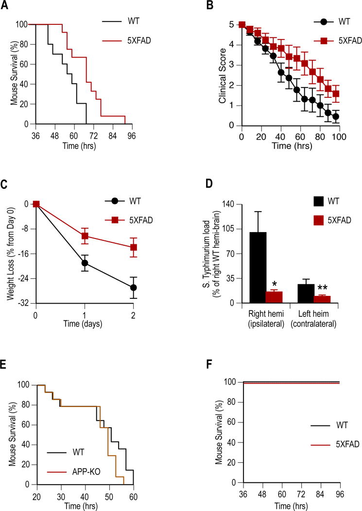
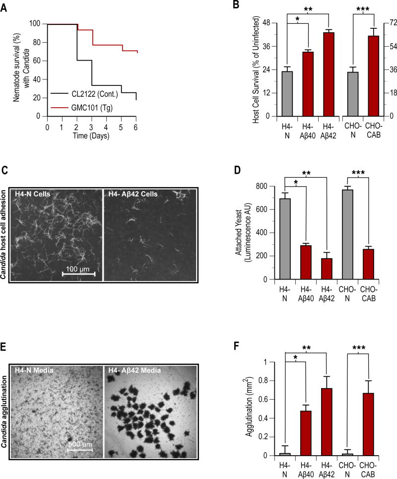
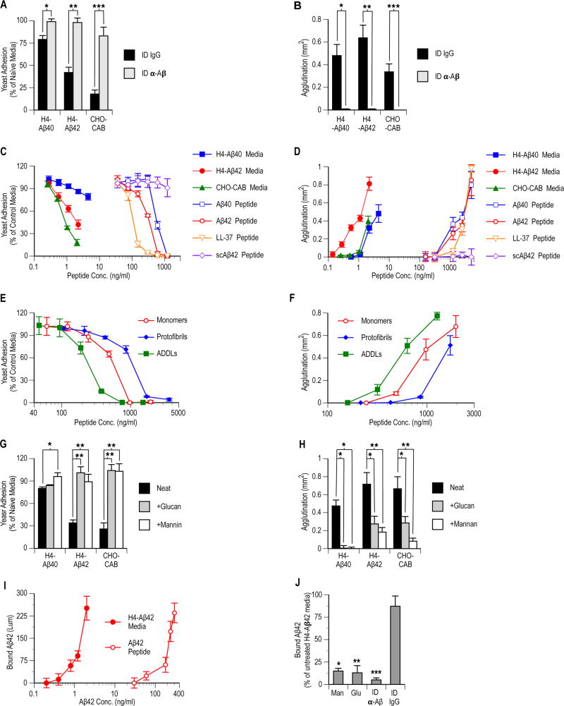
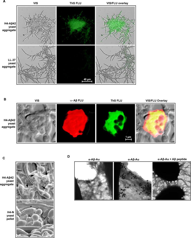
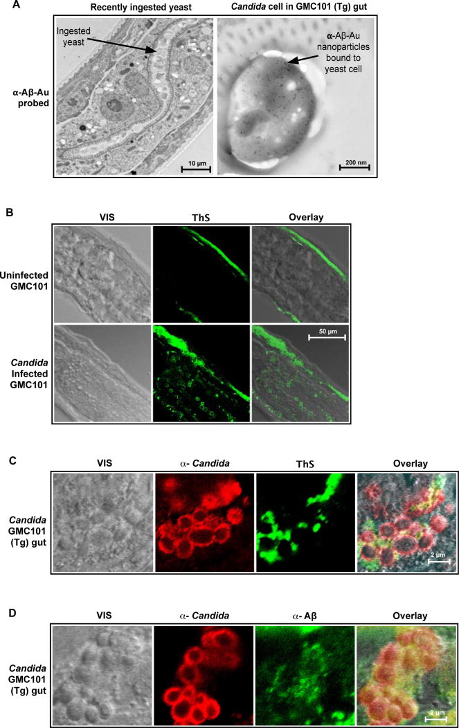
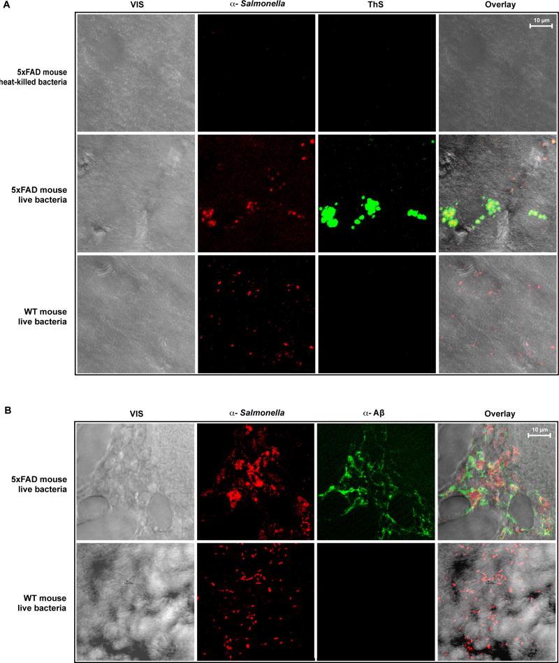
The amyloid-β peptide (Aβ) is a key protein in Alzheimer's disease (AD) pathology. We previously reported in vitro evidence suggesting that Aβ is an antimicrobial peptide. We present in vivo data showing that Aβ expression protects against fungal and bacterial infections in mouse, nematode, and cell culture models of AD. We show that Aβ oligomerization, a behavior traditionally viewed as intrinsically pathological, may be necessary for the antimicrobial activities of the peptide. Collectively, our data are consistent with a model in which soluble Aβ oligomers first bind to microbial cell wall carbohydrates via a heparin-binding domain. Developing protofibrils inhibited pathogen adhesion to host cells. Propagating β-amyloid fibrils mediate agglutination and eventual entrapment of unatttached microbes. Consistent with our model, Salmonella Typhimurium bacterial infection of the brains of transgenic 5XFAD mice resulted in rapid seeding and accelerated β-amyloid deposition, which closely colocalized with the invading bacteria. Our findings raise the intriguing possibility that β-amyloid may play a protective role in innate immunity and infectious or sterile inflammatory stimuli may drive amyloidosis. These data suggest a dual protective/damaging role for Aβ, as has been described for other antimicrobial peptides.


Conflict of interest statement

Competing interests: The authors have no competing interests pertaining to this work.

Figures

Fig. 1
Fig. 1. Aβ expression protects against S. Typhimurium meningitis in genetically modified AD mouse models
Transgenic (5XFAD) mice expressing human Aβ and mice lacking murine APP (APP-KO) were compared to genetically unmodified littermates (WT) for resistance to S. Typhimurium meningitis. One-month old mice received single ipsilaeral intracranial injections of S. Typhimurium and clinical progression was followed to moribundity. (A to C) Performance of 5XFAD (n =12) mice compared to WT (n = 11) are shown following infection for survival (P = 0.009) (A), clinical score (P < 0.0001) (B), percent weight loss (P = 0.0008) (C). (D) S. Typhimurium load 24 hours post-infection in 5XFAD (n = 4) and WT (n = 4) mouse brain hemisphere homogenates shown as mean CFU ± SEM (*P = 0.03 and **P = 0.04). (E) APP-KO mice (n = 15) show a trend (P = 0.104) towards reduced survival compared to WT (n = 15) littermates following infection. (F) No mortality was observed among control sham-infected WT (n = 6) or 5XFAD (n = 6) mice injected with heat-killed S. Typhimurium. Statistical significance was calculated by Log-rank (Mantel-Cox) test for survival (A, E, and F), linear regression for clinical score and weight (B and C), and statistical means compared by t-test (D). For survival and clinical analysis (A to C) data were pooled from three independent experiments.
Fig. 2
Fig. 2. Aβ expression in nematodes and cultured cells increases host resistance to infection by Candida
Aβ-mediated protection against C. albicans (Candida) was characterized in C. elegans and cultured host cell monolayer mycosis models. Experimental nematodes included control non-Aβ expressing (CL2122) and transgenic human Aβ-expressing (GMC101) strains. Host cell lines included control non-transformed (H4-N and CHO-N) and transformed human Aβ-overexpressing (H4-Aβ40, H4-Aβ42, and CHO-CAB) cells. (A) Survival curves for CL2122 (n = 61) and GMC101 (n = 57) nematodes following infection with Candida (P < 0.00001). (B) Viability of non-transformed and transformed host cell monolayers following 36 hours of incubation with Candida. Host cell viability was followed by pre-labeling host cell monolayers with BrdU and then comparing wells for an anti-BrdU signal. Signal of infected wells shown as percentage of uninfected control wells (*P = 0.002, **P = 0.001, and ***P = 0.004). (C) Candida adherence to host cells. Fluorescence micrograph of Calcofluor White stained Candida adhering to control H4-N or transformed H4-Aβ42 host cell monolayers following 2 hours of co-incubation in pre-conditioned culture media. (D) Quantitative analysis of Candida host cell colonization. Adhering Candida were detected using a immunochemical luminescence assay with anti-Candida antibodies (*P = 0.003, **P = 0.001, and ***P = 0.004). Well comparisons use arbitrary luminescence units (AU). (E) Phase contrast micrographs of agglutinated Candida following overnight incubation with H4-N or H4-Aβ42 host cells. (F) Quantitative analysis of Candida agglutination. Wells were compared for yeast aggregate surface area using image analysis software (*P = 0.007, **P = 0.002, and ***P = 0.009). Bars in panel’s (B), (D), and (F) are means of six replicate wells ± SEM. Statistical significance was calculated by Log-rank (Mantel-Cox) test for nematode survival (A) and statistical mean comparisons by t-test (B, D, and F). Micrographs (C and E) are representative of data from three replicate experiments and multiple discrete image fields (table S1A).
Fig. 3
Fig. 3. Aβ's protective actions in cell culture are mediated by adhesion inhibition and agglutination activities against Candida
C. albicans adhesion to abiotic surfaces and agglutination in the bulk phase were characterized in the presence of cell-derived or synthetic Aβ. After 36 hours conditioning, host cell free culture media was collected from control non-transformed (H4-N or CHO-N) or transformed Aβ-overexpressing (H4-Aβ40, H4-Aβ42, or CHO-CAB) cultured cells. Aβ-immunodepleted (ID β-Aβ) and control immunodepleted ([ID IgG (immunoglobulin)] media were prepared by incubation with immobilized anti-Aβ or nonspecific antibodies. Experimental synthetic peptides included Aβ (Aβ40 and Aβ42), AMP positive control (LL-37), and negative control scrambled Aβ42 (scAβ42). (A and B) Comparison of ID β-Aβ and ID IgG media's adhesion inhibition (*P = 0.009, **P = 0.001, and ***P = 0.004) and agglutination (*P = 0.001, **P = 0.0005, and ***P = 0.004) activities. (C and D) Comparison of anti-Candida activities of serially diluted conditioned media and synthetic peptides. (E and F) Activities of synthetic Aβ42 monomer, soluble oligomeric amyloid-β derived diffusible ligands (ADDLs), and protofibril preparations. (G and H) Conditioned culture media adhesion inhibition (*P = 0.003 and **P < 0.0003) and agglutinating (*P < 0.02 and **P < 0.003) source activities alone (Neat) or in the presence of soluble yeast wall carbohydrates (+Glucan or +Mannan). (I) Synthetic monomeric Aβ42 and cell-generated peptide from H4-Aβ42 cells were compared for Candida binding using an Aβ/Candida binding ELISA. (J) Untreated, immunodepleted, or glucan (Glu)- or mannan (Man)-spiked H4-Aβ42 conditioned media were incubated with intact immobilized yeast cells in an Aβ/Candida binding ELISA assay (*P = 0.006, **P = 0.008, and ***P < 0.004). Synthetic peptide incubations (C to F and I) were performed in H4-Aβ42 conditioned culture media pre-treated to remove cell-derived Aβ by α-Aβ immunodepletion. Symbols and bars for (A) to (J) are statistical means of 6 replicate wells ± SEM. Statistical significance was by t-test.
Fig. 4
Fig. 4. β-amyloid fibrils propagate from yeast surfaces and capture Candida in H4-Aβ42 media
Early stage C. albicans aggregates harvested from H4-Aβ42 conditioned media were probed with α-Aβ-Au nanoparticles and analyzed by TEM. (A) Yeast agglutination is mediated by fibrillar structures. Micrograph shows fibrils binding cells within yeast aggregates and linking C. albicans clusters. (B) Fibrillar structures extending from yeast cell surfaces. (C and D) α-Aβ-Au nanoparticle labeling of short fibrillar structures extending from C. albicans surfaces and long fibrils running between yeast clumps. (E) Absorption experiment showing ablated α-Aβ-Au binding of fibrils extending from yeast in the presence of soluble synthetic Aβ peptide. Data are consistent with specific α-Aβ-Au labeling of β-amyloid fibrils. Micrographs are representative of data from three replicate experiments and multiple discrete image fields (table S1A).
Fig. 5
Fig. 5. Candida cells are entrapped by β-amyloid in H4-Aβ42 culture media
Following overnight incubation with H4-Aβ42 media, yeast (C. albicans) aggregates were harvested and probed for β-amyloid markers. (A and B) Visible yeast aggregates (VIS), yeast aggregates stained with green-fluorescence Thioflavin S (Thioflavin S FLU), yeast aggregates stained with red-fluorescence anti-Aβ (α-Aβ FLU); superpositioned images (VIS/FLU overlay). Yeast aggregates generated with the control synthetic LL-37 peptide (A) are negative for Thioflavin S enhanced fluorescence. (B) Yellow denotes co-localization of anti-Aβ and Thioflavin S signals. Co-localization of these signals is the hallmark of β-amyloid. (C) SEM analysis revealed fibrous material in H4-Aβ42 yeast aggregates that is absent from control C. albicans pellets prepared by centrifugation in H4-N media. (D) H4-Aβ42 yeast aggregates incubated with immunogold nanoparticles coated with anti-Aβ antibodies (α-Aβ-Au) and analyzed by TEM. First and second panels show labeling of fibrous material by α-Aβ-Au. Third panel shows inhibition of α-Aβ-Au nanoparticle binding by soluble synthetic Aβ peptide (α-Aβ-Au + Aβ peptide), consistent with specific labeling of β-amyloid. Micrographs are representative of data from two or more replicate experiments and multiple discrete image fields (table S1A).
Fig. 6
Fig. 6. Intestinal infection with Candida induces Aβ fibrillization in transgenic GMC101 nematode gut
Aβ42-expressing GMC101 C. elegans were infected with C. albicans (Candida) and probed for anti-Aβ immunoreactivity and β-amyloid markers using TEM and confocal microscopy CFM. (A) Micrograph shows positive labeling of yeast cell surface in GMC101 worm gut by immunogold nanoparticles coated with anti-Aβ antibodies (α-Aβ-Au) two hours following Candida ingestion. (B and D) Figures show visible (VIS) and fluorescence signals from freeze fracture nematode sections with advanced Candida infections. Figure B compares uninfected and infected worms. Figures C and D show Thioflavin S and anti-Aβ staining for gut yeast aggregates. Signals include anti-Candida immunoreactivity α-Candida), Thioflavin S enhanced fluorescence (ThS), anti-Aβ immunoreactivity (α-Aβ), and superpositioned (Overlay) signals . Yellow denotes signal co-localization. Uninfected and infected CL2122 nematode controls were negative for anti-Aβ immunoreactivity and enhanced Thioflavin S fluorescence (figs. 2S and S8). Micrographs are representative of data from three or more replicate experiments and multiple discrete image fields (Table S1B).
Fig. 7
Fig. 7. Infection-induced β-amyloid deposits co-localize with invading S. Typhimurium cells in 5XFAD mouse brain
Four-week-old wildtype (WT) mice or transgenic 5XFAD animals expressing high levels of human Aβ were injected intracerebrally with viable S. Typhimurium bacteria. Mice were also injected with heat-treated S. Typhimurium cell debris as a negative control for the injection procedure. (A and B) Mouse brain sections were prepared 24 (A) or 48 hours (B) after infection. Signals shown include visible (VIS), anti-Salmonella immunoreactivity (α-Salmonella), enhanced Thioflavin S fluorescence (ThS) or anti-Aβ immunoreactivity (α-Aβ), and superpositioned (Overlay) signals. Panels are representative images of multiple images captured as z-sections using confocal fluorescence microscopy . Yellow denotes signal co-localization. (Z-series projections showing β-amyloid surrounding and entrapping bacteria colonies in a rotating 3-dimension section of 5XFAD mouse brain are also included in video S1). Micrographs are representative of data from three replicate experiments and multiple discrete image fields (table S1C).

Comment in

References

    1. Tanzi RE, Bertram L. Twenty years of the Alzheimer‘s disease amyloid hypothesis: a genetic perspective. Cell. 2005;120:545–555. - PubMed
    1. Luna S, Cameron DJ, Ethell DW. Amyloid-|3 and APP deficiencies cause severe cerebrovascular defects: important work for an old villain. PLoS ONE. 2013;8:e75052. - PMC - PubMed
    1. Soscia SJ, Kirby JE, Washicosky KJ, Tucker SM, Ingelsson M, Hyman B, Burton MA, Goldstein LE, Duong S, Tanzi RE, Moir RD. The Alzheimer‘s disease-associated amyloid-|3 protein is an antimicrobial peptide. PLoS ONE. 2010;5:e9505. - PMC - PubMed
    1. Kagan BL, Jang H, Capone R, Teran Arce F, Ramachandran S, Lal R, Nussinov R. Antimicrobial properties of amyloid peptides. Mol Pharm. 2012;9:708–717. - PMC - PubMed
    1. Wiesner J, Vilcinskas A. Antimicrobial peptides: the ancient arm of the human immune system. Virulence. 2010;1:440–464. - PubMed

Publication types

Substances