. 2016 May 25;8(340):340ra73.
doi: 10.1126/scitranslmed.aad4059.

Dormant breast cancer micrometastases reside in specific bone marrow niches that regulate their transit to and from bone

Affiliations

Dormant breast cancer micrometastases reside in specific bone marrow niches that regulate their transit to and from bone

Trevor T Price et al. Sci Transl Med. .

Abstract

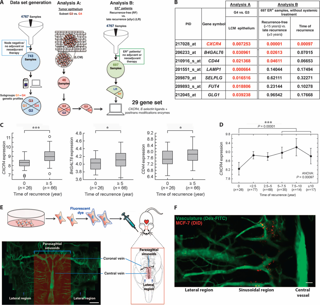
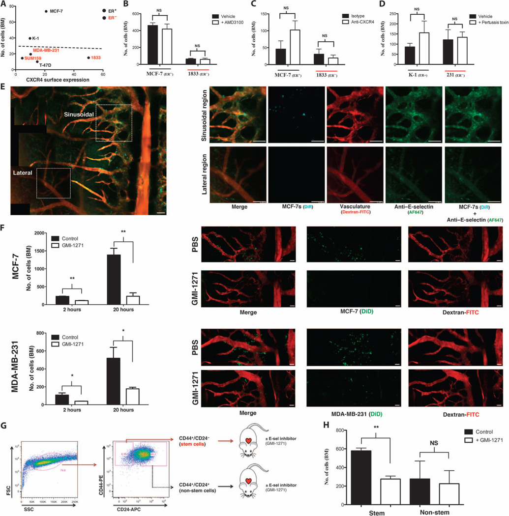
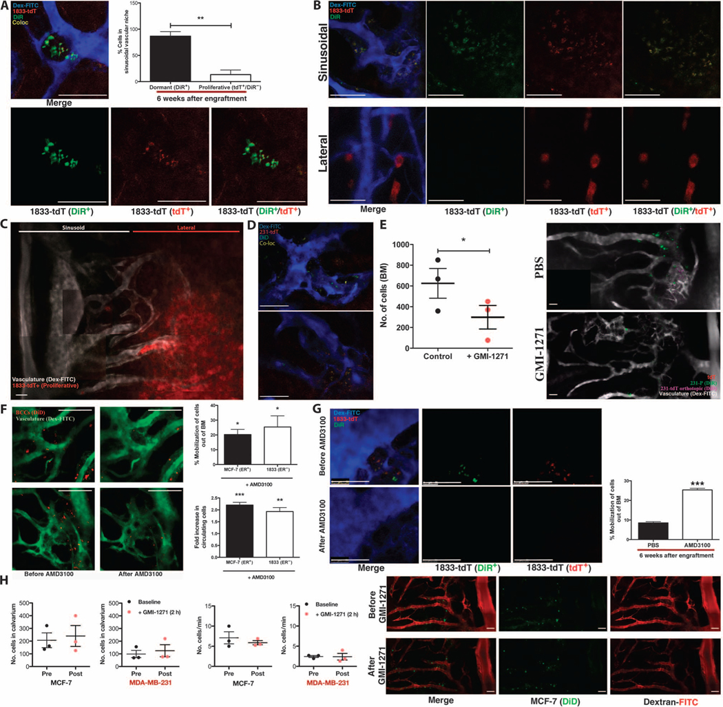
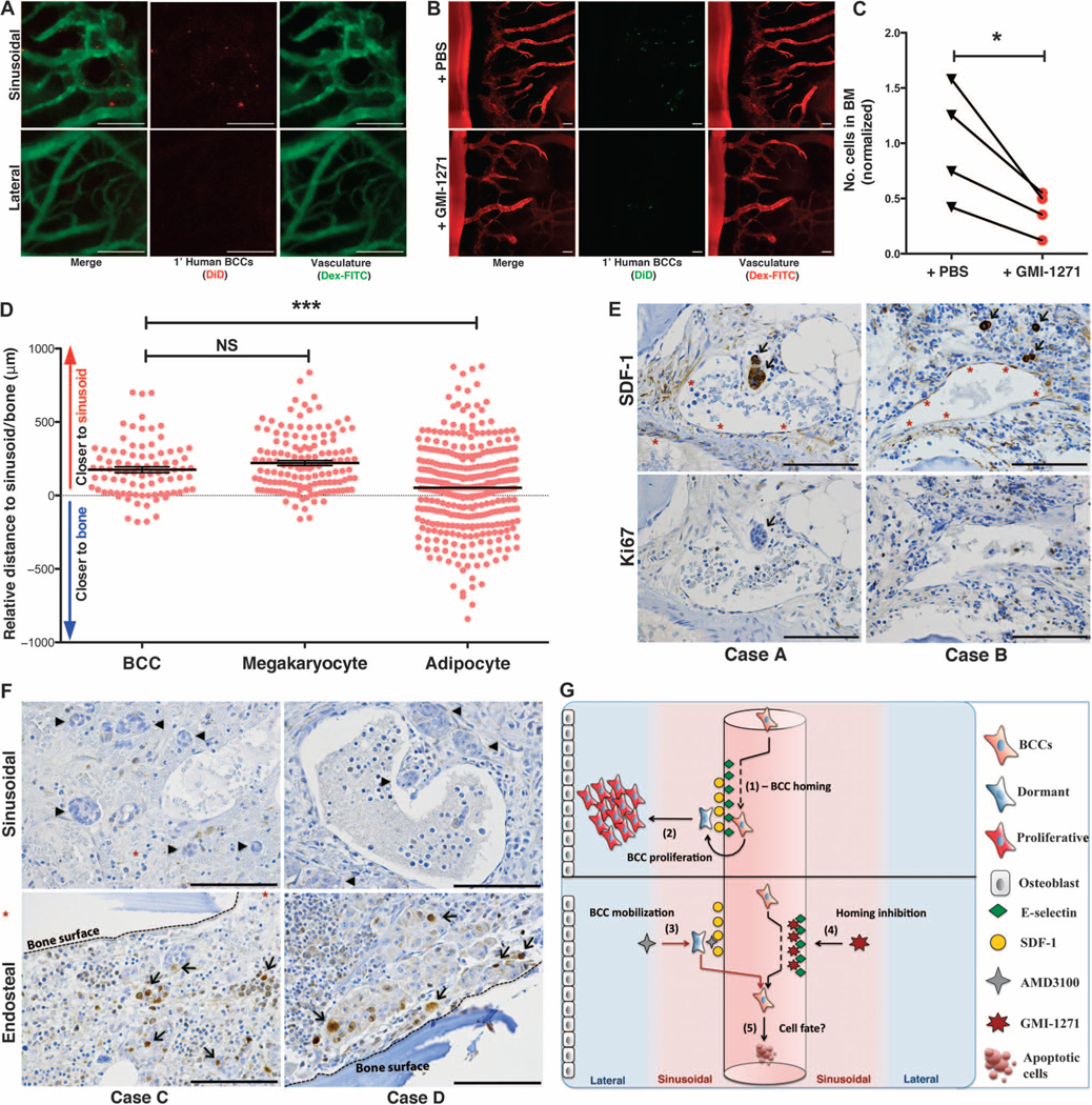
Breast cancer metastatic relapse can occur years after therapy, indicating that disseminated breast cancer cells (BCCs) have a prolonged dormant phase before becoming proliferative. A major site of disease dissemination and relapse is bone, although the critical signals that allow circulating BCCs to identify bone microvasculature, enter tissue, and tether to the microenvironment are poorly understood. Using real-time in vivo microscopy of bone marrow (BM) in a breast cancer xenograft model, we show that dormant and proliferating BCCs occupy distinct areas, with dormant BCCs predominantly found in E-selectin- and stromal cell-derived factor 1 (SDF-1)-rich perisinusoidal vascular regions. We use highly specific inhibitors of E-selectin and C-X-C chemokine receptor type 4 (CXCR4) (SDF-1 receptor) to demonstrate that E-selectin and SDF-1 orchestrate opposing roles in BCC trafficking. Whereas E-selectin interactions are critical for allowing BCC entry into the BM, the SDF-1/CXCR4 interaction anchors BCCs to the microenvironment, and its inhibition induces mobilization of dormant micrometastases into circulation. Homing studies with primary BCCs also demonstrate that E-selectin regulates their entry into bone through the sinusoidal niche, and immunohistochemical staining of patient BMs shows dormant micrometastatic disease adjacent to SDF-1(+) vasculature. These findings shed light on how BCCs traffic within the host, and suggest that simultaneous blockade of CXCR4 and E-selectin in patients could molecularly excise dormant micrometastases from the protective BM environment, preventing their emergence as relapsed disease.


Figures

Fig. 1.
Fig. 1.. BCCs hijack SDF-1+E-selectin+ vascular gateways to enter the bone.
(A) Discovery cohort of untreated, node-negative breast cancer patients revealed four subgroups based on gene expression. Of these, G3 had the highest recurrence-free survival, whereas G4 had an increased risk of late recurrence (≥5 years after diagnosis). In Analysis A, expression of 29 genes representing CXCR4, E-selectin ligands, and enzymes critical for posttranslational processing of E-selectin ligands was analyzed in a data set compiled from 46 microdissected samples (G3, n = 18; G4, n = 10). In Analysis B, these same 29 genes were analyzed in the ER+ subset of our discovery cohort of patients with untreated, node-negative disease [late recurrence (LR) ≥ 5 years, n = 66; recurrence-free (RF) > 15 years, n = 26]. LCM, laser capture microdissection. (B to D) The expression of 7 of 29 genes analyzed was significantly increased in microdissected tumor epithelium of patients with late recurrence (t test). In the ER+ subset of our discovery cohort, 3 of these 29 genes were significantly up-regulated in tumor samples from patients with late recurrence compared to those from patients with recurrence-free survival at ≥15 years (Mann-Whitney U test). (D) CXCR4 expression correlates with time to recurrence in the ER+ subgroup [***P = 0.00097, analysis of variance (ANOVA)] [P values presented in (B); red indicates P < 0.05]. (E) Intravital microscopy was used to image the calvarial BM of mice engrafted with fluorescent membrane dye–labeled BCCs in real time with single-cell resolution. Dextran-FITC (fluorescein isothiocyanate) was used to visualize the blood pool. As shown in the montage image of in vivo micrographs, sinusoidal vasculature, which is known to have a distinct SDF-1+E-selectin+ phenotype, is concentrated in distinct parasagittal locations (montage containing multiple images; scale bar, 200 μm). (F) Fluorescently labeled BCCs were engrafted by intracardiac injection in mice, and the calvarium was imaged in the initial 2 hours after engraftment. BCCs were detected entering the bone specifically in perisinusoidal areas. Cells were not detected in lateral or caudal regions that lack substantial sinusoidal vasculature (montage containing multiple images; scale bar, 100 μm).
Fig. 2.
Fig. 2.. E-selectin regulates entry of BCCs into bone from peripheral circulation.
(A) Cell surface expression of CXCR4 versus day 1 BCC BM homing (R2 = 0.0013, linear regression; n = 3 per cell line). (B) BCC BM homing in mice treated ± CXCR4 inhibitor AMD3100 (n = 3 each cell line). NS, not significant. (C) BCC BM homing in mice treated ± CXCR4 neutralizing antibodies (MCF-7: n = 3; 1833: n = 4). (D) BCC BM homing in mice treated ± pertussis toxin to broadly inhibit chemokine signaling before engraftment (n = 3 each cell line). (E) E-selectin expression (green) in the sinusoidal regions of calvarial BM was detected by in vivo microscopy using anti–E-selectin–Cy5 antibodies and was colocalized with fluorescently labeled BCCs (cyan) imaged simultaneously on day 1 after intracardiac engraftment (dextran-FITC, red; montage contains multiple images). (F) BCC homing was quantified in mice treated ± E-selectin inhibitor GMI-1271 at both initial (2 hours; MCF-7: **P = 0.0068, n = 3; MDA-MB-231: *P = 0.0356, n = 3; unpaired, two-way t test) and late (20 hours; MCF-7: **P = 0.0052, n = 3; MDA-MB-231: *P = 0.0491, n = 3; unpaired, two-way t test) time points. Representative images shown (BCC, green; dextran-FITC, red; montage contains multiple images). (G) Schematic of MCF-7 stem (CD44+CD24−/low) and non-stem (CD44+CD24−/high) cell isolation by flow sorting and engraftment ± GMI-1271 into female severe combined immunodeficient (SCID) mice. SSC, side scatter; FSC, forward scatter. (H) BM homing of MCF-7 stem and non-stem cell populations ± GMI-1271 at 20 hours after engraftment (**P = 0.0026, n = 3; unpaired, two-way t test). Scale bars, 100 μm.
Fig. 3.
Fig. 3.. SDF-1/CXCR4 interactions tether dormant BCCs to the vascular niche.
(A) Six weeks after intracardiac engraftment, dormant BCCs (DiR+tdT+) are detected in clusters localized to the sinusoidal vascular region of calvarial BM. Representative images are shown. The percentage of dormant (DiR+tdT+) versus proliferative (DiRtdT+) BCCs in the sinusoidal niche was calculated (**P = 0.0042, n = 3 mice; unpaired, two-way t test). (B and C) In mice with proliferative bone tumors, rapidly dividing BCCs (DiRtdT+) were found in distinct lateral, nonsinusoidal regions of BM [representative images of n = 4 experiments; (C) is a montage containing multiple images]. (D) Dormant (DiD+tdT+) BCCs that spontaneously metastasized to bone from orthotopic tumors were detected in the sinusoidal vascular niche at 48 to 79 days. (E) Forty-eight to 79 days after orthotopic engraftment of DiD-labeled tdT+ BCCs, mice were treated ± GMI-1271 and engrafted with DiR-labeled BCCs. DiR-BCCs homed to sinusoids in orthotopic tumor-engrafted mice, with a twofold homing reduction in GMI-1271–treated mice (*P = 0.0126, n = 3; unpaired, two-way t test). Representative images are shown (montage containing multiple images). (F) One day after engraftment, mice were treated ± AMD3100, and images of the calvarium were obtained before and 2 hours after treatment to determine the percentage of BCCs mobilized from BM (MCF-7: *P = 0.0455, n = 3; 1833: *P = 0.0300, n = 4; unpaired, two-way t test). Video-rate microscopy was used to assess the fold increase in peripherally circulating BCCs (MCF-7: ***P = 0.0005, n = 3; 1833: **P = 0.0069, n = 4; unpaired, two-way t test). Representative images are shown. (G) Six weeks after engraftment, mice were treated ± AMD3100. The percentage of dormant DiR+tdT+ BCCs mobilized from BM was calculated (***P = 0.0008, n = 3; unpaired, two-way t test). Representative images are shown. (H) One day after engraftment, mice were treated ± GMI-1271. Images before and 2 hours after treatment demonstrated no significant mobilization of BCCs from BM (n = 3 mice for each cell line; unpaired, two-way t test). Representative images are shown (montage containing multiple images). Scale bars, 100 μm.
Fig. 4.
Fig. 4.. Primary human BCCs use E-selectin to enter a dormant sinusoidal perivascular niche in the BM.
(A) Primary patient BCCs were isolated from surgical resection tissue and engrafted in mice. Intravital calvarial imaging revealed that primary cells home specifically to sinusoidal vasculature (n = 4). Representative images are shown. (B and C) Primary human BCCs were engrafted ± GMI-1271, and BM homing was quantified at 20 hours. Representative images are shown. Homing was decreased almost threefold in GMI-1271–treated mice (*P = 0.0393, n = 4; paired, two-way t test). (D) Histologic sections of BM biopsies from patients with micrometastatic involvement of breast cancer were obtained. The distance of each individual micrometastasis from the nearest sinusoid or bone spicule was measured by hand by a hematopathologist. Distance measurements for megakaryocytes, which are intimately associated with the sinusoidal vasculature, and for adipocytes, which are randomly scattered in bone, were also performed. Similar to megakaryocytes, breast cancer micrometastases were identified near sinusoids, with relative distance measurements that were significantly different from randomly distributed adipocytes [***P = 0.0002, data from three separate patient samples (BCCs, n = 86; megakaryocytes, n = 136; adipocytes, n = 348); unpaired, two-way t test]. (E) Immunohistochemical staining of BM biopsies from micrometastatic disease patients shows that BCCs (arrows) adjacent to SDF-1+ vasculature (asterisks) are Ki67. (F) BM biopsy samples from patients with macrometastatic disease were stained for Ki67. Perisinusoidal (asterisks) BCCs were Ki67 (arrowheads), but Ki67+ BCCs (arrows) could be identified near bony spicules (dashed lines). (G) Cartoon model of BCC homing (1) and retention and proliferation (2) mechanisms in BM niches. Forced mobilization of dormant cells (3) combined with simultaneous blockade of BM reentry (4) could cause apoptosis or chemosensitize cells deprived of supportive stroma (5). Scale bars, 100 μm.

References

    1. DeVita VT Jr., Lawrence TS, Rosenberg SA, Eds., DeVita, Hellman, and Rosenberg’s Cancer: Principles & Practice of Oncology (Lippincott, Williams & Wilkins, Philadelphia, PA, 2014).
    1. Braun S, Vogl F, Naume B, Janni W, Osborne MP, Coombes RC, Schlimok G, Diel IJ, Gerber B, Gebauer G, Pierga J-Y, Marth C, Oruzio D, Wiedswang G, Solomayer E-F, Kundt G, Strobl B, Fehm T, Wong GYC, Bliss J, Vincent-Salomon A, Pantel K, A pooled analysis of bone marrow micrometastasis in breast cancer. N. Engl. J. Med 353, 793–802 (2005). - PubMed
    1. Weigelt B, Peterse JL, van’t Veer LJ, Breast cancer metastasis: Markers and models. Nat. Rev. Cancer 5, 591–602 (2005). - PubMed
    1. Rack B, Schindlbeck C, Jückstock J, Andergassen U, Hepp P, Zwingers T, Friedl TWP, Lorenz R, Tesch H, Fasching PA, Fehm T, Schneeweiss A, Lichtenegger W, Beckmann MW, Friese K, Pantel K, Janni W; SUCCESS Study Group, Circulating tumor cells predict survival in early average-to-high risk breast cancer patients. J. Natl. Cancer Inst 106, dju066 (2013). - PMC - PubMed
    1. Serrano MJ, Rovira PS, Martinez-Zubiaurre I, Rodriguez MD, Fernández M, Lorente JA, Dynamics of circulating tumor cells in early breast cancer under neoadjuvant therapy. Exp. Ther. Med 4, 43–48 (2012). - PMC - PubMed

MeSH terms