. 2016 Oct;112(1):443-51.
doi: 10.1093/cvr/cvw111. Epub 2016 May 25.

Elevated ventricular wall stress disrupts cardiomyocyte t-tubule structure and calcium homeostasis

Affiliations

Elevated ventricular wall stress disrupts cardiomyocyte t-tubule structure and calcium homeostasis

Michael Frisk et al. Cardiovasc Res. 2016 Oct.

Abstract

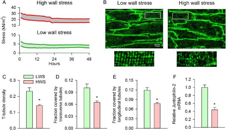
Aims: Invaginations of the cellular membrane called t-tubules are essential for maintaining efficient excitation-contraction coupling in ventricular cardiomyocytes. Disruption of t-tubule structure during heart failure has been linked to dyssynchronous, slowed Ca(2+) release and reduced power of the heartbeat. The underlying mechanism is, however, unknown. We presently investigated whether elevated ventricular wall stress triggers remodelling of t-tubule structure and function.

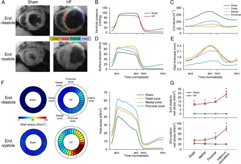
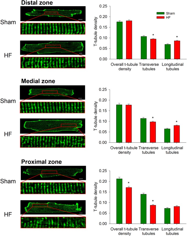
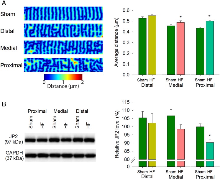
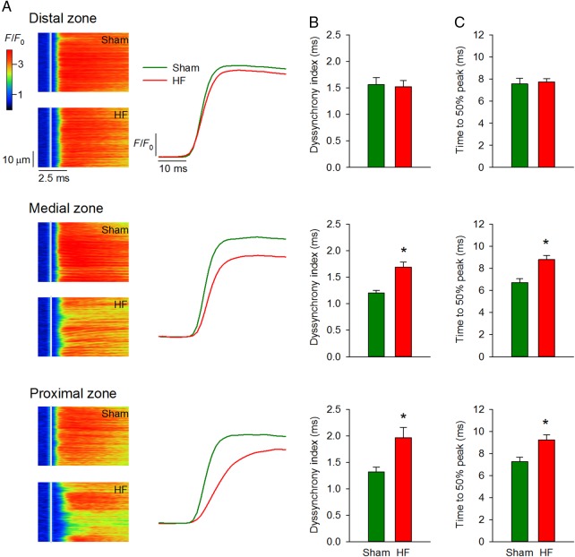
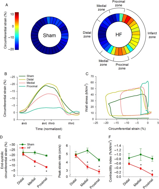
Methods and results: MRI and blood pressure measurements were employed to examine regional wall stress across the left ventricle of sham-operated and failing, post-infarction rat hearts. In failing hearts, elevated left ventricular diastolic pressure and ventricular dilation resulted in markedly increased wall stress, particularly in the thin-walled region proximal to the infarct. High wall stress in this proximal zone was associated with reduced expression of the dyadic anchor junctophilin-2 and disrupted cardiomyocyte t-tubular structure. Indeed, local wall stress measurements predicted t-tubule density across sham and failing hearts. Elevated wall stress and disrupted cardiomyocyte structure in the proximal zone were also associated with desynchronized Ca(2+) release in cardiomyocytes and markedly reduced local contractility in vivo. A causative role of wall stress in promoting t-tubule remodelling was established by applying stretch to papillary muscles ex vivo under culture conditions. Loads comparable to wall stress levels observed in vivo in the proximal zone reduced expression of junctophilin-2 and promoted t-tubule loss.

Conclusion: Elevated wall stress reduces junctophilin-2 expression and disrupts t-tubule integrity, Ca(2+) release, and contractile function. These findings provide new insight into the role of wall stress in promoting heart failure progression.

Keywords: Ca2+ homeostasis; Heart failure; Junctophilin-2; Left ventricular wall stress; T-tubules.


Figures

Figure 1
Figure 1
Graded elevation of wall stress across the post-infarction, failing heart. MRI recordings showed left ventricular dilation in post-infarction rats with heart failure (HF) compared with sham-operated controls (A), and haemodynamic measurements indicated elevation of diastolic blood pressure (B). Division of failing hearts into equally sized zones proximal, medial, and distal to the infarct revealed reduction in curvature across the failing heart relative to Sham (C), and increased surface tension throughout the cardiac cycle (D). Due to a thinner ventricular wall in the proximal zone (E), wall stress was markedly elevated in this region (F). Wall stress declined with increasing distance from the infarct but was elevated in all regions relative to Sham, particularly during diastole (G). avo, aortic valve opening; avc, aortic valve closing; mvo, mitral valve opening; mvc, mitral valve closing. (nSham hearts= 3; nHF hearts= 4; *P < 0.05 vs. Sham).
Figure 2
Figure 2
T-tubules are most markedly disrupted proximal to the infarction. di-8-ANEPPS stains were employed to examine t-tubule density and organization. In comparison with sham-operated controls, overall t-tubule density was only reduced in the proximal zone of failing hearts. However, transverse elements were observed to be lost in all regions, but compensated by growth of longitudinal tubules in the medial and distal zones. (nSham cells = 183 from 3 hearts, nHF cells= 246 from 4 hearts; *P < 0.05 vs. Sham).
Figure 3
Figure 3
T-tubule disruption is associated with reduced junctophilin-2 expression. Images of di-8-ANEPPS-stained cardiomyocytes were used to create ‘distance maps’ (A, left panel), indicating distance from all points in the cytosol to the nearest t-tubule or surface sarcolemma. Average distance was increased with greater proximity to the infarction (right panel). (nSham cells = 183 from 3 hearts, nHF cells= 246 from four hearts). (B) Junctophilin-2 (JP2) protein abundance was assessed by western blotting and normalized to GAPDH. Mean JP2 levels were significantly down-regulated in the proximal zone only. (nhearts= 6 in Sham and HF). (*P < 0.05 vs. Sham).
Figure 4
Figure 4
Ca2+ transients are progressively desynchronized with greater proximity to the infarct. As predicted by distance maps in Figure 3, confocal line-scan images (A) showed progressively more dyssynchronous and slowed Ca2+ transients approaching the infarct. Mean measurements of dyssynchrony index and time to 50% rise of the Ca2+ transient are shown in (B and C), respectively (nSham cells = 82 from three hearts, nHF cells= 83 from four hearts) (*P < 0.05 vs. Sham).
Figure 5
Figure 5
High wall stress induces t-tubule disruption and loss in vitro. Papillary muscles were isolated, mounted in a Myobath system, and maintained under culture conditions for 48 h during 0.5 Hz stimulation. Muscles were exposed to modest pre-load (diastolic stress ≈4 kN/m2) or elevated diastolic stress approximating that observed during end-diastole in the proximal zone (15–20 kN/m2). (A) Representative recordings of diastolic and systolic stress for the two treatment groups during the protocol. Visualization of t-tubules by caveolin-3 immunostaining after the 48 h incubation period revealed well-maintained t-tubular structure in the low-wall stress group, but marked t-tubule disruption following high wall stress (B). Overall t-tubule density was reduced by exposure to high wall stress (C) and included loss of both transverse and longitudinal elements [D and E, low wall stress: n = 67 cells from five muscles (4 hearts), high wall stress: n = 64 cells from five muscles (three hearts)]. T-tubule loss was associated with a marked decrease in relative junctophilin-2 mRNA expression in the high-wall stress group (F, nmuscles = 4, 3 in low, high wall stress groups, respectively). LWS, low wall stress; HWS, high wall stress (*P < 0.05 vs. low wall stress).
Figure 6
Figure 6
Local in vivo contractility is reduced in regions with disrupted cardiomyocyte structure and Ca2+ handling. (A) Representative measurements of MRI-determined circumferential strain show graded contraction across the failing heart. Mean strain and peak strain rate were markedly reduced in the proximal zone but near-normal in the distal zone (B, D and E). (C) Wall stress–strain loops were employed to estimate a ‘contractility index’, calculated as end-systolic strain/stress. In comparison with Sham, the contractility index was significantly reduced in regions of failing hearts near the infarction (F, nSham hearts= 3, nHF hearts= 4). avo, aortic valve opening; avc, aortic valve closing; mvo, mitral valve opening; mvc, mitral valve closing. (*P < 0.05 vs. Sham).

Comment in

References

    1. Franzini-Armstrong C, Protasi F, Ramesh V. Shape, size, and distribution of Ca2+ release units and couplons in skeletal and cardiac muscles. Biophys J 1999;77:1528–1539. - PMC - PubMed
    1. Guo A, Zhang C, Wei S, Chen B, Song LS. Emerging mechanisms of T-tubule remodelling in heart failure. Cardiovasc Res 2013;98:204–215. - PMC - PubMed
    1. Louch WE, Sejersted OM, Swift F. There goes the neighborhood: pathological alterations in T-tubule morphology and consequences for cardiomyocyte Ca2+ handling. J Biomed Biotechnol 2010;2010:503906. - PMC - PubMed
    1. Roe AT, Frisk M, Louch WE. Targeting cardiomyocyte Ca2+ homeostasis in heart failure. Curr Pharm Des 2014;21:431–448. - PMC - PubMed
    1. Gomez AM, Valdivia HH, Cheng H, Lederer MR, Santana LF, Cannell MB, McCune SA, Altschuld RA, Lederer WJ. Defective excitation–contraction coupling in experimental cardiac hypertrophy and heart failure. Science 1997;276:800–806. - PubMed

Publication types

MeSH terms