

Dynamic enhancement patterns of intrahepatic cholangiocarcinoma in cirrhosis on contrast-enhanced computed tomography: risk of misdiagnosis as hepatocellular carcinoma
- PMID: 27226026
- PMCID: PMC4880940
- DOI: 10.1038/srep26772
Dynamic enhancement patterns of intrahepatic cholangiocarcinoma in cirrhosis on contrast-enhanced computed tomography: risk of misdiagnosis as hepatocellular carcinoma
Abstract
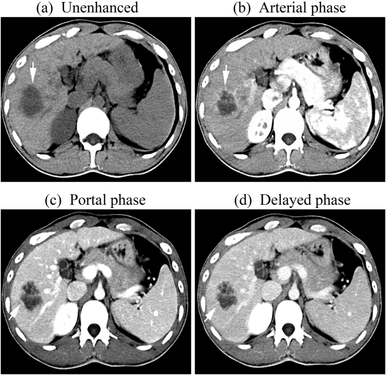
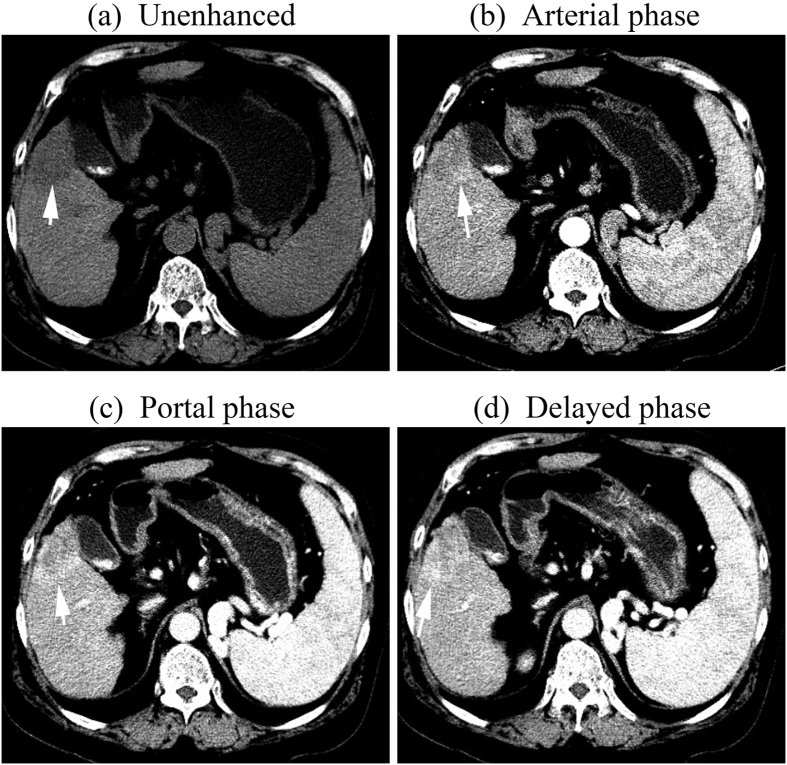
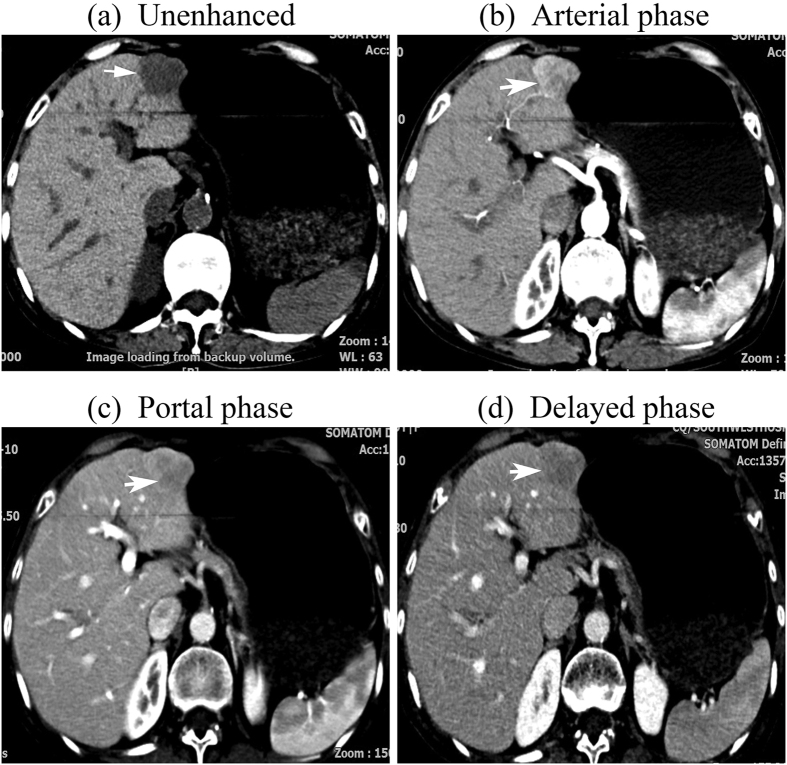
This study aimed to assess the features of intrahepatic cholangiocarcinoma (ICC) at computerized tomography (CT) and verify the risk of misdiagnosis of ICC as hepatocellular carcinoma (HCC) in cirrhosis. CT appearances of 98 histologically confirmed ICC nodules from 84 cirrhotic patients were retrospectively reviewed, taking into consideration the pattern and dynamic contrast uptake during the arterial, portal venous and delayed phases. During the arterial phase, 53 nodules (54.1%) showed peripheral rim-like enhancement, 35 (35.7%) hyperenhancement, 9 (9.2%) hypoenhancement and 1 (1.0%) isoenhancement. The ICC nodules showed heterogeneous dynamic contrast patterns, being progressive enhancement in 35 nodules (35.7%), stable enhancement in 28 nodules (28.6%), wash-in and wash-out pattern in 15 nodules (15.3%) and all other enhancement patterns in 20 nodules (20.4%). There were no significant differences in the dynamic vascular patterns of ICC according to nodule size (p > 0.05). ICC in cirrhosis has varied enhancement patterns at contrast-enhanced multiphase multidetector CT. Though the majority of ICC did not display typical radiological hallmarks of HCC, if dynamic CT scan was used as the sole modality for the non-invasive diagnosis of nodules in cirrhosis, the risk of misdiagnosis of ICC for HCC is not negligible.
Figures
References
-
- Patel T. Increasing incidence and mortality of primary intrahepatic cholangiocarcinoma in the United States. Hepatology. 33, 1353–1357 (2001). - PubMed
-
- Khan S. A. et al.. Changing international trends in mortality rates for liver, biliary and pancreatic tumours. J Hepatol. 37, 806–813 (2002). - PubMed
-
- Khan S. A., Thomas H. C., Davidson B. R. & Taylor-Robinson S. D. Cholangiocarcinoma. Lancet. 366, 1303–1314 (2005). - PubMed
-
- Shaib Y. & El-Serag H. B. The epidemiology of cholangiocarcinoma. Semin Liver Dis. 24, 115–125 (2004). - PubMed
-
- Shaib Y. H., El-Serag H. B., Davila J. A., Morgan R. & McGlynn K. A. Risk factors of intrahepatic cholangiocarcinoma in the United States: a case-control study. Gastroenterology. 128, 620–626 (2005). - PubMed
Publication types
MeSH terms
Substances
LinkOut - more resources
Full Text Sources
Other Literature Sources
Medical
