. 2016 Jun 7;113(23):E3260-9.
doi: 10.1073/pnas.1522811113. Epub 2016 May 25.

Coxiella burnetii effector CvpB modulates phosphoinositide metabolism for optimal vacuole development

Affiliations

Coxiella burnetii effector CvpB modulates phosphoinositide metabolism for optimal vacuole development

Eric Martinez et al. Proc Natl Acad Sci U S A. .

Abstract

The Q fever bacterium Coxiella burnetii replicates inside host cells within a large Coxiella-containing vacuole (CCV) whose biogenesis relies on the Dot/Icm-dependent secretion of bacterial effectors. Several membrane trafficking pathways contribute membranes, proteins, and lipids for CCV biogenesis. These include the endocytic and autophagy pathways, which are characterized by phosphatidylinositol 3-phosphate [PI(3)P]-positive membranes. Here we show that the C. burnetii secreted effector Coxiella vacuolar protein B (CvpB) binds PI(3)P and phosphatidylserine (PS) on CCVs and early endosomal compartments and perturbs the activity of the phosphatidylinositol 5-kinase PIKfyve to manipulate PI(3)P metabolism. CvpB association to early endosome triggers vacuolation and clustering, leading to the channeling of large PI(3)P-positive membranes to CCVs for vacuole expansion. At CCVs, CvpB binding to early endosome- and autophagy-derived PI(3)P and the concomitant inhibition of PIKfyve favor the association of the autophagosomal machinery to CCVs for optimal homotypic fusion of the Coxiella-containing compartments. The importance of manipulating PI(3)P metabolism is highlighted by mutations in cvpB resulting in a multivacuolar phenotype, rescuable by gene complementation, indicative of a defect in CCV biogenesis. Using the insect model Galleria mellonella, we demonstrate the in vivo relevance of defective CCV biogenesis by highlighting an attenuated virulence phenotype associated with cvpB mutations.

Keywords: Coxiella burnetii; host–pathogen interactions; phosphoinositides.


Conflict of interest statement

The authors declare no conflict of interest.

Figures
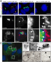
Fig. 1.
Fig. 1.
CvpB localizes at host membranes in Coxiella-infected and noninfected cells. (A) U2OS cells were challenged with cvpB::Tn 4xHAcvpB in the presence (Left) or absence (Middle) of aTc or with dotA::Tn 4xHA-cvpB in the presence of aTc (Right). Cells were fixed 24 h post infection, and translocated CvpB was labeled by using an anti-HA antibody (red), bacteria were visualized using GFP (green), and host cell nuclei were visualized using Hoechst 33258 (blue). (B) U2OS cells were infected for 3 d with Coxiella Tn1832 and transfected with pLVX-CvpB-mCherry. At 12 h post transfection, cells were fixed, and Coxiella colonies, CvpB, and LAMP1 were detected by using green fluorescence (green), mCherry fluorescence (red), and anti-LAMP1 coupled to Alexa Fluor 647 (blue), respectively. White arrows indicate discrete regions of the Coxiella vacuole where CvpB and LAMP1 colocalize. (C, Left and Top Right) U2OS cells transiently transfected with pLVX-CvpB-mCherry (green) were fixed and labeled with Hoechst 33258 (blue). CvpB is found on large vacuolar structures (1), tubules (2), and peripheral clusters of smaller vesicles (3). (Insets) Images shown converted to inverted grayscale. (C, Bottom Right) U2OS cells transiently transfected with CvpB-HA were fixed and processed for immuno-EM with the HA tag labeled using Nanogold particles. CvpB could be detected in clusters of small vesicles (arrow), enlarged vesicles (filled arrow), or plasma membrane protrusions (arrowheads). (Scale bars: AC, Left and Top Right, 10 μm; C, Bottom Right, 1 μm).
Fig. 2.
Fig. 2.
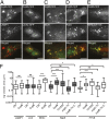
CvpB colocalizes with early endosome markers triggering their clustering and enlargement. U2OS (AD) or U2OS GFP-2xFYVE (E) transiently transfected with pLVX-CvpB-mCherry (red) and LC3C-GFP (B) were fixed and labeled with anti-LAMP1 (A), anti-EEA1 (C), or anti-Rab5 (D) coupled to Alexa Fluor 488 (green). (Scale bars: 10 μm.) (F) The median area of 100 vesicles was calculated in cells transfected with pLVX-mCherry (Ctrl) or pLVX-CvpB-mCherry (CvpB) and in cells infected with Coxiella Tn1832, the cvpB::Tn mutant, or its complemented strain (cvpB::Tn C.; ***P < 0.0001, *P < 0.05; ns, not significant, one-way ANOVA, Bonferroni’s multiple comparison test).
Fig. 3.
Fig. 3.
CvpB interacts with PI3P and PS via its N-terminal domain. Representative protein/lipid overlay assays performed, incubating GST-CvpB with PIP Strips (A), whereby several lipids are spotted at 100 pmol per spot, or with PIP Arrays (B), whereby PIs are spotted at decreasing concentrations. (C) Representative Western blot of cosedimentation assays in which histidine-tagged CvpB (His-CvpB), its 1–500-aa N-terminal domain (His-CvpB1–500), or its 500–809-aa C-terminal domain (His-CvpB500–809) were incubated with LUVs containing 100% PC, 70% PC and 30% PS (PC/PS), 98% PC and 2% PI(3)P [PC/PI(3)P], or 68% PC, 30% PS, and 2% PI(3)P [PC/PS/PI(3)P]. Following LUV centrifugation, anti-Histidine antibodies were used to detect CvpB in the pellet (P; bound to LUVs) or the supernatant (SN; unbound to LUVs) fraction of samples. (D, Left) Schematic representation of the CvpB fragments ectopically expressed as mCherry fusion proteins. (Right) U2OS GFP-2xFYVE (green) cells were transiently transfected with pLVX-CvpB-mCherry (1–809) or CvpB fragments cloned into pLVX-mCherry (red). (Scale bars: 10 μm.)
Fig. 4.
Fig. 4.
CvpB induces the presence of PI(3)P at CCV. U2OS mCherry-2xFYVE cells were infected with Coxiella Tn1832 (A), the cvpB::Tn mutant (B), or the complemented strain (C) for 3 d. Coxiella colonies, FYVE-positive vesicles, and LAMP1 were detected by using GFP (green), mCherry (red), and an anti-LAMP1 antibody coupled to Alexa Fluor 647 (blue), respectively. (D) Representative images from Movie S3 illustrating surface rendering of mCherry-2xFYVE (red) surrounding Coxiella Tn1832 colonies (green). Filled arrows point at PI(3)P-positive tubules connected with the CCV. Simple arrows follow a representative fusion event between a large PI(3)P-positive vesicle and the PI(3)P-enriched CCV (Movie S3). (Scale bars: AC, 10 μm; D, 5 μm.)
Fig. 5.
Fig. 5.
Ectopic expression of CvpB or PIKfyve inhibition counters the effect of PI 3-kinase inhibitors. U2OS GFP-2xFYVE cells were incubated with DMSO (Top) or the PI3-kinase inhibitor LY294002 (Bottom) for 4 h and transfected with pLVX-mCherry (A) or pLVX-CvpB-mCherry (B) for 12 h. The redistribution of the GFP-2xFYVE probe into the cytoplasm was measured as the vesicle-to-background signal ratio of GFP-2xFYVE from 20 treated cells for each condition (C) (SI Appendix, Fig. S7). (D) The PI3-kinase activity of CvpB at 780 nM (light red bars) or 1,560 nM (dark red bars) was measured at room temperature (RT) or 37 °C and compared with no-lipid (white bars) or no-enzyme (gray bars) conditions as negative controls and purified Vps34 at 780 nM (light blue bars) or 1,560 nM (dark blue bars) as positive controls. (E) U2OS GFP-2xFYVE cells were incubated with DMSO (control) or YM201636 for 4 h or transfected with pLVX-CvpB-mCherry for 12 h. (F) The size of GFP-2xFYVE–positive vesicles in U2OS GFP-2xFYVE cells treated as in D was calculated. (G) U2OS GFP-2xFYVE cells were incubated with DMSO or the PI3-kinase inhibitor LY294002 for 4 h. The PIKfyve inhibitor YM201636 was then added to cells treated with LY294002, and cells were incubated for 4 h. Values in C and D are means ± SEM of triplicate experiments (ns, nonsignificant; ***P < 0.0001, two-way ANOVA, Bonferroni’s multiple comparison test). Values in F are calculated from 500 vesicles measured for each condition (***P < 0.0001, one-way ANOVA). (Scale bars: 10 μm.)
Fig. 6.
Fig. 6.
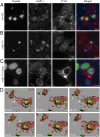
CvpB perturbs PIKfyve activity by interfering with its recruitment to PI(3)P-positive membranes. U2OS cells were transfected with pGFP-PIKfyve (PIKfyve-GFP) in combination with pLVX-mCherry (A), pLVX-CvpB-mCherry (B), or pLVX-CvpB100–809-mCherry, pLVX-CvpB1–400-mCherry, and pLVX-CvpB1–500-mCherry (C). pLVX-CvpB-mCherry(1–500) (red) and PIKfyve (green) are merged in C to illustrate colocalization (arrows and Inset). (D) The number of PIKfyve-positive puncta (arrows) per cell was calculated by using ICY from 42 cells for each condition. Red bars indicate medians (***P < 0.0001; *P < 0.05; ns, not significant, one-way ANOVA, Dunnett’s multiple comparison test). (Scale bars: 10 μm.)
Fig. 7.
Fig. 7.
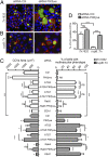
CvpB-mediated inhibition of PIKfyve promotes homotypic fusion of CCVs via autophagy. U2OS cells were treated with control siRNA (Left) or siRNA targeting PIKfyve (Right) for 24 h before being challenged with Coxiella Tn1832 (A) or the cvpB::Tn mutant (B) for 5 d. Cells were then fixed, and LAMP1 and DNA were labeled by using an anti-LAMP1 antibody coupled to Alexa Fluor 555 (red) or Hoechst 33258 (blue), respectively. Coxiella colonies were detected by using green Fluorescence (green). (C) U2OS cells transfected with the indicated siRNA sequences were challenged with Coxiella Tn1832 (white bars) or the cvpB::Tn mutant (gray bars). The median area of CCVs and the percentage of cells presenting a multivacuolar phenotype (CCVs per cell > 3) were calculated for each condition by using CellProfiler. (D) The percentage of CCVs positive for LC3 in cells treated as in A and B was assessed for each condition. Values are means ± SEM of triplicate experiments in which 200 CCVs or 200 cells were analyzed for each condition (***P < 0.0001; **P < 0.01; *P < 0.05; ns, not significant, one-way ANOVA, Bonferroni’s multiple comparison test). (Scale bars: 10 μm.)

References

    1. van Schaik EJ, Chen C, Mertens K, Weber MM, Samuel JE. Molecular pathogenesis of the obligate intracellular bacterium Coxiella burnetii. Nat Rev Microbiol. 2013;11(8):561–573. - PMC - PubMed
    1. Madariaga MG, Rezai K, Trenholme GM, Weinstein RA. Q fever: A biological weapon in your backyard. Lancet Infect Dis. 2003;3(11):709–721. - PubMed
    1. Newton HJ, McDonough JA, Roy CR. Effector protein translocation by the Coxiella burnetii Dot/Icm type IV secretion system requires endocytic maturation of the pathogen-occupied vacuole. PLoS One. 2013;8(1):e54566–e54569. - PMC - PubMed
    1. Moffatt JH, Newton P, Newton HJ. Coxiella burnetii: Turning hostility into a home. Cell Microbiol. 2015;17(5):621–631. - PubMed
    1. Campoy EM, Mansilla ME, Colombo MI. Endocytic SNAREs are involved in optimal Coxiella burnetii vacuole development. Cell Microbiol. 2013;15(6):922–941. - PubMed

Publication types

Substances

LinkOut - more resources