

New paradigm to assess brain cell morphology by diffusion-weighted MR spectroscopy in vivo
- PMID: 27226303
- PMCID: PMC4914152
- DOI: 10.1073/pnas.1504327113
New paradigm to assess brain cell morphology by diffusion-weighted MR spectroscopy in vivo
Abstract
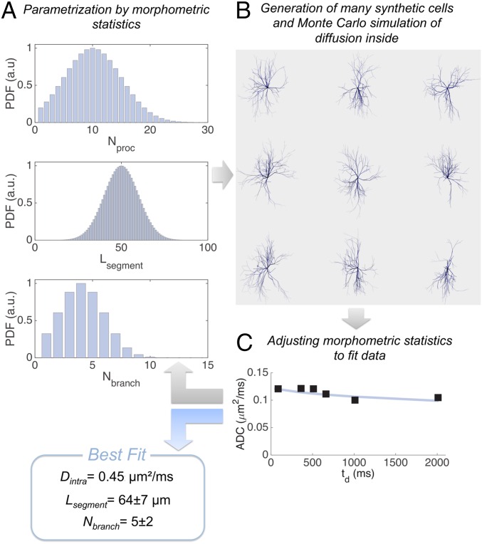
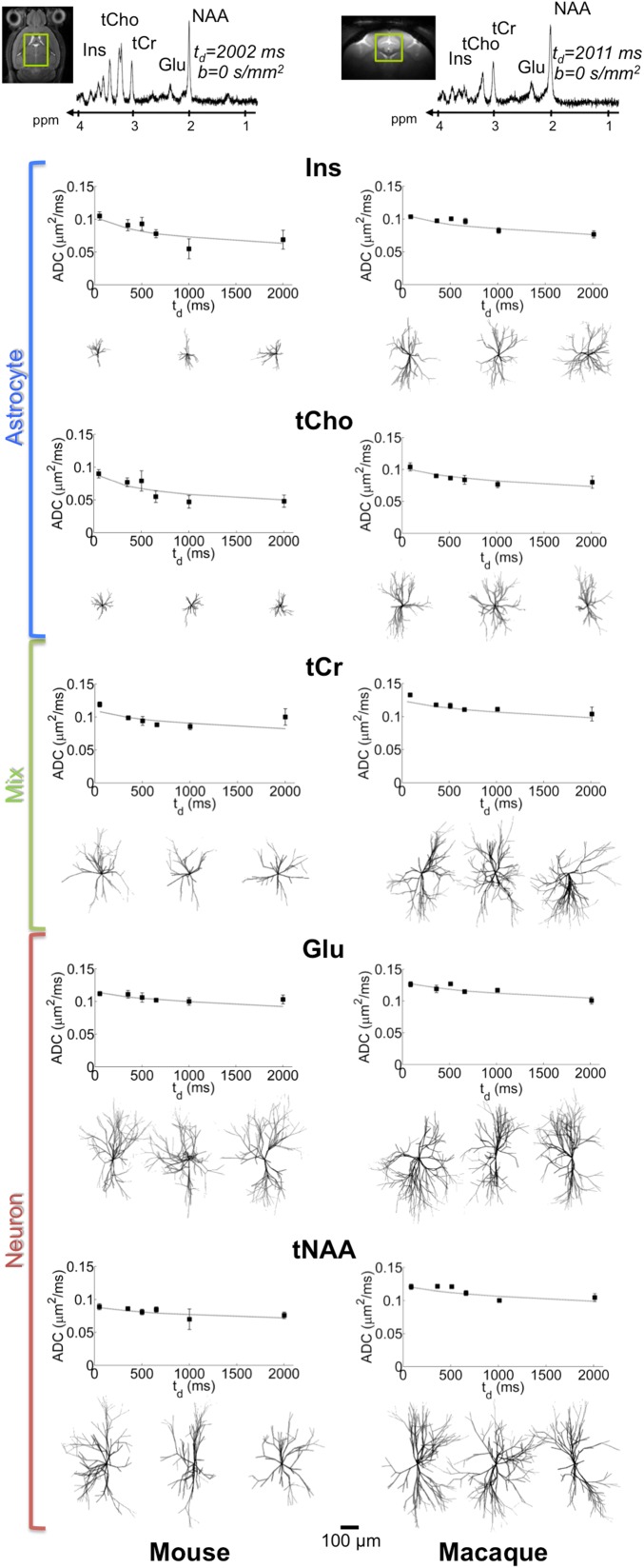
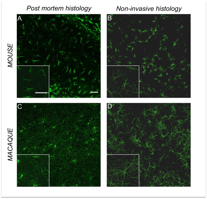
The brain is one of the most complex organs, and tools are lacking to assess its cellular morphology in vivo. Here we combine original diffusion-weighted magnetic resonance (MR) spectroscopy acquisition and novel modeling strategies to explore the possibility of quantifying brain cell morphology noninvasively. First, the diffusion of cell-specific metabolites is measured at ultra-long diffusion times in the rodent and primate brain in vivo to observe how cell long-range morphology constrains metabolite diffusion. Massive simulations of particles diffusing in synthetic cells parameterized by morphometric statistics are then iterated to fit experimental data. This method yields synthetic cells (tentatively neurons and astrocytes) that exhibit striking qualitative and quantitative similarities with histology (e.g., using Sholl analysis). With our approach, we measure major interspecies difference regarding astrocytes, whereas dendritic organization appears better conserved throughout species. This work suggests that the time dependence of metabolite diffusion coefficient allows distinguishing and quantitatively characterizing brain cell morphologies noninvasively.
Keywords: cell morphology; diffusion-weighted NMR spectroscopy; metabolites; noninvasive histology; numerical simulations.
Conflict of interest statement
The authors declare no conflict of interest.
Figures
References
-
- Beaulieu C. The basis of anisotropic water diffusion in the nervous system: A technical review. NMR Biomed. 2002;15(7-8):435–455. - PubMed
-
- Basser PJ, Pierpaoli C. Microstructural and physiological features of tissues elucidated by quantitative-diffusion-tensor MRI. 1996. J Magn Reson. 2011;213(2):560–570. - PubMed
-
- Norris DG. The effects of microscopic tissue parameters on the diffusion weighted magnetic resonance imaging experiment. NMR Biomed. 2001;14(2):77–93. - PubMed
Publication types
MeSH terms
Grants and funding
LinkOut - more resources
Full Text Sources
Other Literature Sources
