. 2016 Jul 1;143(13):2356-66.
doi: 10.1242/dev.135913. Epub 2016 May 25.

Adenomatous polyposis coli regulates radial axonal sorting and myelination in the PNS

Affiliations

Adenomatous polyposis coli regulates radial axonal sorting and myelination in the PNS

Benayahu Elbaz et al. Development. .

Abstract

The tumor suppressor protein adenomatous polyposis coli (APC) is multifunctional - it participates in the canonical Wnt/β-catenin signal transduction pathway as well as modulating cytoskeleton function. Although APC is expressed by Schwann cells, the role that it plays in these cells and in the myelination of the peripheral nervous system (PNS) is unknown. Therefore, we used the Cre-lox approach to generate a mouse model in which APC expression is specifically eliminated from Schwann cells. These mice display hindlimb weakness and impaired axonal conduction in sciatic nerves. Detailed morphological analyses revealed that APC loss delays radial axonal sorting and PNS myelination. Furthermore, APC loss delays Schwann cell differentiation in vivo, which correlates with persistent activation of the Wnt signaling pathway and results in perturbed extension of Schwann cell processes and disrupted lamellipodia formation. In addition, APC-deficient Schwann cells display a transient diminution of proliferative capacity. Our data indicate that APC is required by Schwann cells for their timely differentiation to mature, myelinating cells and plays a crucial role in radial axonal sorting and PNS myelination.

Keywords: Adenomatous polyposis coli; PNS; Radial axonal sorting; Schwann cells; Wnt signaling.


Conflict of interest statement

Competing interests

The authors declare no competing or financial interests.

Figures

Fig. 1.
Fig. 1.
Apclox/lox;P0-Cre mice exhibit hindlimb weakness and impaired axonal conduction in the sciatic nerve. (A) Excision of Apc exon 14 was verified in sciatic nerves of P7 Apclox/lox;P0-Cre mice using genomic DNA and primers that recognize the excised allele only: both the Apclox/lox;P0-Cre and Apclox/lox (used as control) carry the Apc exon 14 loxP-flanked allele (top panel), whereas the excised allele can be detected in sciatic nerve of Apclox/lox;P0-Cre mice only (bottom panel). (B) The excised Apc allele was also detected at different time points by quantitative PCR analysis on genomic DNA extracted from sciatic nerves of Apclox/lox;P0-Cre and Apclox/lox (control) mice. Il2 was used as an internal control gene for normalization. (C) In order to study the recombination efficiency of the P0-Cre line, we generated a ROSA26-stop-EYFP;P0-Cre reporter line. YFP+ cells were labeled in sciatic nerves of ROSA26-stop-EYFP;P0-Cre mice at different ages with an anti-GFP antibody and the percentage of DAPI+ cells expressing the YPF reporter gene protein was quantified. (D) Apclox/lox;P0-Cre mice exhibit hindlimb clenching when suspended by the tail, a common neuropathic phenotype. (E) Apclox/lox;P0-Cre mice also suffer from hindlimb weakness as detected by the grip strength assay. (F) The conduction velocity and the amplitude of compound muscle action potential were both significantly reduced in sciatic nerves of the Apclox/lox;P0-Cre mice. For detection of the excised allele, three to four animals were taken per each time point. The excised allele was not detected in the Apclox/lox mice at any time point. For reporter line assay, sciatic nerves from three to four animals were harvested per each time point. No YFP-positive cells were detected in control littermate ROSA26-stop-EYFP mice (Cre negative) at any time point. For grip strength assay: Apclox/lox, n=11 mice and Apclox/lox;P0-Cre, n=16 mice at P60. For electrophysiological studies: Apclox/lox, n=7 mice and Apclox/lox;P0-Cre, n=6 mice at P60. DL, distal latency; CV, conduction velocity; dAmp, distal amplitude. *P<0.05, **P<0.001.
Fig. 2.
Fig. 2.
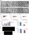
Loss of APC disrupts PNS myelination. The sciatic nerves of Apclox/lox;P0-Cre mutant and Apclox/lox control mice were analyzed by electron microscopy (EM). (A) The number of myelinated fibers is significantly reduced in the sciatic nerves of the Apclox/lox;P0-Cre mice at P1, P4 and P7 compared with control mice. Scale bars: 2 µm. (B-D) Apclox/lox;P0-Cre mice have a thinner myelin (higher g-ratio) compared with controls at P7 (B), P14 (C) and P60 (D). At P7, the mean g-ratio in the Apclox/lox;P0-Cre mice was 0.782 (±0.004) compared with 0.73 (±0.007) in the Apclox/lox mice, **P<0.001. At P14, the mean g-ratio in the Apclox/lox;P0-Cre mice was 0.72 (±0.006) compared to 0.68 (±0.009) in the Apclox/lox mice, **P<0.001. At P60, the mean g-ratio in the Apclox/lox;P0-Cre mice was 0.73 (±0.007) compared to 0.68 (±0.007) in the Apclox/lox mice, **P<0.001. (E) Apclox/lox;P0-Cre sciatic nerve contained fewer myelinated axons at P1, P4 and P7. *P<0.05, **P<0.001. (F) Three-dimensional EM images were acquired from sciatic nerve serial sections of Apclox/lox;P0-Cre and Apclox/lox (control) mice at P7, and individual nerve fibers were traced and analyzed in reconstructed EM images of both genotypes. The Apclox/lox;P0-Cre sciatic nerve contained much shorter internodes compared with controls. (G) Sciatic nerves of Apclox/lox;P0-Cre and Apclox/lox control mice at P60 were teased and immunolabeled for paranodin/Caspr, a marker of paranodal junctions to measure internodal length in adulthood. (H) Quantitative analysis showed that the Apclox/lox;P0-Cre sciatic nerves contained much shorter internodes compared with controls. For EM analysis, at least three mice were taken for each time point, per genotype. For g-ratio analysis, at least 50 axons from at least three mice per genotype were analyzed. The number of internodes analyzed in vivo in P7 animals was: Apclox/lox, n=9; Apclox/lox;P0-Cre, n=8; *P<0.05. The number of internodes analyzed in teased fibers of P60 animals was: Apclox/lox, n=46; Apclox/lox;P0-Cre, n=37; *P<0.05. Internodes from at least three mice per genotype were analyzed.
Fig. 3.
Fig. 3.
APC loss disrupts the radial axonal sorting process in the PNS. The sciatic nerves of Apclox/lox;P0-Cre and control Apclox/lox mice were analyzed morphometrically on Toluidine Blue-stained semi-thin sections (A,B) and by EM (C,D). (A,B) The sciatic nerves of the Apclox/lox;P0-Cre mice contained large bundles of axons compared with control mice. Black arrows indicate bundles of axons. Scale bars: 10 µm. (C,D) Apclox/lox;P0-Cre bundles of axons contained large- and small-caliber axons (D) in contrast to the bundles found in control mice (C) that contained small-caliber axons only. White arrows indicate bundles of axons. Scale bars: 5 µm. (E) The large bundles of axons were ∼3 times larger than the bundles of the control mice at P1, P4, P7 and P14. (F) Apclox/lox;P0-Cre large bundles of axons contained large- and small-caliber axons in contrast to the bundles found in control mice. Box plots show the 25–75% range (box) and 2.5–97.5% range (whiskers). (G) The bundles of unsorted axons in the Apclox/lox;P0-Cre mice contained a higher percentage of large-caliber axons (diameter >1 µm). (H) The density of the axons was not statistically different between Apclox/lox;P0-Cre mice and the control mice at P7. The images in A-D were taken from P7 mice. Axon bundles were analyzed from at least three mice per genotype. Axon diameter was analyzed from at least three mice per genotype at P7. **P<0.001. Large-caliber axons were defined here as axons with diameter of 1 µm and above. Low-caliber axons were defined here as axons with diameter less than 1 µm.
Fig. 4.
Fig. 4.
Delayed differentiation of APC-deficient Schwann cells. (A) Apclox/lox;P0-Cre sciatic nerve has increased numbers of premyelinating Scip+ Schwann cells. (C) Apclox/lox;P0-Cre sciatic nerve has reduced numbers of myelinating Krox20+ Schwann cells. (E) Ablation of APC results in a slight, non-statistically significant, reduction in cell proliferation. Graphs in B,D,F show quantitative analysis of data in A,C,E, respectively. Apclox/lox, n=4 mice; Apclox/lox;P0-Cre, n=3 mice at P7; *P<0.005.
Fig. 5.
Fig. 5.
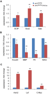
Loss of APC results in upregulation of Wnt signaling target genes and delayed differentiation of Schwann cells. (A) Expression of immature Schwann cell genes Scip, Sox2 and Galc is upregulated. (B) The expression of mature myelinating Schwann cell genes Krox20, Mbp, P0 and Mag is downregulated in Apclox/lox;P0-Cre sciatic nerves compared with controls at P7. (C) Expression of the Wnt signaling target genes Axin2, Myc and Lef is upregulated in the sciatic nerves of Apclox/lox;P0-Cre mice compared with controls at P7. Apclox/lox, n=3 mice; Apclox/lox;P0-Cre, n=3 mice. *P<0.05, **P<0.001.
Fig. 6.
Fig. 6.
Loss of APC results in perturbed Schwann cell process extension and lamellipodia formation. 3DEM images of serial sections were acquired from sciatic nerves of Apclox/lox;P0-Cre and Apclox/lox (control) mice at P7 and individual Schwann cells traced and analyzed in both genotypes. (A) Detailed analysis of a single Schwann cell in the 3DEM images of sciatic nerves revealed the presence of shorter cell processes in Apclox/lox;P0-Cre mice. (B) Primary Schwann cells were cultured and stained for S100, a Schwann cell marker. The Schwann cells derived from the Apclox/lox;P0-Cre sciatic nerves were much shorter (quantified results in C). Increased numbers of lamellipodia per cell were detected in the Schwann cells derived from the Apclox/lox;P0-Cre sciatic nerves (quantified results in D). Yellow arrows indicate lamellipodia. (E) No significant changes in the number of cell processes were found in the mutant cells compared with controls. Number of Schwann cells traced and analyzed in vivo in the sciatic nerves: Apclox/lox, n=5; Apclox/lox;P0-Cre, n=27; **P<0.001. Number of cells analyzed in vitro (primary culture): Apclox/lox, n=414; Apclox/lox;P0-Cre, n=469; **P<0.001.
Fig. 7.
Fig. 7.
Role of the Wnt signaling pathway in lamellipodia formation in Schwann cells. (A) Primary Schwann cells were cultured on laminin-coated plates in the presence of either the Wnt signaling activator CHIR99021 or the Wnt signaling inhibitor XAV939 or with DMSO (used as control; CON). The cells were fixed and stained for S100, a Schwann cell marker. (B) The Schwann cells derived from Apclox/lox;P0-Cre sciatic nerves were shorter, regardless of treatment. (C) Lamellipodia formation was increased in the Apclox/lox-derived cells upon CHIR99021 treatment, and decreased upon XAV939 treatment. The treatments did not affect lamellipodia formation in the cells derived from the Apclox/lox;P0-Cre mice. (D) No significant changes were detected in the number of processes per cell in either genotype. Number of cells analyzed in vitro (primary culture): DMSO control: Apclox/lox, n=736; Apclox/lox;P0-Cre, n=534; CHIR99021: Apclox/lox, n=625; Apclox/lox;P0-Cre, n=552; XAV939: Apclox/lox, n=558; Apclox/lox;P0-Cre, n=684; *P<0.05, **P<0.001. Yellow arrows indicate lamellipodia.

Similar articles

Cited by

References

    1. Auer R. N. (1994). Automated nerve fibre size and myelin sheath measurement using microcomputer-based digital image analysis: theory, method and results. J. Neurosci. Methods 51, 229-238. 10.1016/0165-0270(94)90015-9 - DOI - PubMed
    1. Bermingham J. R. Jr, Scherer S. S., O'Connell S., Arroyo E., Kalla K. A., Powell F. L. and Rosenfeld M. G. (1996). Tst-1/Oct-6/SCIP regulates a unique step in peripheral myelination and is required for normal respiration. Genes Dev. 10, 1751-1762. 10.1101/gad.10.14.1751 - DOI - PubMed
    1. Birchmeier C. and Nave K.-A. (2008). Neuregulin-1, a key axonal signal that drives Schwann cell growth and differentiation. Glia 56, 1491-1497. 10.1002/glia.20753 - DOI - PubMed
    1. Brosius Lutz A. (2014). Purification of Schwann cells from the neonatal and injured adult mouse peripheral nerve. In Purifying and Culturing Neural Cells: A Laboratory Manual (ed. Ben B. S. and Barres A.), pp. 177-188. Cold Spring Harbor Laboratory Press (in print). - PubMed
    1. Eccleston P. A., Mirsky R., Jessen K. R., Sommer I. and Schachner M. (1987). Postnatal development of rat peripheral nerves: an immunohistochemical study of membrane lipids common to non-myelin forming Schwann cells, myelin forming Schwann cells and oligodendrocytes. Brain Res. Dev Brain Res. 432, 249-256. 10.1016/0165-3806(87)90049-6 - DOI - PubMed

Publication types

Substances

LinkOut - more resources