. 2016 Jul 15;27(14):2161-71.
doi: 10.1091/mbc.E15-12-0865. Epub 2016 May 25.

Specific requirements of nonbilayer phospholipids in mitochondrial respiratory chain function and formation

Affiliations

Specific requirements of nonbilayer phospholipids in mitochondrial respiratory chain function and formation

Charli D Baker et al. Mol Biol Cell. .

Abstract

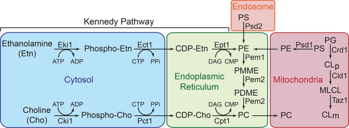
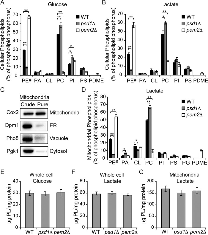
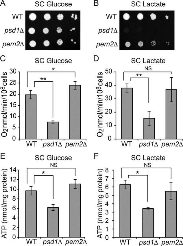
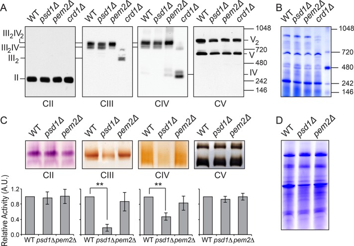
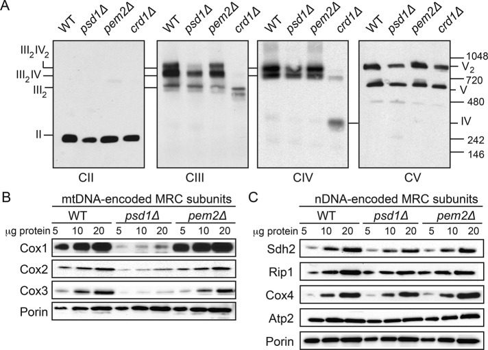
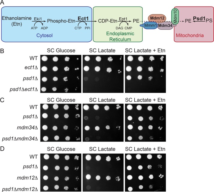
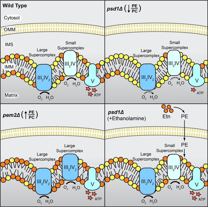
Mitochondrial membrane phospholipid composition affects mitochondrial function by influencing the assembly of the mitochondrial respiratory chain (MRC) complexes into supercomplexes. For example, the loss of cardiolipin (CL), a signature non-bilayer-forming phospholipid of mitochondria, results in disruption of MRC supercomplexes. However, the functions of the most abundant mitochondrial phospholipids, bilayer-forming phosphatidylcholine (PC) and non-bilayer-forming phosphatidylethanolamine (PE), are not clearly defined. Using yeast mutants of PE and PC biosynthetic pathways, we show a specific requirement for mitochondrial PE in MRC complex III and IV activities but not for their formation, whereas loss of PC does not affect MRC function or formation. Unlike CL, mitochondrial PE or PC is not required for MRC supercomplex formation, emphasizing the specific requirement of CL in supercomplex assembly. Of interest, PE biosynthesized in the endoplasmic reticulum (ER) can functionally substitute for the lack of mitochondrial PE biosynthesis, suggesting the existence of PE transport pathway from ER to mitochondria. To understand the mechanism of PE transport, we disrupted ER-mitochondrial contact sites formed by the ERMES complex and found that, although not essential for PE transport, ERMES facilitates the efficient rescue of mitochondrial PE deficiency. Our work highlights specific roles of non-bilayer-forming phospholipids in MRC function and formation.


Figures

FIGURE 1:
FIGURE 1:
Aminoglycerophospholipids and CL biosynthetic pathways in yeast. PE biosynthesis in yeast is accomplished by three major pathways: 1) Psd1-catalyzed decarboxylation of PS in the mitochondria, 2) Psd2-catalyzed decarboxylation of PS in endosomal compartments, and 3) incorporation of Etn via the cytosolic/ER Kennedy pathway. PC is produced by two major pathways: 1) formation of Pem1 and Pem2 PC by successive methylation of PE, and 2) incorporation of choline via the Kennedy pathway. CL biosynthesis occurs exclusively in the mitochondria, where premature cardiolipin (CLp) is synthesized from phosphatidylglycerol (PG) by Crd1. The resulting CLp is deacylated by the phospholipase Cld1 to produce monolysocardiolipin (MLCL) and reacylated by Taz1 to form mature cardiolipin (CLm). CDP, cytidine diphosphate; CTP, cytidine triphosphate; CMP, cytidine monophosphate; DAG, diacylglycerol; PA, phosphatidic acid; PI, phosphatidylinositol; PDME, phosphatidyl­dimethylethanolamine; PPi, inorganic pyrophosphate.
FIGURE 2:
FIGURE 2:
Cellular and mitochondrial phospholipid composition of psd1Δ and pem2Δ cells. The whole-cell phospholipid composition of WT, psd1Δ, and pem2Δ cells grown in (A) SC glucose and (B) SC lactate. Phospholipid levels are expressed as the percentage of total phospholipid phosphorus in each phospholipid class. PE# represents the sum of PE and PMME in pem2Δ cells. Data are expressed as mean ± SD (n = 3); **p < 0.005, *p < 0.05. (C) Western blot analysis of crude and sucrose-gradient purified mitochondria from WT cells. Cox2, Dpm1, Pho8, and Pgk1 are used as markers of the yeast mitochondria, ER, vacuole, and cytoplasm, respectively. (D) Phospholipid composition of sucrose gradient–purified mitochondria from WT, psd1Δ, and pem2Δ cells grown in SC lactate. Data are expressed as mean ± SD (n = 3); **p < 0.005, *p < 0.05. (E, F) Total phospholipid content of whole-cell homogenates of WT, psd1Δ, and pem2Δ cells grown in (E) SC glucose or (F) SC lactate. (G) Total phospholipid content of mitochondria from SC lactate–grown cells. Data are expressed as mean ± SD (n = 3).
FIGURE 3:
FIGURE 3:
Mitochondrial respiration is dependent on PE but not PC levels. Tenfold serial dilutions of WT, psd1Δ, and pem2Δ cells were spotted onto (A) SC glucose and (B) SC lactate plates, and images were captured after 2 (SC glucose) or 5 d (SC lactate) of growth at 30°C. Data are representative of at least three independent experiments. (C, D) WT, psd1Δ, and pem2Δ cells were grown in (C) SC glucose or (D) SC lactate to late log phase, and the rate of oxygen consumption was measured. Data are expressed as mean ± SD (n = 6); *p < 0.05, **p < 0.005. (E, F) Cellular ATP levels of WT, psd1Δ, and pem2Δ cells cultured in (E) SC glucose or (F) SC lactate. Data are expressed as mean ± SD (n = 3); *p < 0.05.
FIGURE 4:
FIGURE 4:
Mitochondrial PE is required for MRC complex III and IV activities but not MRC supercomplex formation. (A) Mitochondria from SC lactate–grown cells were solubilized by 1% digitonin and subjected to BN–PAGE/Western blot, and complexes II–V were detected by Sdh2, Rip1, Cox2, and Atp2 antibodies, respectively. Mitochondria from CL-deficient crd1Δ cells were used as positive control to demonstrate loss of supercomplexes (III2IV2, large supercomplex; III2IV, small supercomplex) under identical conditions. (B) Samples from A were stained with Coomassie blue to demonstrate equal loading. (C) Digitonin-solubilized mitochondrial complexes from WT, psd1Δ, and pem2Δ cells were separated by CN-PAGE, followed by in-gel activity staining for complexes II–V. In-gel activities of MRC complexes were quantified by densitometric analysis, and relative activities were plotted for complexes II–V. Data were normalized to WT cells and expressed as mean ± SD (n = 3); **p < 0.005. (D) Samples from C were stained with Coomassie blue, and total protein, quantified using densitometric analysis, was used to normalize activity staining.
FIGURE 5:
FIGURE 5:
Depletion of mitochondrial PE in glucose-grown psd1Δ cells results in a specific loss of mtDNA-encoded MRC subunits. (A) Digitonin-solubilized mitochondria from SC glucose–grown WT, psd1Δ, and pem2Δ cells were subjected to BN–PAGE/Western blot. Complexes II–V were detected by Sdh2, Rip1, Cox2, and Atp2 antibodies, respectively. Data are representative of at least three independent experiments. (B) Mitochondria from SC glucose–grown WT, psd1Δ, and pem2Δ cells were subjected to SDS–PAGE, and mtDNA-encoded subunits were probed using Cox1, Cox2, and Cox3 antibodies. (C) Nuclear-encoded subunits were probed using Sdh2, Rip1, Cox4, and Atp2. Porin was used as a loading control. Data are representative of at least three independent experiments.
FIGURE 6:
FIGURE 6:
Ethanolamine supplementation rescues respiratory defects of psd1Δ cells by restoring mitochondrial PE levels. (A) Cellular and (B) mitochondrial phospholipid composition of WT cells grown in SC lactate and psd1Δ cells grown in SC lactate with and without 2 mM Etn. Phospholipid levels are expressed as percentage of total phospholipid phosphorus in each phospholipid class. Data are expressed as mean ± SD (n = 3); **p < 0.005, *p < 0.05. Tenfold serial dilutions of WT and psd1Δ cells were spotted onto (C) SC lactate and (D) SC lactate + 2 mM Etn plates, and images were captured after 4 d of growth at 30°C. Data are representative of at least three independent trials. (E) Rate of oxygen consumption and (F) total cellular ATP levels of WT and psd1Δ cells grown in SC lactate ± 2 mM Etn to late logarithmic phase were quantified. Data are expressed as mean ± SD (n = 3); *p < 0.05, **p < 0.005 (G, H) Digitonin- solubilized mitochondrial complexes from WT and psd1Δ cells grown in SC lactate ± 2 mM Etn were separated by CN-PAGE, followed by in-gel activity staining for (G) complex III and (H) complex IV. Densitometric quantifications of relative in-gel activities for complexes III and IV. Data were normalized to WT cells and are expressed as mean ± SD (n = 3); **p < 0.005, *p < 0.05. A.U., arbitrary units.
FIGURE 7:
FIGURE 7:
PE synthesized by the Kennedy pathway requires ERMES for complete rescue of mitochondrial PE deficiency. (A) Schematic representation of the Kennedy pathway of PE biosynthesis and the ERMES complex. The Kennedy pathway enzyme Ect1, mitochondrial Psd1, and Mdm34 and Mdm12 of the ERMES complex are depicted in boldface to indicate that these genes are targeted to construct double-knockout strains. Tenfold serial dilutions of (B) WT, ect1Δ, psd1Δ, and psd1Δect1Δ, (C) WT, psd1Δ, mdm34Δ, and psd1Δmdm34Δ, and (D) WT, psd1Δ, mdm12Δ, and psd1Δmdm12Δ cells were spotted onto SC glucose and SC lactate ± Etn plates, and images were captured after 2 (SC glucose) or 5 d (SC lactate ± Etn) of growth at 30°C. Data are representative of at least three independent experiments.
FIGURE 8:
FIGURE 8:
Model depicting the specific roles of mitochondrial PE and PC in MRC complex activity and assembly. Reduced PE/PC ratio in mitochondrial PE–depleted psd1Δ cells leads to decreased MRC complex III and IV activities without affecting supercomplex formation. Increased PE/PC ratio in mitochondrial PC–depleted pem2Δ cells results in enhanced formation of the larger supercomplex (III2IV2) without altering the activities of complexes. PE synthesized in ER by exogenous supplementation of Etn is transported into mitochondria and completely restores MRC supercomplex activities in PE-deficient psd1Δ cells. IMM, inner mitochondrial membrane; IMS, intermembrane space; OMM, outer mitochondrial membrane.

References

    1. Acín-Pérez R, Fernández-Silva P, Peleato ML, Pérez-Martos A, Enriquez JA. Respiratory active mitochondrial supercomplexes. Mol Cell. 2008;32:529–539. - PubMed
    1. AhYoung AP, Jiang J, Zhang J, Khoi Dang X, Loo JA, Zhou ZH, Egea PF. Conserved SMP domains of the ERMES complex bind phospholipids and mediate tether assembly. Proc Natl Acad Sci USA. 2015;112:3179–3188. - PMC - PubMed
    1. Aktas M, Wessel M, Hacker S, Klusener S, Gleichenhagen J, Narberhaus F. Phosphatidylcholine biosynthesis and its significance in bacteria interacting with eukaryotic cells. Eur J Cell Biol. 2010;89:888–894. - PubMed
    1. Amberg DC, Burke DJ, Strathern JN. Methods in Yeast Genetics. A Cold Spring Harbor Laboratory Course Manual. Cold Spring Harbor, NY: Cold Spring Harbor Laboratory Press; 2005.
    1. Bartlett GR. Phosphorus assay in column chromatography. J Biol Chem. 1959;234:466–468. - PubMed

MeSH terms

LinkOut - more resources