. 2016 Jul 1;291(27):14324-14339.
doi: 10.1074/jbc.M115.712026. Epub 2016 May 11.

Biochemical Characterization of the Human Mitochondrial Replicative Twinkle Helicase: SUBSTRATE SPECIFICITY, DNA BRANCH MIGRATION, AND ABILITY TO OVERCOME BLOCKADES TO DNA UNWINDING

Affiliations

Biochemical Characterization of the Human Mitochondrial Replicative Twinkle Helicase: SUBSTRATE SPECIFICITY, DNA BRANCH MIGRATION, AND ABILITY TO OVERCOME BLOCKADES TO DNA UNWINDING

Irfan Khan et al. J Biol Chem. .

Abstract

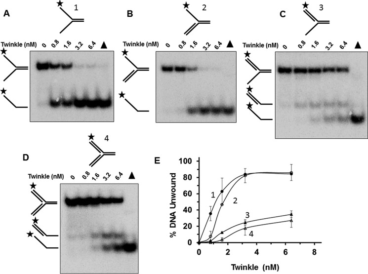
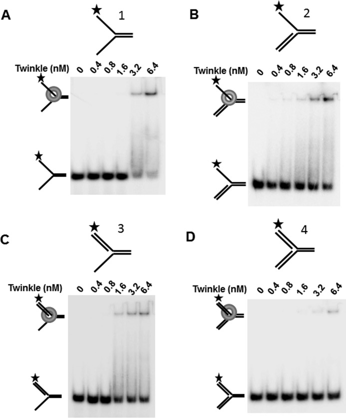
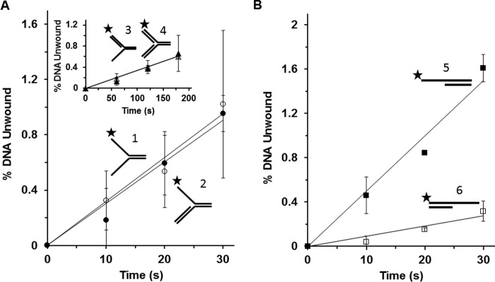
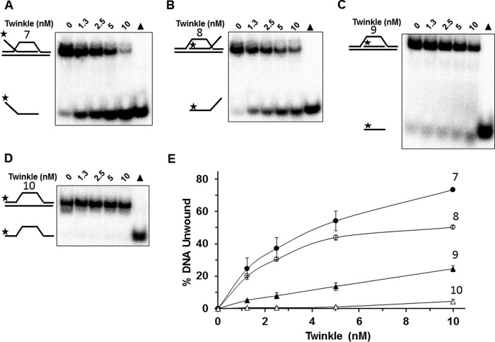
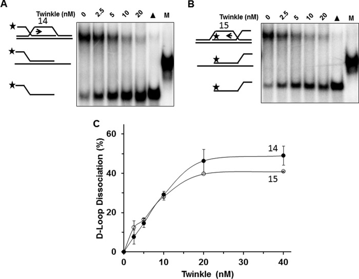
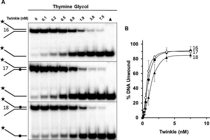
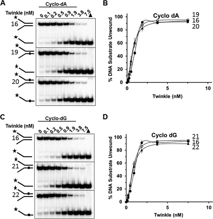
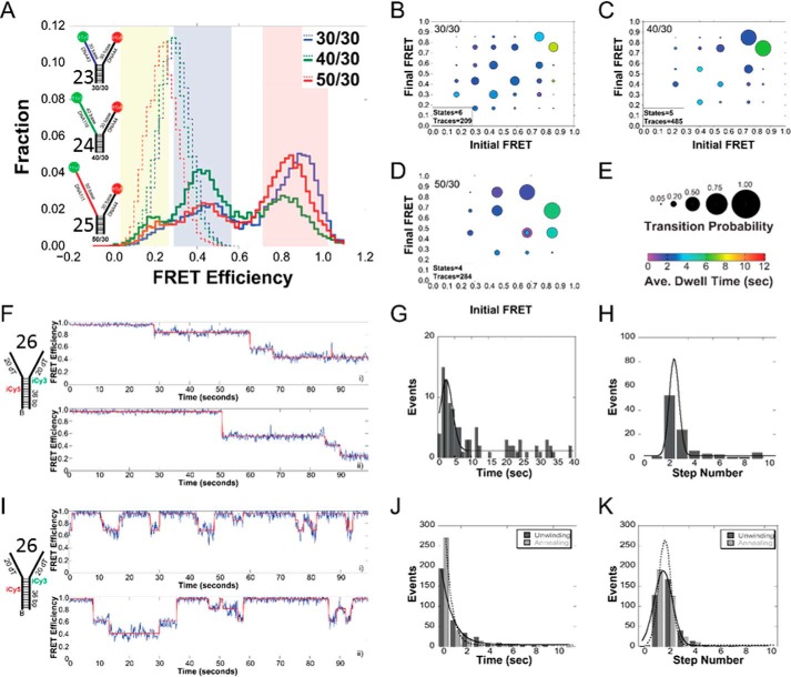
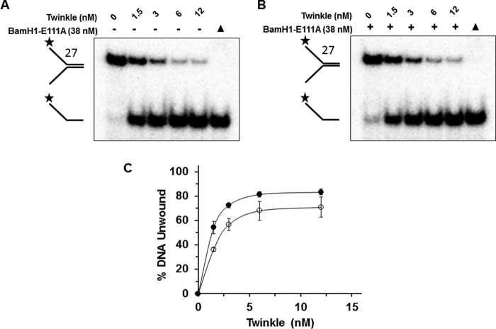
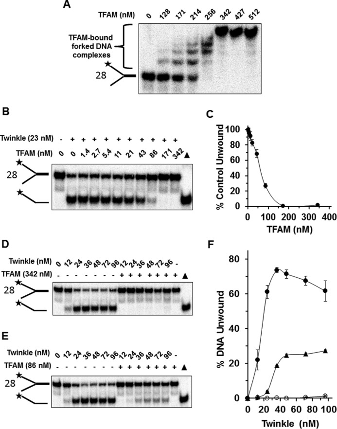
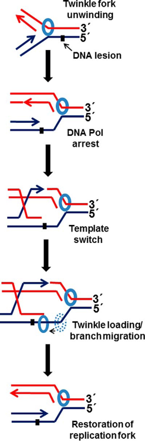
Mutations in the c10orf2 gene encoding the human mitochondrial DNA replicative helicase Twinkle are linked to several rare genetic diseases characterized by mitochondrial defects. In this study, we have examined the catalytic activity of Twinkle helicase on model replication fork and DNA repair structures. Although Twinkle behaves as a traditional 5' to 3' helicase on conventional forked duplex substrates, the enzyme efficiently dissociates D-loop DNA substrates irrespective of whether it possesses a 5' or 3' single-stranded tailed invading strand. In contrast, we report for the first time that Twinkle branch-migrates an open-ended mobile three-stranded DNA structure with a strong 5' to 3' directionality preference. To determine how well Twinkle handles potential roadblocks to mtDNA replication, we tested the ability of the helicase to unwind substrates with site-specific oxidative DNA lesions or bound by the mitochondrial transcription factor A. Twinkle helicase is inhibited by DNA damage in a unique manner that is dependent on the type of oxidative lesion and the strand in which it resides. Novel single molecule FRET binding and unwinding assays show an interaction of the excluded strand with Twinkle as well as events corresponding to stepwise unwinding and annealing. TFAM inhibits Twinkle unwinding, suggesting other replisome proteins may be required for efficient removal. These studies shed new insight on the catalytic functions of Twinkle on the key DNA structures it would encounter during replication or possibly repair of the mitochondrial genome and how well it tolerates potential roadblocks to DNA unwinding.

Keywords: DNA helicase; DNA repair; DNA replication; Twinkle; branch migration; genetic disease; helicase; mitochondria.


Figures

FIGURE 1.
FIGURE 1.
Twinkle unwinding activity on DNA replication structures. A–D, purified recombinant Twinkle helicase, designated in hexamer concentration, was incubated with the indicated replication fork DNA substrates 1–4 (0.5 nm) at 37 °C for 30 min as described under “Experimental Procedures.” Proteinase K-digested reaction products were resolved on non-denaturing 12% polyacrylamide gels. Representative gel images from at least three independent experiments are shown. Filled triangle, heat-denatured DNA substrate control. E, quantitative analysis of percent DNA substrate unwound from A to D. Fork with single-stranded 5′ and 3′ arms, filled circle; fork with 5′ single-stranded arm, open circle; fork with 3′ single strand arm, filled triangle; fork with double-stranded 5′ and 3′ arms, open triangle. Average values of at least three independent experiments with standard deviations indicated by error bars are shown.
FIGURE 2.
FIGURE 2.
Twinkle DNA binding to replication fork structures. A–D, indicated concentration of Twinkle hexamer was incubated with the indicated replication fork DNA substrates 1–4 (0.5 nm) at 37 °C for 30 min as described under “Experimental Procedures.” DNA species from binding mixtures were resolved on non-denaturing 5% polyacrylamide gels. Representative images from EMSA of at least three independent experiments are shown.
FIGURE 3.
FIGURE 3.
Single-turnover kinetics of Twinkle helicase activity on replication fork structures. Twinkle hexamer (3.2 nm) was pre-incubated with the indicated radiolabeled DNA substrate 1–6 (2.5 nm) prior to simultaneous addition of ATP and 100-fold excess of dT200, followed by incubation at specified time points as described under “Experimental Procedures” for single-turnover kinetic assays. Reaction products were resolved on native 12% polymacrylamide gels and analyzed. A, Twinkle (3.2 nm hexamer) unwinding kinetics on forked duplex substrate with single-stranded 5′ and 3′ arms (substrate 1) (filled circle) or 5′ flap substrate (substrate 2) (open circle). Inset, 3′ flap substrate (substrate 3) (filled triangle) or synthetic replication fork with duplex leading and lagging strand arms (substrate 4) (open triangle). B, Twinkle (6.4 nm hexamer) unwinding kinetics on 5′ single strand tailed duplex (substrate 5) (filled square) or 3′ single strand tailed duplex (substrate 6) (open square). Average values of at least three independent experiments with standard deviations indicated by error bars are shown.
FIGURE 4.
FIGURE 4.
Twinkle helicase activity on immobile D-loop DNA substrates. A–D, indicated concentrations of Twinkle helicase were incubated with the specified immobile D-loop (substrates 7–9) or bubble (substrate 10) DNA substrates (0.5 nm) at 37 °C for 30 min as described under “Experimental Procedures.” Reaction products were resolved on non-denaturing 12% polyacrylamide gels. Representative gel images from at least three independent experiments are shown. E, quantitative analysis of percent DNA substrate unwound from A to D. Immobile D-loop with single-stranded 5′ tail (substrate 7), filled circle; immobile D-loop with single-stranded 3′ tail (substrate 8), open circle; immobile D-loop with flush invading strand (substrate 9), filled triangle; bubble DNA substrate (substrate 10), open triangle. Average values of at least three independent experiments with standard deviations indicated by error bars are shown.
FIGURE 5.
FIGURE 5.
Twinkle helicase promotes branch migration of mobile three-way junction DNA structure preferentially in the 5′ to 3′ direction. A and B, indicated concentrations of Twinkle helicase hexamer were incubated with the specified DNA substrate (0.5 nm) at 37 °C for 30 min as described under “Experimental Procedures.” Reaction products were resolved on non-denaturing 12% polyacrylamide gels. Representative gel images from at least three independent experiments are shown. C, quantitative analysis of percent branch migration product in the 5′ to 3′ direction (substrate 11) (A, filled circle) or 3′ to 5′ direction (substrate 13) (B, open circle). Average values of at least three independent experiments with standard deviations indicated by error bars are shown. D, Twinkle-catalyzed branch migration is dependent on hydrolyzable ATP. Reaction mixtures containing 5 nm Twinkle and the 5′ to 3′ mobile three-way junction DNA substrate (substrate 11) (0.5 nm, A) were incubated and analyzed as described above. E, indicated concentrations of Twinkle helicase hexamer were incubated with a forked duplex DNA substrate (substrate 12) (0.5 nm) at 37 °C for 30 min as described under “Experimental Procedures.” The third oligonucleotide added to the reaction mixture requires helicase-catalyzed unwinding of the duplex region to anneal to one of the two unwound strands. Reaction products were resolved on non-denaturing 12% polyacrylamide gels. Representative gel images from at least three independent experiments are shown. F, quantitative analysis of percent helicase product. Average values of at least three independent experiments with standard deviations indicated by error bars are shown.
FIGURE 6.
FIGURE 6.
Twinkle helicase dissociates mobile D-loop DNA structures. A and B, indicated Twinkle helicase hexamer concentrations were incubated with the specified mobile D-loop DNA substrate (0.5 nm) at 37 °C for 30 min as described under “Experimental Procedures.” Reaction products were resolved on non-denaturing 12% polyacrylamide gels. Representative gel images from at least three independent experiments are shown. C, quantitative analysis of percent D-loop dissociation in the 5′ to 3′ direction (substrate 14) or 3′ to 5′ direction (substrate 15) (A, filled circle (substrate 14); B, open circle (substrate 15)). Average values of at least three independent experiments with standard deviations indicated by error bars are shown.
FIGURE 7.
FIGURE 7.
Twinkle is sensitive to a single thymine glycol in the helicase translocating strand. A, indicated Twinkle helicase hexamer concentrations were incubated with the specified thymine glycol containing DNA substrates (0.5 nm) at 37 °C for 30 min as described under “Experimental Procedures.” Reaction products were resolved on non-denaturing 12% polyacrylamide gels. Representative gel images from at least three independent experiments are shown. B, quantitative analysis of percent DNA substrate unwound from A. Undamaged substrate (substrate 16), filled circle; bottom (non-translocating) strand thymine glycol substrate (substrate 17), open circle; top (translocating) strand thymine glycol substrate (substrate 18), filled triangle. Average values of at least three independent experiments with standard deviations indicated by error bars are shown.
FIGURE 8.
FIGURE 8.
Twinkle tolerates a single cyclopurine in the helicase translocating or non-translocating strands. A and C, indicated Twinkle helicase hexamer concentrations were incubated with the specified cyclo-dA (A) containing DNA substrates (substrates 19, 20) or cyclo-dG (C) containing DNA substrates (substrates 21 and 22) (0.5 nm) at 37 °C for 30 min as described under “Experimental Procedures.” Reaction products were resolved on non-denaturing 12% polyacrylamide gels. Representative gel images from at least three independent experiments are shown. B and D, quantitative analysis of percent DNA substrate unwound from A and C, respectively. Average values of at least three independent experiments with standard deviations indicated by error bars are shown. Undamaged substrate (substrate 16), filled circle; bottom (non-translocating) strand cPu substrate (substrates 19, 21), open circle; top (translocating) strand cPu substrate (substrates 20, 22), filled triangle.
FIGURE 9.
FIGURE 9.
Histograms and ExPRT plots of Twinkle bound to DNA forks. A, histogram of the FRET signals from the 30/30 DNA fork substrate (substrate 23) (blue), 40/30 DNA fork substrate (substrate 24) (green), and 50/30 DNA fork substrate (substrate 25) (red), DNA fork substrates alone (dashed lines) and after addition of 25 nm Twinkle (solid lines). The length of the 5′-translocating strand was kept constant at 30 (dT) nucleotides, and the length of the 3′ excluded strand was varied from 30 to 50 (dT) nucleotides. Yellow, blue, and red regions indicate low, medium, and high FRET states, respectively. ExPRT plots for Twinkle bound to the 30/30 (B), 40/30 (C), and 50/30 (D) DNA forks. Each marker on the ExPRT plot represents a transition from the initial FRET state on the x axis to the final FRET state on the y axis. E, size of the marker corresponds to the fraction of analyzed traces that exhibit that particular transition (transition probability), and the color represents the dwell time (seconds) of the initial state. Representative traces from smFRET experiments showing undirectional unwinding (F) or alternating unwinding and reannealing (I) for substrate 26. The calculated FRET signal is shown in blue, and the fit to ideal states is overlaid in red. A global quantification of the unwinding time or number of steps for unidirectional unwinding (G and H) or alternating unwinding and reannealing (J and K). For multistate smFRET traces, the individual unwinding (dark gray) and annealing (light gray) times and steps are quantified individually. The lines correspond to fits to a Gaussian equation.
FIGURE 10.
FIGURE 10.
Twinkle displaces BamHI-E111A bound to forked duplex DNA substrate and unwinds the forked duplex. A and B, indicated concentrations of Twinkle (hexamer) were incubated at 37 °C for 30 min with a BamHI forked duplex substrate (substrate 27) (0.5 nm) in the absence (A) or presence (B) of catalytically inactive BamHI-E111A restriction endonuclease (38 nm) as described under “Experimental Procedures.” Proteinase K-digested products were electrophoresed on non-denaturing 12% polyacrylamide gels. Representative gel image from at least three independent experiments is shown. C, quantitative analysis of percent DNA substrate unwound from A and B. Naked forked duplex, filled circles; BamHI-E111A-bound forked duplex, open circles. Average values of at least three independent experiments with standard deviations indicated by error bars are shown.
FIGURE 11.
FIGURE 11.
TFAM bound to forked duplex DNA substrate inhibits DNA unwinding by Twinkle helicase. A, TFAM binding to forked duplex. The indicated concentration of TFAM was incubated with the TFAM forked duplex DNA substrate (substrate 28) (0.5 nm) at 24 °C for 30 min as described under “Experimental Procedures.” DNA species from binding mixtures were resolved on non-denaturing 5% polyacrylamide gels. Representative image from EMSA of at least three independent experiments is shown. B, Twinkle helicase (23 nm hexamer) was incubated at 37 °C for 30 min with a forked duplex substrate (0.5 nm) that had been pre-bound with the indicated concentrations of TFAM as described under “Experimental Procedures.” Proteinase K-digested products were electrophoresed on non-denaturing 12% polyacrylamide gels. Representative gel image from at least three independent experiments is shown. C, quantitative analysis of forked duplex DNA substrate unwound (percent control activity compared with the naked forked duplex in which TFAM was omitted from pre-binding step) from experiments as conducted in B. D and E, indicated concentrations of Twinkle hexamer were incubated at 37 °C for 30 min with forked duplex DNA substrate (0.5 nm) that had been pre-incubated in the absence or presence of 342 nm TFAM (D) or 86 nm TFAM (E). Proteinase K-digested products were electrophoresed on non-denaturing 12% polyacrylamide gels. Representative gel image from at least three independent experiments is shown. F, quantitative analysis of forked duplex DNA substrate unwound from experiments as conducted in D and E. Naked forked duplex, filled circles; TFAM (342 nm)-bound forked duplex, open circles; TFAM (86 nm)-bound forked duplex, filled triangles. Average values of at least three independent experiments with standard deviations indicated by error bars are shown.
FIGURE 12.
FIGURE 12.
Proposed model for Twinkle branch migration activity at a stalled mitochondrial DNA replication fork. See text for details.

References

    1. Rajala N., Gerhold J. M., Martinsson P., Klymov A., and Spelbrink J. N. (2014) Replication factors transiently associate with mtDNA at the mitochondrial inner membrane to facilitate replication. Nucleic Acids Res. 42, 952–967 - PMC - PubMed
    1. Copeland W. C. (2012) Defects in mitochondrial DNA replication and human disease. Crit. Rev. Biochem. Mol. Biol. 47, 64–74 - PMC - PubMed
    1. Korhonen J. A., Pham X. H., Pellegrini M., and Falkenberg M. (2004) Reconstitution of a minimal mtDNA replisome in vitro. EMBO J. 23, 2423–2429 - PMC - PubMed
    1. McKinney E. A., and Oliveira M. T. (2013) Replicating animal mitochondrial DNA. Genet. Mol. Biol. 36, 308–315 - PMC - PubMed
    1. Shutt T. E., and Gray M. W. (2006) Twinkle, the mitochondrial replicative DNA helicase, is widespread in the eukaryotic radiation and may also be the mitochondrial DNA primase in most eukaryotes. J. Mol. Evol. 62, 588–599 - PubMed

Publication types