. 2016 Jul 22;291(30):15896-907.
doi: 10.1074/jbc.M115.713107. Epub 2016 May 19.

S100A1 Protein Does Not Compete with Calmodulin for Ryanodine Receptor Binding but Structurally Alters the Ryanodine Receptor·Calmodulin Complex

Affiliations

S100A1 Protein Does Not Compete with Calmodulin for Ryanodine Receptor Binding but Structurally Alters the Ryanodine Receptor·Calmodulin Complex

Robyn T Rebbeck et al. J Biol Chem. .

Abstract

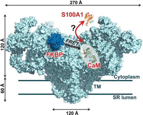
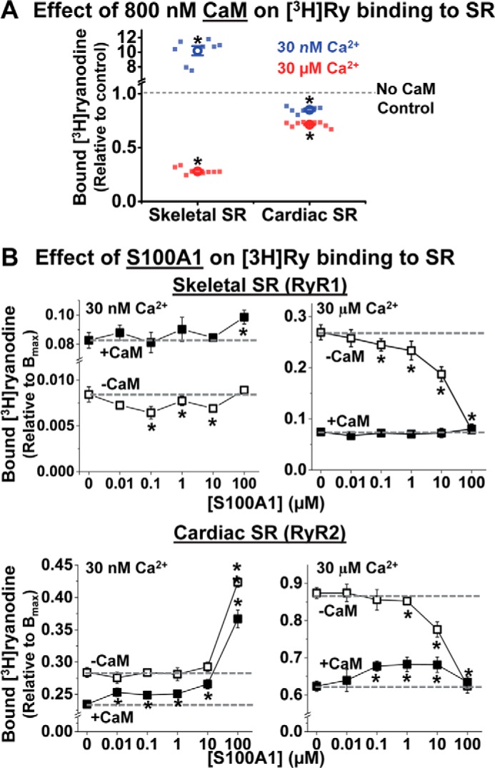
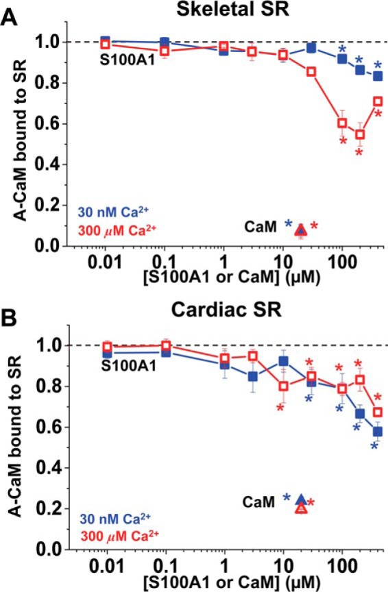
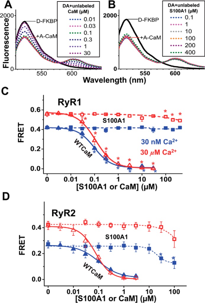
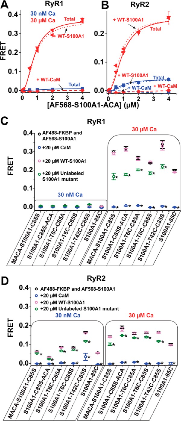
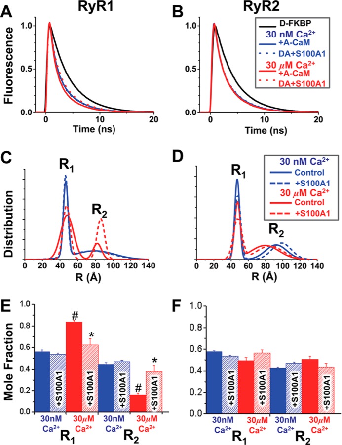
S100A1 has been suggested as a therapeutic agent to enhance myocyte Ca(2+) cycling in heart failure, but its molecular mode of action is poorly understood. Using FRET, we tested the hypothesis that S100A1 directly competes with calmodulin (CaM) for binding to intact, functional ryanodine receptors type I (RyR1) and II (RyR2) from skeletal and cardiac muscle, respectively. Our FRET readout provides an index of acceptor-labeled CaM binding near donor-labeled FKBP (FK506-binding protein 12.6) on the cytoplasmic domain of RyR in isolated sarcoplasmic reticulum vesicles. S100A1 (0.01-400 μm) partially inhibited FRET (i.e. CaM binding), with Ki > 10 μm, for both RyR1 and RyR2. The high [S100A1] required for partial effects on FRET indicates a lack of competition by S100A1 on CaM/RyR binding under normal physiological conditions. High-resolution analysis of time-resolved FRET detects two structural states of RyR-bound CaM, which respond to [Ca(2+)] and are isoform-specific. The distribution of these structural states was perturbed only by high micromolar [S100A1], which promoted a shift of bound CaM to a lower FRET orientation (without altering the amount of CaM bound to RyR). Thus, high micromolar S100A1 does alter the CaM/RyR interaction, without involving competition. Nevertheless, submicromolar S100A1 can alter RyR function, an effect that is influenced by both [Ca(2+)] and [CaM]. We conclude that CaM and S100A1 can concurrently bind to and functionally modulate RyR1 and RyR2, but this does not involve direct competition at the RyR CaM binding site.

Keywords: FKBP12.6; calcium channel; calmodulin (CaM); fluorescence lifetime; fluorescence resonance energy transfer (FRET); sarcoplasmic reticulum (SR); structure-function.


Figures

FIGURE 1.
FIGURE 1.
Hypothesis tested and RyR-targeted FRET molecular toolkit. S100A1 (orange) competes with CaM (green) binding to RyR (light blue). The RyR1 model (cyan) is based on a published cryo-EM density map (Protein Data Bank code 3J8H) (42) with docked FKBP12 (dark blue) and suggested location of the CaM binding site (18) indicated (shaded oval). FKBP and CaM N-lobe bind to locations that are within ∼50 Å from each other in the RyR complex, a feature that is exploited by our FRET-based system to investigate the RyR structure and binding events in vitro and in situ (18–20).
FIGURE 2.
FIGURE 2.
CaM alters the effect of S100A1 on [3H]ryanodine binding to RyR1 and RyR2. SR vesicles from skeletal or cardiac muscle were incubated with S100A1 (0–100 μm) and CaM (0 or 800 nm) at 30 nm or 30 μm Ca2+ in the presence of [3H]ryanodine. A, as a control, the functional effect of 800 nm CaM was tested on [3H]ryanodine binding to skeletal (left) and cardiac (right) SR vesicles in 30 nm (blue) or 30 μm (red) Ca2+. Data are shown as individual data points (square) and mean ± S.E. (open circle). The fraction of [3H]ryanodine binding is relative to “no CaM” control. B, effect of S100A1 on [3H]ryanodine binding to skeletal SR (top panels) and cardiac SR (bottom panels) at 30 nm Ca2+ (left) and 30 μm Ca2+ (right) and in the absence of CaM (hollow squares) and in the presence of 800 nm CaM (solid squares). Control levels (absence of S100A1) are indicated by dashed lines. The fraction of [3H]ryanodine bound to SR vesicles is relative to maximum binding capacity (Bmax). Data are shown as mean ± S.E. (n = 6–8). *, significant differences relative to no-S100A1 controls; p < 0.05, as determined by paired Student's t test.
FIGURE 3.
FIGURE 3.
Competitive inhibition of CaM binding to SR vesicles by S100A1. SRs from skeletal (A) or cardiac (B) muscle were preincubated with WT-S100A1 or WT-CaM with 30 nm (blue solid symbol) or 300 μm (red open symbol) Ca2+ and then incubated with 0.1 μm A-CaM before SR membrane sedimentation by centrifugation. Data are normalized to no-S100A1 or no-CaM controls (black dotted lines) and expressed as means ± S.E. (n = 4). *, significantly different from control, p < 0.05 as determined by paired Student's t test.
FIGURE 4.
FIGURE 4.
Competitive inhibition of A-CaM binding to RyR1 and RyR2 by S100A1. RyR in native SR membranes isolated from skeletal or cardiac muscle was labeled with D-FKBP, preincubated with S100A1 (0–400 μm), and then incubated with 0.1 μm A-CaM (labeled via T34C mutation). The inhibition of CaM bound to RyR was calculated as the fractional decrease of FRET between D-FKBP and A-CaM, based on the fluorescence intensity readout. Shown are representative fluorescence spectra of samples preincubated with unlabeled CaM (A) or S100A1 (B). Such measurements are plotted as competition curves for samples consisting of skeletal (C) and cardiac (D) SR. Data are shown as mean FRET ± S.E. (n = 4). *, significantly different from no-S100A1 controls, as determined by paired Student's t test.
FIGURE 5.
FIGURE 5.
Competition of A-S100A1 by unlabeled S100A1 and CaM for binding to RyR. RyRs in SR membranes isolated from skeletal or cardiac muscle were labeled with D-FKBP and incubated with A-S100A1. Binding specificity of A-S100A1 to RyR was investigated by the addition of a >10-fold molar excess of unlabeled S100A1 or CaM. Saturation binding of AF568-S100A1-C85S-ACA to RyR from skeletal (A) and cardiac (B) SR is indicated as Total (solid lines) at 30 nm Ca2+ (blue) and 30 μm Ca2+ (red). Total FRET is compared with the saturation binding curve in the presence of 40 μm WT-S100A1 (dotted lines) or 40 μm WT-CaM (dashed lines). Binding specificities of several A-S100A1 variants to RyR from skeletal SR (C) or cardiac SR (D) were tested by the addition of 20 μm CaM (blue circles), WT-S100A1 (pink circles), or unlabeled S100A1 (green circles). Data are expressed as means ± S.E. (n = 4). *, significantly different from respective control (black circles), p < 0.05, as determined by analysis of variance with Fisher's post hoc test.
FIGURE 6.
FIGURE 6.
TR-FRET detection of S100A1 structural effect on the CaM·RyR complex. SR membranes from skeletal (A, C, and E) or cardiac (B, D, and F) muscle were labeled with D-FKBP, preincubated with 200 μm S100A1, and then incubated with 800 nm CaM labeled with acceptor probe at the N-lobe residue via T34C (AN-CaM). A and B, fluorescence decays of D-FKBP with AN-CaM in low (blue) and high Ca2+ (red), with (dotted lines) and without (solid lines) S100A1. C and D, multiexponential analysis of the TR-FRET data yielded a two-distance (R1 and R2) Gaussian distribution model of the separation between D-FKBP and AN-CaM within RyR. E and F, molar fractions of the populations associated with each of the two structural states characterized by distances R1 and R2. The S100A1 effects are shown in hatched bars. *, significantly different from the no-S100A1 control. #, significantly different from 30 nm Ca2+, p < 0.05, as determined by analysis of variance with Fisher's post hoc test.

References

    1. Buratti R., Prestipino G., Menegazzi P., Treves S., and Zorzato F. (1995) Calcium dependent activation of skeletal muscle Ca2+ release channel (ryanodine receptor) by calmodulin. Biochem. Biophys. Res. Commun. 213, 1082–1090 - PubMed
    1. Tripathy A., Xu L., Mann G., and Meissner G. (1995) Calmodulin activation and inhibition of skeletal muscle Ca2+ release channel (ryanodine receptor). Biophys. J. 69, 106–119 - PMC - PubMed
    1. Treves S., Scutari E., Robert M., Groh S., Ottolia M., Prestipino G., Ronjat M., and Zorzato F. (1997) Interaction of S100A1 with the Ca2+ release channel (ryanodine receptor) of skeletal muscle. Biochemistry 36, 11496–11503 - PubMed
    1. Kettlewell S., Most P., Currie S., Koch W. J., and Smith G. L. (2005) S100A1 increases the gain of excitation-contraction coupling in isolated rabbit ventricular cardiomyocytes. J. Mol. Cell Cardiol. 39, 900–910 - PubMed
    1. Völkers M., Loughrey C. M., Macquaide N., Remppis A., DeGeorge B. R. Jr., Wegner F. V., Friedrich O., Fink R. H., Koch W. J., Smith G. L., and Most P. (2007) S100A1 decreases calcium spark frequency and alters their spatial characteristics in permeabilized adult ventricular cardiomyocytes. Cell Calcium 41, 135–143 - PubMed

Publication types

MeSH terms

Associated data