. 2016 Jul 15;291(29):15267-81.
doi: 10.1074/jbc.M116.714824. Epub 2016 May 12.

Neutralization of RANTES and Eotaxin Prevents the Loss of Dopaminergic Neurons in a Mouse Model of Parkinson Disease

Affiliations

Neutralization of RANTES and Eotaxin Prevents the Loss of Dopaminergic Neurons in a Mouse Model of Parkinson Disease

Goutam Chandra et al. J Biol Chem. .

Retraction in

Abstract

Parkinson disease (PD) is second only to Alzheimer disease as the most common human neurodegenerative disorder. Despite intense investigation, no interdictive therapy is available for PD. Recent studies indicate that both innate and adaptive immune processes are active in PD. Accordingly, we found a rapid increase in RANTES (regulated on activation normal T cell expressed and secreted) and eotaxin, chemokines that are involved in T cell trafficking, in vivo in the substantia nigra pars compacta and the serum of 1-methyl-4-phenyl-1,2,3,6-tetrahydropyridine (MPTP)-intoxicated mice. RANTES and eotaxin were also up-regulated in the substantia nigra pars compacta of post-mortem PD brains as compared with age-matched controls. Therefore, we investigated whether neutralization of RANTES and eotaxin could protect against nigrostriatal degeneration in MPTP-intoxicated mice. Interestingly, after peripheral administration, functional blocking antibodies against RANTES and eotaxin reduced the infiltration of CD4(+) and CD8(+) T cells into the nigra, attenuated nigral expression of proinflammatory molecules, and suppressed nigral activation of glial cells. These findings paralleled dopaminergic neuronal protection, normalized striatal neurotransmitters, and improved motor functions in MPTP-intoxicated mice. Therefore, we conclude that attenuation of the chemokine-dependent adaptive immune response may be of therapeutic benefit for PD patients.

Keywords: Parkinson disease; T-cell; animal model; glial cell; neurodegeneration; neuroinflammation.


Figures

FIGURE 1.
FIGURE 1.
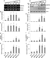
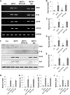
Rapid up-regulation of RANTES and eotaxin in nigra and serum of MPTP-intoxicated mice. Male C57/BL6 mice (6–8 weeks old) were insulted with 20 mg/kg of body weight MPTP (four injections at every 2-h interval). After 4, 8, 12, 24, and 72 h of MPTP intoxication, the mRNA expression of RANTES and eotaxin in nigra was monitored by semiquantitative RT-PCR (A) and real time PCR (B, RANTES; C, eotaxin). The protein expression of RANTES and eotaxin was monitored by Western blotting (D). Actin was run as a control. Bands were scanned and values (E, RANTES/actin; F, eotaxin/actin) are presented as relative to control (Con). Levels of RANTES and eotaxin were also measured in nigral homogenates by ELISA (G, RANTES; H, eotaxin). Levels of RANTES and eotaxin were also measured in serum by ELISA (I, RANTES; J, eotaxin). Results are mean ± S.E. of four mice (n = 4) per group. a, p < 0.001 versus control; b, p < 0.05 versus control. Error bars represent S.E.
FIGURE 2.
FIGURE 2.
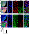
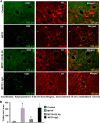
Glial expression of RANTES in the SNpc of MPTP-intoxicated mice. A, male C57/BL6 mice (6–8 weeks old) were insulted with 20 mg/kg of body weight MPTP (four injections at every 2-h interval). After 1 day, nigral sections were double labeled (A, Iba1 and RANTES; B, GFAP and RANTES). Cells positive for RANTES (C) were counted in two nigral sections (two images per slide) of each of five mice (n = 5) per group as described under “Materials and Methods.” a, p < 0.001 versus control. Error bars represent S.E.
FIGURE 3.
FIGURE 3.
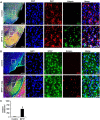
Induction of eotaxin in the SNpc of MPTP-intoxicated mice. A, male C57/BL6 mice (6–8 weeks old) were insulted with 20 mg/kg of body weight MPTP (four injections at every 2-h interval). After 1 day, nigral sections were double labeled (A, Iba1 and eotaxin; B, GFAP and eotaxin). Cells positive for eotaxin (C) were counted in two nigral sections (two images per slide) of each of five mice (n = 5) per group. a, p < 0.001 versus control. Error bars represent S.E.
FIGURE 4.
FIGURE 4.
Presence of RANTES and eotaxin in the SNpc of post-mortem PD brains. Midbrain sections of post-mortem PD brains and age-matched controls were double labeled (A, Iba1 and RANTES; B, Iba1 and eotaxin). Cells positive for RANTES (C) and eotaxin (D) were counted in two nigral sections (two images per slide) of each of four brains (n = 4) per group. a, p < 0.001 versus control. Error bars represent S.E.
FIGURE 5.
FIGURE 5.
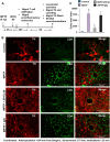
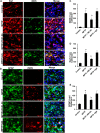
Functional blocking antibodies against RANTES and eotaxin inhibit the infiltration of CD4+ T cells into the nigra of MPTP-intoxicated mice. A, schematic presentation of treatment of MPTP-intoxicated mice with antibodies and related experiments. B, male C57/BL6 mice (6–8 weeks old) were insulted with 20 mg/kg of body weight MPTP (four injections at every 2-h interval). 2 h after the last injection of MPTP, animals were treated with a combination of 20 μg/mouse anti-RANTES (R) Ab and 20 μg/mouse anti-eotaxin (E) Ab via i.p. injection. 1 day after the last injection of MPTP, ventral midbrain sections (coordinates: anteroposterior, −4.04 mm from bregma; dorsoventral, 3.75 mm; mediolateral, 1.25 mm) were double labeled for CD4 and TH. C, CD4-positive cells were counted in two nigral sections (two images per slide) of each of five mice (n = 5) per group. a, p < 0.001 versus control; b, p < 0.001 versus MPTP. Error bars represent S.E.
FIGURE 6.
FIGURE 6.
Functional blocking antibodies against RANTES and eotaxin inhibit the infiltration of CD8+ T cells into the nigra of MPTP-intoxicated mice. A, male C57/BL6 mice (6–8 weeks old) were insulted with 20 mg/kg of body weight MPTP (four injections at every 2-h interval). 2 h after the last injection of MPTP, animals were treated with a combination of 20 μg/mouse anti-RANTES (R) Ab and 20 μg/mouse anti-eotaxin (E) Ab via i.p. injection. 1 day after the last injection of MPTP, ventral midbrain sections (coordinates: anteroposterior, −4.04 mm from bregma; dorsoventral, 3.75 mm; mediolateral, 1.25 mm) were double labeled for CD8 and TH. C, CD8-positive cells were counted in two nigral sections (two images per slide) of each of five mice (n = 5) per group. a, p < 0.001 versus control; b, p < 0.001 versus MPTP. Error bars represent S.E.
FIGURE 7.
FIGURE 7.
Neutralization of RANTES and eotaxin reduces the expression of proinflammatory molecules in the nigra of MPTP-intoxicated mice. Male C57/BL6 mice (6–8 weeks old) were insulted with 20 mg/kg of body weight MPTP (four injections at every 2-h interval). 2 h after the last injection of MPTP, animals were treated with a combination of 20 μg/mouse anti-RANTES (R) Ab and 20 μg/mouse anti-eotaxin (E) Ab via i.p. injection. 1 day after the last injection of MPTP, the mRNA expression of iNOS, IL-1β, GFAP, and CD11b was monitored in the nigra by RT-PCR (A) and real time PCR (B, iNOS; C, IL-1β; D, GFAP; E, CD11b). Results are mean ± S.E. of four mice (n = 4) per group. a, p < 0.001 versus control; b, p < 0.001 versus MPTP. 1 day after the last injection of MPTP, the protein expression of iNOS, IL-1β, GFAP, and Iba1 was monitored in the nigra by Western blotting (F). Actin was run as a control. Bands were scanned and values (G, iNOS/actin; H, IL-1β/actin; I, GFAP/actin; J, Iba1/actin) are presented as relative to control (Con). Results are mean ± S.E. of four mice (n = 4) per group. a, p < 0.001 versus control; b, p < 0.001 versus MPTP. Error bars represent S.E.
FIGURE 8.
FIGURE 8.
Neutralization of RANTES and eotaxin decreases glial activation in the nigra of MPTP-intoxicated mice. Male C57/BL6 mice (6–8 weeks old) were insulted with 20 mg/kg of body weight MPTP (four injections at every 2-h interval). 2 h after the last injection of MPTP, animals were treated with a combination of 20 μg/mouse anti-RANTES (R) Ab and 20 μg/mouse anti-eotaxin (E) Ab via i.p. injection. 1 day after the last injection of MPTP, nigral sections were double labeled (A, Iba1 and iNOS; B, GFAP and iNOS). Cells positive for iNOS (C), Iba1 (D), and GFAP (E) were counted in two nigral sections (two images per slide) of each of five mice (n = 5) per group. a, p < 0.001 versus control; b, p < 0.001 versus control. Error bars represent S.E.
FIGURE 9.
FIGURE 9.
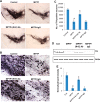
Neutralization of RANTES and eotaxin protects dopaminergic neurons in the nigra of MPTP-intoxicated mice. Male C57/BL6 mice (6–8 weeks old) were insulted with 20 mg/kg of body weight MPTP (four injections at every 2-h interval). 2 h after the last injection of MPTP, animals were treated with a combination of 20 μg/mouse anti-RANTES (R) Ab and 20 μg/mouse anti-eotaxin (E) Ab via i.p. injection. 7 days after the last injection of MPTP, nigral sections were stained for TH (A). A magnified image of TH-stained SNpc is shown in B. TH neurons were counted by stereology using the STEREO INVESTIGATOR software (C). Results are mean ± S.E. of five mice (n = 5) per group. a, p < 0.001 versus control; b, p < 0.001 versus MPTP. Nigral homogenates were immunoblotted for TH (D). Actin was run as a control. Bands were scanned and values (TH/actin) are presented as relative to control (Con) (E). Results are mean ± S.E. of four mice (n = 4) per group. a, p < 0.001 versus control; b, p < 0.001 versus MPTP. Error bars represent S.E.
FIGURE 10.
FIGURE 10.
Neutralization of RANTES and eotaxin protects TH fibers and restores neurotransmitters in the striatum of MPTP-intoxicated mice. Male C57/BL6 mice (6–8 weeks old) were insulted with 20 mg/kg of body weight MPTP (four injections at every 2-h interval). 2 h after the last injection of MPTP, animals were treated with a combination of 20 μg/mouse anti-RANTES (R) Ab and 20 μg/mouse anti-eotaxin (E) Ab via i.p. injection. 7 days after the last injection of MPTP, striatal sections were stained for TH (A) followed by quantification of TH-positive fibers (B). Concentrations of dopamine (C), DOPAC (D), and HVA (E) were measured in the striatum by HPLC. Results are mean ± S.E. of five mice (n = 5) per group. a, p < 0.001 versus control (Con); b, p < 0.001 versus MPTP. A.U., arbitrary units. Error bars represent S.E.
FIGURE 11.
FIGURE 11.
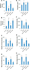
Neutralization of RANTES and eotaxin improves motor functions in MPTP-intoxicated mice. Male C57/BL6 mice (6–8 weeks old) were insulted with 20 mg/kg of body weight MPTP (four injections at every 2-h interval). 2 h after the last injection of MPTP, animals were treated with a combination of 20 μg/mouse anti-RANTES (R) Ab and 20 μg/mouse anti-eotaxin (E) Ab via i.p. injection. 7 days after the last injection of MPTP, mice were tested for motor functions (A, rotarod; B, pole test; C, number of movements; D, movement time; E, rest time; F, horizontal activity; G, total distance; H, stereotypy). Data are means ± S.E. of nine mice per group. a, p < 0.001 versus control (Con); c, p < 0.05 versus control; b, p < 0.001 versus MPTP; d, p < 0.05 versus MPTP. Error bars represent S.E.

References

    1. Vila M., and Przedborski S. (2004) Genetic clues to the pathogenesis of Parkinson's disease. Nat. Med. 10, (suppl.) S58–S62 - PubMed
    1. Olanow C. W., and Tatton W. G. (1999) Etiology and pathogenesis of Parkinson's disease. Annu. Rev. Neurosci. 22, 123–144 - PubMed
    1. Dauer W., and Przedborski S. (2003) Parkinson's disease: mechanisms and models. Neuron 39, 889–909 - PubMed
    1. Martin H. L., Santoro M., Mustafa S., Riedel G., Forrester J. V., and Teismann P. (2016) Evidence for a role of adaptive immune response in the disease pathogenesis of the MPTP mouse model of Parkinson's disease. Glia 64, 386–395 - PMC - PubMed
    1. Brochard V., Combadière B., Prigent A., Laouar Y., Perrin A., Beray-Berthat V., Bonduelle O., Alvarez-Fischer D., Callebert J., Launay J. M., Duyckaerts C., Flavell R. A., Hirsch E. C., and Hunot S. (2009) Infiltration of CD4+ lymphocytes into the brain contributes to neurodegeneration in a mouse model of Parkinson disease. J. Clin. Investig. 119, 182–192 - PMC - PubMed

Publication types

MeSH terms