. 2016 Jul 29;291(31):16048-58.
doi: 10.1074/jbc.M116.715748. Epub 2016 May 24.

Krüppel-like Factor 3 (KLF3/BKLF) Is Required for Widespread Repression of the Inflammatory Modulator Galectin-3 (Lgals3)

Affiliations

Krüppel-like Factor 3 (KLF3/BKLF) Is Required for Widespread Repression of the Inflammatory Modulator Galectin-3 (Lgals3)

Alexander J Knights et al. J Biol Chem. .

Abstract

The Lgals3 gene encodes a multifunctional β-galactoside-binding protein, galectin-3. Galectin-3 has been implicated in a broad range of biological processes from chemotaxis and inflammation to fibrosis and apoptosis. The role of galectin-3 as a modulator of inflammation has been studied intensively, and recent evidence suggests that it may serve as a protective factor in obesity and other metabolic disorders. Despite considerable interest in galectin-3, little is known about its physiological regulation at the transcriptional level. Here, using knockout mice, chromatin immunoprecipitations, and cellular and molecular analyses, we show that the zinc finger transcription factor Krüppel-like factor 3 (KLF3) directly represses galectin-3 transcription. We find that galectin-3 is broadly up-regulated in KLF3-deficient mouse tissues, that KLF3 occupies regulatory regions of the Lgals3 gene, and that KLF3 directly binds its cognate elements (CACCC boxes) in the galectin-3 promoter and represses its activation in cellular assays. We also provide mechanistic insights into the regulation of Lgals3, demonstrating that C-terminal binding protein (CtBP) is required to drive optimal KLF3-mediated silencing. These findings help to enhance our understanding of how expression of the inflammatory modulator galectin-3 is controlled, opening up avenues for potential therapeutic interventions in the future.

Keywords: Krüppel-like factor (KLF); Krüppel-like factor 3 (KLF3); adipose tissue; galectin; galectin-3 (Lgals3); gene expression; gene regulation; inflammation; metabolism; transcription regulation.


Figures

FIGURE 1.
FIGURE 1.
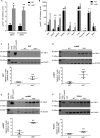
Lgals3 is up-regulated in the absence of KLF3. A and B, real-time quantitative PCR analysis of Lgals3 mRNA levels in WT and Klf3−/− MEFs (A) and primary tissues (B). Lgals3 expression was normalized with 18S rRNA levels and normalized again to the condition with the lowest mean Lgals3/18S expression, which was set to 1 (n = 3 for A and n = 4–5 for B). C–F, Western blots showing galectin-3 protein expression in WT and Klf3−/− BAT (C), scWAT (D), epiWAT (E), and spleen (F) with densitometry analysis (n = 3–4). 15 μg of total protein was loaded per lane, alongside a Rainbow molecular weight marker (Amersham Biosciences) used as a protein size standard (Size Std.). Negative controls (nuclear extract from untransfected COS cells) and positive controls (nuclear extracts from COS cells expressing Gal-3) were included to show the expected migration pattern and test the specificity of the antibody for Gal-3. Error bars represent mean ± S.E., and Student's t tests were conducted to determine significance. *, p < 0.05; **, p < 0.01.
FIGURE 2.
FIGURE 2.
Galectin-3 levels are elevated in Klf3−/− adipose tissue. A, immunofluorescent staining of scWAT to assess galectin-3 levels, showing two representative WT and Klf3−/− pairs. DAPI and wheat germ agglutinin (WGA) were used to stain the nuclei and cell membranes, respectively. B, corrected total galectin-3 fluorescence using WT and Klf3−/− images from the Gal-3 column in A. C, levels of galectin-3 secreted after 2 h from WT and Klf3−/− epiWAT explants were measured using ELISA (n = 7 for WT and 5 for Klf3−/−). D, the merged images from A were further magnified to assess galectin-3 up-regulation in adipocytes and the stromal vascular fraction of Klf3−/− scWAT. Up-regulation in both compartments of the scWAT was confirmed by real-time quantitative PCR analysis of Lgals3 mRNA levels (n = 3) E, Lgals3 levels were also assessed in cultured bone marrow-derived macrophages (n = 4). SVF, stromal vascular fraction. F, Lgals3 expression was normalized with 18S expression levels and normalized again to the condition with the lowest mean Lgals3/18S expression, which was set to 1. For C, E, and F, error bars represent mean ± S.E., and Student's t tests were conducted to determine significance. *, p < 0.05; ***, p < 0.001.
FIGURE 3.
FIGURE 3.
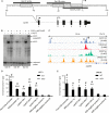
KLF3 binds to the Lgals3 promoter in vitro and in vivo. A, Lgals3 promoter region proximal to the transcription start site (+1) showing three KLF3 consensus binding sites (boxed) and their corresponding EMSA probes (Gal-3A, Gal-3B, and Gal-3C; black bars). The Lgals3 sequence was derived from the mm9 Mus musculus genome (NCBI reference sequence NM_001145953). B, nuclear extracts were prepared from COS-7 cells transfected with 5 μg of pMT3-Klf3. These were used in EMSA to assess KLF3 binding of 32P-radiolabeled probes Gal-3A, Gal-3B, and Gal-3C, each corresponding to KLF3 consensus motifs in the Lgals3 proximal promoter from A. The addition of anti-KLF3 antibody was used to confirm the identity of the protein bound to the probes by supershift. C, KLF3 binding at Lgals3 in MEFs from previously published KLF3-V5 ChIP-seq data available from GEO (accession# GSE44748) (29). KLF3 enrichment peaks are aligned with histone 3 lysine 4 trimethylation (accession no. GSM769029) and histone 3 lysine 27 acetylation (accession no. GSM1000139). ChIP-seq data from MEFs and a DNaseI hypersensitivity (accession no. GSM1014199) dataset were produced from murine fibroblasts (48–50). Significant KLF3 binding peaks at Lgals3 are denoted by i, ii, and iii. D and E, in vivo occupancy of KLF3 at these sites (i, ii, and iii) in Lgals3 was assessed in WT and Klf3−/− MEFs (D) and bone marrow-derived macrophages (E) (n = 3–4 for WT and 2–3 for Klf3−/−). Sites i, ii, and iii from C were interrogated by quantitative PCR following chromatin immunoprecipitation with Pierce anti-KLF3 antibody (PA5-18030) or normal goat IgG. Fam132a and Klf8 promoter 1a were used as positive control sites and Klf8 negative controls A and B as negative control loci, as described previously (14, 15, 23). Error bars represent the mean ± S.E. Student's t tests were used to determine significance. *, p < 0.05; **, p < 0.01; ***p < 0.001; WT versus WT Klf8-negative control B (D) or Klf8 negative control A (E). #, p < 0.05, WT versus Klf3−/− (D and E).
FIGURE 4.
FIGURE 4.
Optimal KLF3-mediated repression of Lgals3 is partially dependent on CtBP and requires the KLF3 functional domain. A, SL2 cells were transfected with 1 μg of pGL4.10[luc2] containing the core Lgals3 promoter (−190 to +34, relative to the transcription start site). 1 μg of pGL4.74[hRLuc] was used as a transfection control. Empty pPac was added to assess transactivation in the absence of KLF1, KLF3, and KLF3-ΔDL, followed by addition of 50, 100, and 250 ng of pPac-Klf1. With activation of the construct driven by 250 ng of KLF1, increasing amounts of pPac-Klf3 or pPac-Klf3-ΔDL were added to assess repression (5, 10, and 25 ng). The average of three replicates per condition is shown. B, real-time quantitative PCR analysis of Lgals3 expression in WT, Klf3−/−, and rescued Klf3−/− MEF cell lines (n = 3). Error bars represent the mean ± S.E., and Student's t tests were conducted to determine significance between conditions, shown in adjacent tables as p values (p < 0.05).

References

    1. Dagher S. F., Wang J. L., and Patterson R. J. (1995) Identification of galectin-3 as a factor in pre-mRNA splicing. Proc. Natl. Acad. Sci. U.S.A. 92, 1213–1217 - PMC - PubMed
    1. MacKinnon A. C., Farnworth S. L., Hodkinson P. S., Henderson N. C., Atkinson K. M., Leffler H., Nilsson U. J., Haslett C., Forbes S. J., and Sethi T. (2008) Regulation of alternative macrophage activation by galectin-3. J. Immunol. 180, 2650–2658 - PubMed
    1. Elad-Sfadia G., Haklai R., Balan E., and Kloog Y. (2004) Galectin-3 augments K-Ras activation and triggers a Ras signal that attenuates ERK but not phosphoinositide 3-kinase activity. J. Biol. Chem. 279, 34922–34930 - PubMed
    1. Yang R. Y., Hsu D. K., and Liu F. T. (1996) Expression of galectin-3 modulates T-cell growth and apoptosis. Proc. Natl. Acad. Sci. U.S.A. 93, 6737–6742 - PMC - PubMed
    1. de Boer R. A., Voors A. A., Muntendam P., van Gilst W. H., and van Veldhuisen D. J. (2009) Galectin-3: a novel mediator of heart failure development and progression. Eur. J. Heart Fail. 11, 811–817 - PubMed

Publication types

LinkOut - more resources