Comparative Study
. 2016 Jul 22;291(30):15881-95.
doi: 10.1074/jbc.M116.717751. Epub 2016 May 19.

Macrophage Migration Inhibitory Factor-CXCR4 Receptor Interactions: EVIDENCE FOR PARTIAL ALLOSTERIC AGONISM IN COMPARISON WITH CXCL12 CHEMOKINE

Affiliations
Comparative Study

Macrophage Migration Inhibitory Factor-CXCR4 Receptor Interactions: EVIDENCE FOR PARTIAL ALLOSTERIC AGONISM IN COMPARISON WITH CXCL12 CHEMOKINE

Deepa Rajasekaran et al. J Biol Chem. .

Abstract

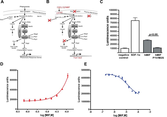
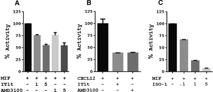
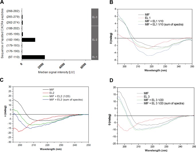
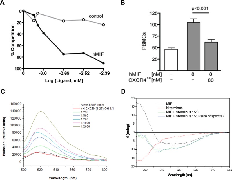
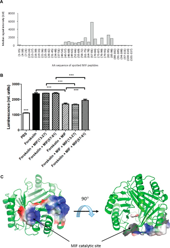
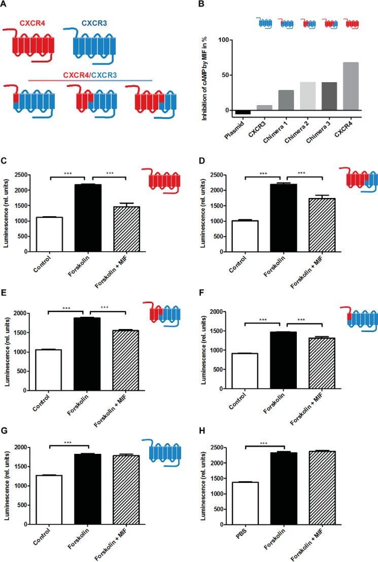
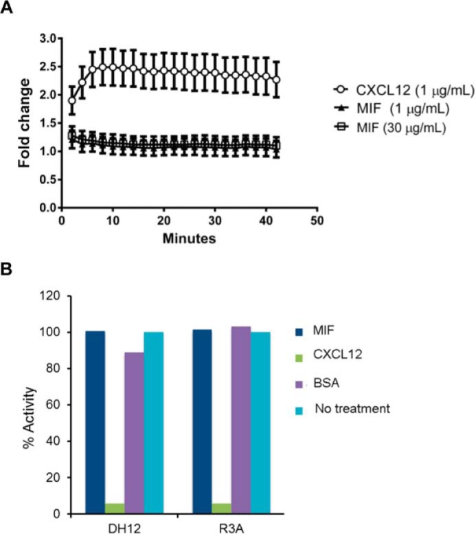
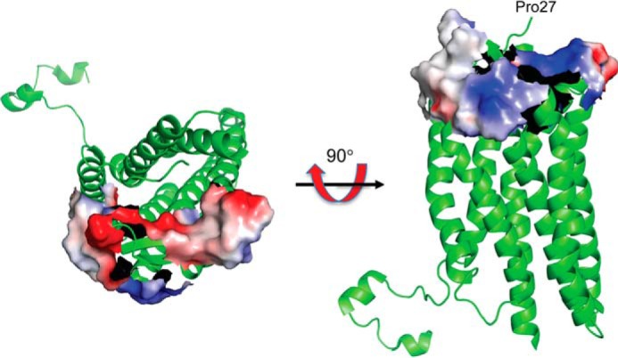
An emerging number of non-chemokine mediators are found to bind to classical chemokine receptors and to elicit critical biological responses. Macrophage migration inhibitory factor (MIF) is an inflammatory cytokine that exhibits chemokine-like activities through non-cognate interactions with the chemokine receptors CXCR2 and CXCR4, in addition to activating the type II receptor CD74. Activation of the MIF-CXCR2 and -CXCR4 axes promotes leukocyte recruitment, mediating the exacerbating role of MIF in atherosclerosis and contributing to the wealth of other MIF biological activities. Although the structural basis of the MIF-CXCR2 interaction has been well studied and was found to engage a pseudo-ELR and an N-like loop motif, nothing is known about the regions of CXCR4 and MIF that are involved in binding to each other. Using a genetic strain of Saccharomyces cerevisiae that expresses a functional CXCR4 receptor, site-specific mutagenesis, hybrid CXCR3/CXCR4 receptors, pharmacological reagents, peptide array analysis, chemotaxis, fluorescence spectroscopy, and circular dichroism, we provide novel molecular information about the structural elements that govern the interaction between MIF and CXCR4. The data identify similarities with classical chemokine-receptor interactions but also provide evidence for a partial allosteric agonist compared with CXCL12 that is possible due to the two binding sites of CXCR4.

Keywords: G protein-coupled receptor (GPCR); chemokine; cytokine; protein mapping; protein-protein interaction; structure-function.


Figures

FIGURE 1.
FIGURE 1.
MIF signaling of CXCR4 in S. cerevisiae. A, pheromone response pathway in S. cerevisiae. Activation of the Ste 2 receptor by pheromone leads to a signaling cascade resulting in transcription of the pheromone response genes. B, CXCR4 replaces the Ste 2 receptor. Gpa1 is modified such that it can couple with CXCR4. Ste 14 and Far 2 are deleted to lead to a more robust signaling response. To measure the robustness of the response, the pheromone response genes are substituted with the lacZ gene, which is produced, and enzymatic activity is measured. C, comparison of the effects of co-expression of CXCR4 with CXCL12/SDF-1α, wild-type MIF, and the double mutant P1V/M2S MIF. D, dose-response effect of exogenous MIF added to CXCR4-expressing S. cerevisiae. The EC50 values cannot be measured because a concentration that reaches the maximum signaling cannot be obtained. E, functional competition between MIF and CXCL12 in activating CXCR4. Dose response of MIF in the presence of a constant concentration of CXCL12 (2 μm) results in a decrease in signaling due to the displacement of CXCL12 by the higher concentrations of the less potent MIF.
FIGURE 2.
FIGURE 2.
Signaling effects (β-galactosidase activity) of CXCR4 antagonists on MIF or CXCL12 agonism and of the MIF inhibitor ISO-1 on CXCR4 signaling. A, 1- or 5-fold excess concentration of IT1t and AMD3100 relative to the MIF concentration shows a dose-response effect that is moderate compared with B, where there is greater response for CXCL12 at equivalent concentrations to IT1t and AMD3100. C, MIF active site inhibitor ISO-1 has a clear dose-response effect at 0.1-, 1-, and 5-fold excess of MIF on CXCR4 signaling, indicating that the active site is involved in binding and/or signaling.
FIGURE 3.
FIGURE 3.
Characterization of CXCR4 extracellular regions interacting with MIF. A, peptide microarray analysis indicates that MIF interacts with extracellular loops (EL) EL1 and EL2 of CXCR4, while EL3 is not involved in the interaction. The interaction of biotinylated full-length recombinant MIF with glass slide-immobilized (“spotted”) peptides corresponding to the indicated sequences of the CXCR4 extracellular regions is shown as relative signal intensity. Gray vertical bar on the right axis indicates to which EL the spotted sequences correspond. B–D, circular dichroism spectropolarimetry confirms the role EL1 (B) and EL2 (C) in the binding interface with MIF, whereas no indication for a role of EL3 (D) was obtained. Recombinant MIF and extracellular loop peptides of CXCR4 were mixed in solution at 1:10 (for EL1) or 1:20 (for EL2 and EL3) molar ratios and spectra compared with the mathematical addition of the individual spectra (“sum of spectra”). Spectra of the individual peptides/proteins and mixtures are presented according to the indicated color code. Conformations and conformational changes in the CD spectra were measured as raw ellipticity versus the wavelength in the far-UV range.
FIGURE 4.
FIGURE 4.
Binding and functional assays between MIF and CXCR4(1–27). A, CXCR4(1–27) coated on 96-well plates and incubated with 150 μm biotinylated MIF was competed with increasing concentrations of MIF or lysozyme control. These results demonstrate a direct and specific interaction. B, CXCR4(1–27) blocks MIF-induced mononuclear cell migration. 1 × 10⁶ cells/ml PBMCs were placed in the upper chamber of a 24-well cell culture insert. 8 nm hMIF were placed in the lower chamber with or without CXCR4(1–27). After 3 h of incubation, the transmigrated cells were fixed, stained, and counted. C, solution mixture of CXCR4(1–27) peptide with Alexa Fluor-labeled full-length human MIF evokes a conformational change in MIF leading to a change in Alexa fluorescence emission. Peptide was added at 1:1 molar ratio and at an excess of 250-, 500-, 750-, 1000-, and 2000-fold as indicated by color code, and fluorescence spectra were recorded between wavelengths 500 and 600 nm. D, no changes in the secondary structure of MIF and CXCR4(1–27) upon mixing the two molecules. CXCR4(1–27) was mixed with full-length human MIF, and potential changes in secondary structure were assessed by far-UV circular dichroism spectroscopy. The spectrum of the mixed molecules does not differ from that of the mathematical addition of the separate CD spectra.
FIGURE 5.
FIGURE 5.
Identification of MIF binding regions to the N-terminal peptide of CXCR4. A, MIF residues 43–98, in particular 67–81, are involved in MIF-CXCR4(1–27) interactions. Peptide array technology was used with immobilized 15-mer MIF peptides, with each peptide positionally shifted by three residues to produce peptides that covered the entire MIF sequence. Peptides were probed for binding to biotinylated CXCR4(1–27). B, cell-based signaling assay confirms a role for residues 67–81 in the MIF-CXCR4 interaction. The ability of peptide 67–81 versus control peptide (residues 13–27) to reverse the inhibitory activity of MIF on forskolin-triggered cAMP production in stable CHO-CXCR4 transfectants was measured by Hit Hunter commercial cAMP test and readout as relative luminescence units. C, residues mapped on the ribbon structure of MIF identified by biochemical experiments in A and B to interact with CXCR4 mapped on a single subunit of MIF (for clarity) with the related electrostatic potential. The identified sites are the catalytic cavity and adjacent region involving part of the second α-helix. ***, p < 0.05.
FIGURE 6.
FIGURE 6.
Confirmation of the interaction sites in CXCR4 using cell-expressed CXCR4/CXCR3 chimeras. A, scheme summarizing the chimeras (N terminus of CXCR4, CXCR4 N terminus through part of helix 3, including EL1, and CXCR4 N terminus through part of helix 5 (including EL1 and EL2)) as well as all-CXCR4 and all-CXCR3 wild-type receptors. B–H, inhibitory effect of MIF on forskolin-induced cAMP production in CHO transfectants expressing various CXCR4/CXCR3 receptor chimeras as indicated and comparison with all-CXCR4 and all-CXCR3 wild-type receptors and empty plasmid-transfected controls. B, summary of experiments represented as relative inhibition of cAMP by MIF. C–H, inhibitory effect of MIF on cAMP production for individual receptor chimera or controls expressed as relative luminescence. C, all-CXCR4 wild-type receptor; D, CXCR4 N terminus through part of helix 5 (including EL1 and EL2); E, CXCR4 N terminus through part of helix 3, including EL1; F, N terminus of CXCR4; G, all-CXCR3 wild-type receptor; H, empty plasmid control. Control refers to CHO-transfectants without forskolin treatment. ***, p < 0.05.
FIGURE 7.
FIGURE 7.
MIF does not re-capitulate CXCR4-mediated β-arrestin activation or CXCL12 inhibition of HIV-1 T-cell entry. A, click beetle luciferase complementation assay for recruitment of β-arrestin 2 to CXCR4 reveals that MIF does not promote interaction of these proteins, whereas CXCL12 drives association of CXCR4 with β-arrestin 2. B, inhibition of U87.CD4.U87 viral entry by MIF, CXCL12 (positive control), and bovine serum albumin (negative control) compared with untreated controls. Cells infected with the viral pseudotypes of the dual-tropic env strains DH12 or R3A with a luciferase reporter gene that is activated upon infection is inhibited by CXCL12 CXCR4 chemokine agonist but not by MIF or the bovine serum control. Results are plotted against the percentage luciferase activity with 100% for the untreated controls for each pseudotype.
FIGURE 8.
FIGURE 8.
Regions of CXCR4 that interact with MIF mapped on the ribbon diagram of CXCR4. Only the electrostatic potential of extracellular loop 1 and residues 182–196 from extracellular loop 2 are shown because the structure of CXCR4(1–27) is not visible in any of the CXCR4 structures. The two orientations show both positive and negative potentials. The negative potential is complementary to the positive potential shown in Fig. 5C. Note that the CXCR4(1–27) has a net charge of −6 without accounting for the three tyrosine residues that are sulfated (45).

References

    1. Katritch V., Cherezov V., and Stevens R. C. (2013) Structure-function of the G protein-coupled receptor superfamily. Annu. Rev. Pharmacol. Toxicol. 53, 531–556 - PMC - PubMed
    1. Thelen M. (2001) Dancing to the tune of chemokines. Nat. Immunol. 2, 129–134 - PubMed
    1. Charo I. F., and Ransohoff R. M. (2006) The many roles of chemokines and chemokine receptors in inflammation. N. Engl. J. Med. 354, 610–621 - PubMed
    1. Moser B., and Loetscher P. (2001) Lymphocyte traffic control by chemokines. Nat. Immunol. 2, 123–128 - PubMed
    1. Bleul C. C., Farzan M., Choe H., Parolin C., Clark-Lewis I., Sodroski J., and Springer T. A. (1996) The lymphocyte chemoattractant SDF-1 is a ligand for LESTR/fusin and blocks HIV-1 entry. Nature 382, 829–833 - PubMed

Publication types

MeSH terms