. 2016 Jul 8;291(28):14677-94.
doi: 10.1074/jbc.M116.726307. Epub 2016 May 18.

Insights into Lysine Deacetylation of Natively Folded Substrate Proteins by Sirtuins

Affiliations

Insights into Lysine Deacetylation of Natively Folded Substrate Proteins by Sirtuins

Philipp Knyphausen et al. J Biol Chem. .

Abstract

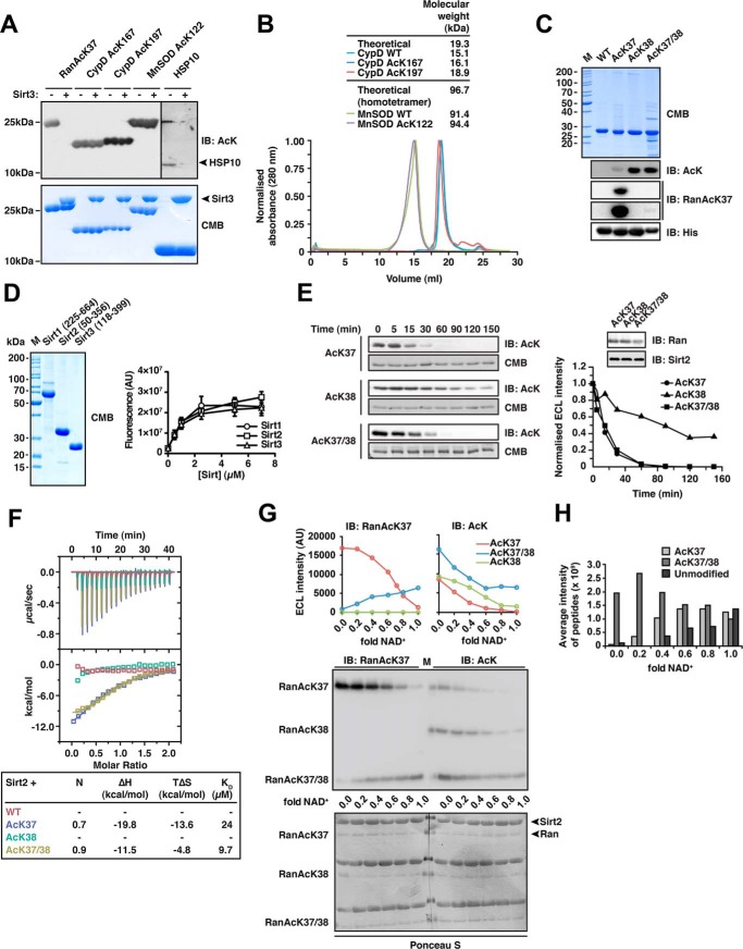
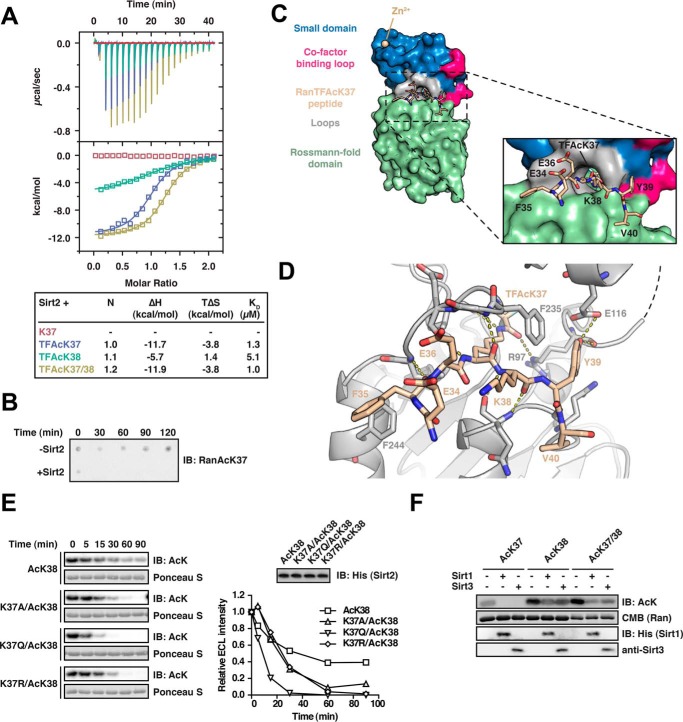
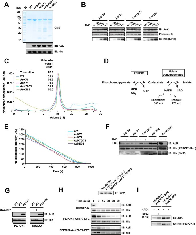
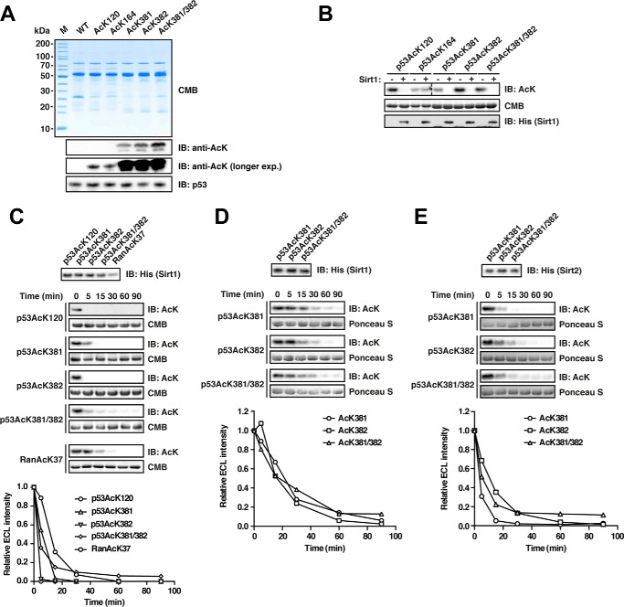
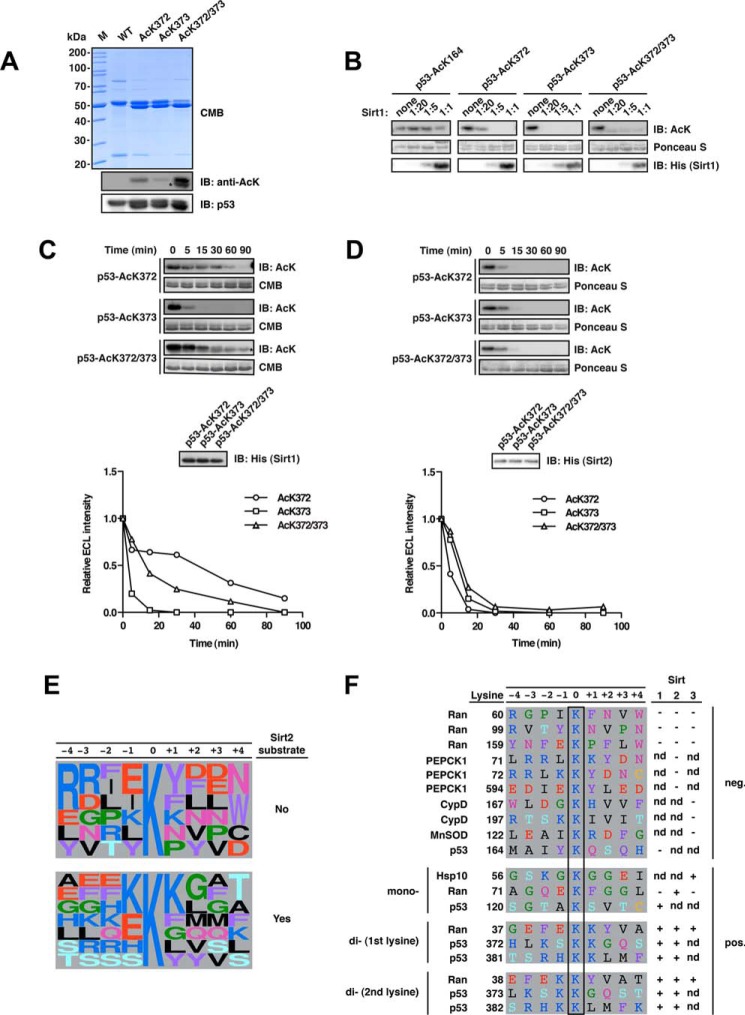
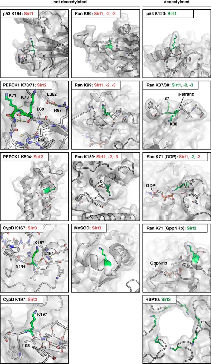
Sirtuins are NAD(+)-dependent lysine deacylases, regulating a variety of cellular processes. The nuclear Sirt1, the cytosolic Sirt2, and the mitochondrial Sirt3 are robust deacetylases, whereas the other sirtuins have preferences for longer acyl chains. Most previous studies investigated sirtuin-catalyzed deacylation on peptide substrates only. We used the genetic code expansion concept to produce natively folded, site-specific, and lysine-acetylated Sirt1-3 substrate proteins, namely Ras-related nuclear, p53, PEPCK1, superoxide dismutase, cyclophilin D, and Hsp10, and analyzed the deacetylation reaction. Some acetylated proteins such as Ras-related nuclear, p53, and Hsp10 were robustly deacetylated by Sirt1-3. However, other reported sirtuin substrate proteins such as cyclophilin D, superoxide dismutase, and PEPCK1 were not deacetylated. Using a structural and functional approach, we describe the ability of Sirt1-3 to deacetylate two adjacent acetylated lysine residues. The dynamics of this process have implications for the lifetime of acetyl modifications on di-lysine acetylation sites and thus constitute a new mechanism for the regulation of proteins by acetylation. Our studies support that, besides the primary sequence context, the protein structure is a major determinant of sirtuin substrate specificity.

Keywords: GTPase; acetylation; p53; sirtuin; synthetic biology.


Figures

FIGURE 1.
FIGURE 1.
Sirt2 can deacetylate di-acetylated Ran. A, deacetylation of selected substrate proteins by Sirt3. We prepared site-specifically acetylated Ran AcK37, CypD AcK167, and AcK197, MnSOD AcK122, and Hsp10 AcK56 using the genetic code expansion concept and analyzed its deacetylation by Sirt3 by immunoblotting using an anti-acetyl-lysine antibody (anti-AcK-AB). Sirt3 was used in equimolar ratio with the respective protein substrate (40 μm), except for Hsp10 (40 μm Sirt3 and 80 μm Hsp10). Coomassie Brilliant (CMB) blue staining is shown as loading control. B, analytical size exclusion chromatography of CypD and MnSOD proteins used in this study (Superdex 200 10/300). To assess the quality of the non-acetylated and site-specifically lysine-acetylated CypD (WT, AcK167, and AcK197) and MnSOD (WT and MnSOD AcK122) proteins used here, we performed analytical SEC experiments. All CypD proteins eluted as apparent monomer from the SEC column. MnSOD behaved as apparent tetramer on SEC, as reported earlier. For both, MnSOD and CypD, all acetylated proteins behaved as the non-acetylated proteins showing that lysine acetylation did not affect protein folding or oligomerization. C, Ran was lysine-acetylated at Lys-38 and at Lys-37/Lys-38 using the genetic code expansion concept. Coomassie Brilliant Blue staining of an SDS-PAGE with 5 μg of protein shows the final purity. Anti-AcK-AB was used to show the successful incorporation of acetyl-l-lysine into Ran. The anti-AcK-AB detects acetyl-l-lysine with different sensitivity depending on the sequence context. Detection with anti-His6-AB serves as a loading control. An antibody raised against a Ran AcK37-derived 11-mer peptide (see “Experimental Procedures”) is specific for Ran AcK37, showing only a very weak signal for RanAcK37/38 after long exposure to x-ray film and does not detect Ran AcK38. D, final purity of the Sirt1, Sirt2, and Sirt3 enzymes used in this study. 5 μg of each sirtuin was separated by SDS-PAGE and the gel Coomassie Brilliant Blue-stained to assess the final purity. The enzymatic activities were tested with a fluor-de-lys assay. All enzymes are active and show a similar activity. Error bars represent the standard error of the mean for three independent measurements. E, kinetics of Ran AcK37, AcK38, and AcK37/38 deacetylation by Sirt2. 12 μm site-specifically lysine-acetylated Ran protein was incubated with 0.14 μm Sirt2 for the indicated times. The level of Ran acetylation was assessed by immunoblotting using a specific anti-AcK-AB (left). Ran AcK37 and di-acetylated Ran AcK37/38 were deacetylated with similar kinetics, whereas Ran AcK38 shows a strongly reduced Sirt2-catalyzed deacetylation rate. The SDS-polyacrylamide gels corresponding to the Western blots were stained with Coomassie Brilliant Blue and serve as a loading control. The reactions were quantified densitometrically using ImageJ software (right). F, Ran AcK37 and AcK37/38 bind to Sirt2 as shown by isothermal titration calorimetry. 45 μm Ran-WT, Ran AcK37, AcK38, or AcK37/38 protein was titrated with 450 μm Sirt2 (50–356, non-His6-tagged). Ran AcK37 and RanAcK37/38 bind with affinities of 24 and 9.7 μm, respectively. Both reactions are solely driven by the reaction enthalpy (Ran AcK37 ΔH, −19.8 kcal/mol; TΔS, −13.6 kcal/mol; Ran AcK37/38 ΔH,−11.5 kcal/mol; TΔS, −4.8 kcal/mol). No binding heat was observed under these conditions for Ran-WT and Ran AcK38. G, deacetylation of Ran AcK38 by Sirt2 is favored over AcK37 in di-acetylated Ran AcK37/38.12 μm Ran AcK37. AcK38 and AcK37/38 were used in Sirt2-mediated deacetylation reactions with increasing amounts of the essential cofactor NAD+ (Sirt2 concentration, 24 μm, molar ratio Ran AcK/NAD+, 0–1.0). Using the anti-Ran AcK37 AB, we observed a linear decrease of acetylated Ran AcK37. In contrast, the signal increased for di-acetylated AcK37/38 as a function of the NAD+ concentration. This suggests that AcK38 in the context of Ran AcK37/38 is deacetylated preferentially by Sirt2. As expected, AcK38 shows no signal using the anti-Ran AcK37 AB. Using the pan-anti-AcK-AB, all acetylated Ran proteins showed a linear decrease in the acetylation level. However, although AcK37 and AcK38 were nearly completely deacetylated at a molar Ran AcK/Sirt2 ratio of 1:1, this was not the case for di-acetylated AcK37/38R. Detection with an anti-Ran-AB was used as a loading control. Densitometric analyses of the signals obtained with the anti-AcK-AB and anti-Ran AcK37 are shown at the top. H, mass spectrometric analysis of the experiment is shown in F. The reactions were analyzed by digest with Glu-C followed by ESI-MS/MS. Without NAD+, there is only di-acetylated Ran present. With increasing NAD+ concentrations, the average intensity of peptides corresponding to di-acetylated Ran decreases and that of the non-acetylated Ran increases. The amount of Ran AcK37 peptide increases with increasing NAD+ concentrations suggesting that Ran AcK38 is deacetylated first. IB, immunoblot.
FIGURE 2.
FIGURE 2.
Characterization of the Ran-Sirt2 interaction using trifluoroacetyl-lysine peptides and Ran mutants. A, Ran TFAcK37, TFAcK38, and TFAcK37/38 13-mer peptides bind to Sirt2 in the micromolar range as shown by isothermal titration calorimetry. 30 μm Sirt2 (50–356, non-His6-tagged) was titrated with 300 μm trifluorolysine-acetylated Ran 13-mer peptide (Ran amino acids 31–43). The reactions are all driven by a favorable reaction enthalpy, ΔH, whereas the reaction of the TFAcK38 peptide is the only one driven by both favorable enthalpy and favorable entropy, TΔS. This shows that the TFAcK38 peptide uses a distinct mechanism of binding. B, Ran 13-mer AcK37 peptide is deacetylated by Sirt2. 133 μm of Ran AcK37 13-mer peptide (Ran amino acids 31–43) was spotted on a nitrocellulose membrane. Addition of 0.5 μm Sirt2 leads to a complete deacetylation already after 30 min, compared with the non-enzyme control (−Sirt2). The specific anti-Ran AcK37 AB was used for detection by immunoblotting. C, crystal structure of Sirt2 (amino acids 50–356) in complex with the TFAcK37 13-mer Ran peptide (amino acids 31–43). The structure was solved at a resolution of 3 Å. Left, overall Sirt2·Ran(31–43) TFAcK37 structure. Sirt2 is shown as a surface representation. Blue, small Zn2+-binding domain; red, cofactor binding loop; green, Rossmann-fold domain; beige, Ran(31–43) TFAcK37 peptide. Right, close-up of the Ran(31–43) TFAcK37 peptide and Sirt2. Three charged residues Glu-34R, Glu-36R, and the Lys-38R are solvent-exposed. The hydrophobic tunnel leading to the Sirt2 active site is too narrow to simultaneously adopt two acetylated lysine residues. The TFAcK37 penetrates into a hydrophobic tunnel formed by the small subdomain and the Rossmann-fold domain. D, two aromatic residues Phe-35R and Tyr-39R of the Ran(31–43) 13-mer TFAcK37 peptide act as anchor points through the formation of stacking interactions with the aromatic residues in Phe-244 and Phe-235 of Sirt2, respectively. Furthermore, the peptide forms several main chain hydrogen bonds with Sirt2, highlighted with the dotted yellow lines, and the hydroxyl group of Tyr-39R forms a hydrogen bond with the main chain carbonyl-oxygen of Arg-97 of Sirt2. The peptide adopts a crescent- and w-shaped conformation. E, effects of Ran mutations on the kinetics of Sirt2-mediated di-deacetylation of Ran AcK38. To assess whether electrostatic or steric properties of AcK37R increase the deacetylation rate of AcK38R, we created Lys-37R mutant proteins in the Ran AcK38 background. The non-mutated mono-acetylated Ran AcK38 shows the slowest Sirt2-catalyzed deacetylation of the proteins compared. All mutant variants (RanK37A/AcK38, K37R/AcK38, and K37Q/AcK38) show an accelerated Sirt2-catalyzed deacetylation compared with the mono-acetylated Ran AcK38 protein (12 μm Ran and 0.06 μm Sirt2). This shows that both electrostatic and steric properties of AcK37R contribute to the observed accelerated Sirt2-mediated deacetylation of AcK38R. F, Sirt1 and Sirt3 are able to di-deacetylate Ran AcK37/38. Ran AcK37, Ran AcK38, and di-acetylated Ran AcK37/38 were incubated with Sirt1 or Sirt3, and the acetylation level was assessed by immunoblotting (IB) using an anti-AcK antibody (12 μm Ran was incubated with 0.8 μm of the indicated Sirtuin for 30 min). Ran AcK38 shows a relatively strong remaining anti-AcK signal compared with the di-acetylated AcK37/38R, suggesting that, as observed for Sirt2, the presence of AcK37R stimulates the deacetylation of AcK38R by Sirt1 and Sirt3. Coomassie (CMB) staining is shown as loading control for Ran. Sirtuins were detected with the indicated antibodies.
FIGURE 3.
FIGURE 3.
PEPCK1 is not directly deacetylated by Sirt2 in vitro but can be converted into a Sirt2 substrate. A, final purity and quality of PEPCK1-WT and the acetylated PEPCK1 AcK70, AcK71, AcK70/71, and AcK594 proteins. For details on the experimental procedures see Fig. 1A. B, PEPCK1 is not directly deacetylated by Sirt2 at AcK70, AcK71, AcK70/71, or AcK594 in vitro. PEPCK1 was site-specifically lysine-acetylated and used as substrate for Sirt2-catalyzed deacetylation. Deacetylation assay was performed with increasing concentrations of Sirt2 for 2 h at 23 °C. Molar ratios of Sirt2/PEPCK1 are indicated. The concentration of PEPCK1 was 12 μm. Ponceau S staining was used as a loading control, Sirt2 was detected using an anti-His6 antibody. C, analytical size exclusion chromatography of PEPCK1 variants. The molecular mass calculated from the elution volume using a standard curve is indicated together with the theoretical mass of ∼70.9 kDa. All PEPCK1 proteins used in this study behave as the non-acetylated wild type PEPCK1 and elute as monomers from the analytical SEC column (Superdex 200 10/300 GL). D, reaction scheme of a fluorescence-based coupled enzymatic assay to analyze PEPCK1 activity. In the first and rate-limiting reaction step, PEPCK1 catalyzes the conversion of phosphoenolpyruvate to oxaloacetate under the consumption of GDP and CO2. In the second reaction step, oxaloacetate is converted to malate by malate dehydrogenase, which involves the oxidation of NADH to NAD+. The accompanied drop in fluorescence upon oxidation of NADH to NAD+ (excitation, 345 nm; emission, 470 nm) served as an indirect measure of PEPCK1 activity. E, acetylated PEPCK1 is active as determined by a coupled enzyme assay as described in B. The activity of PEPCK1-WT and its acetylated versions (AcK70, AcK71, AcK70/71, and AcK594) was analyzed as described (concentration, 25 nm). All enzymes showed an activity comparable with the wild type, non-acetylated protein. F, PEPCK1 is not deacetylated by Sirt2 in vitro at lower concentrations and in an optimized PEPCK1 buffer. Site-specifically lysine-acetylated PEPCK1 (0.6 μm) was used as a substrate for Sirt2-catalyzed deacetylation (0.6 μm). None of the acetylated PEPCK1 (AcK70, AcK71, AcK70/71, and AcK594) variants was directly deacetylated. Ran AcK37 (12 μm) was used as a positive control, showing complete deacetylation under the assay conditions. Acetylation levels were detected with an anti-AcK AB. An anti-His6 antibody was used detect PEPCK1, Ran, and Sirt2. G, anti-acetyl-lysine immunoreactivity of PEPCK1-WT (0.6 μm) after incubation with 2 mm 2′/3′-O-acetyl-ADP-ribose for 2 h at 23 °C. Immunoblotting using an anti-AcK antibody was used to detect the acetylation level of PEPCK1. An anti-His6 antibody was used to stain for PEPCK1. H, replacing the PEPCK1 sequence 67RRLKKY72 by 67EFEKKY72 (corresponding to the sequence N-terminal to the Ran di-acetylation site) converts PEPCK1 into a Sirt2 substrate. To analyze what are the determinants for Sirt2 activity on PEPCK1, we replaced the sequence preceding the Lys-70 (67RRL69) for the corresponding sequence in Ran AcK37 (34EFE36). This replacement is sufficient to convert PEPCK1 into a Sirt2 substrate. PEPCK1 AcK70 and AcK70/71 67EFE69 mutants (indicated as PEPCK1-EFE) were incubated with 0.06 μm Sirt2 for the indicated time. Ran AcK serves as a positive control and is completely deacetylated already after less than 5 min. PEPCK1 was deacetylated after 90 min. All substrates were used at 0.6 μm. The di-acetylated PEPCK1 AcK70/71 shows a different running behavior than the non-acetylated variant as seen in the respective anti-His6 loading control. I, deacetylation assay with PEPCK1-EFE mutants in the absence of NAD+. The loss of anti-AcK-immunoreactivity of PEPCK1 (0.6 μm) is dependent on the presence of the sirtuin cofactor NAD+, as the signal remains constant after 90 min of incubation with Sirt2 (0.06 μm). CMB, Coomassie Brilliant Blue; IB, immunoblot.
FIGURE 4.
FIGURE 4.
Deacetylation of p53 by Sirt1 and Sirt2. A, p53 was site-specifically lysine-acetylated at several sites (AcK120, AcK164, AcK381, AcK382, and AcK381/382). For details on the experimental procedures, see Fig. 1A. An anti-p53 antibody was used to detect p53. B, p53 is deacetylated by Sirt1 at AcK120, AcK381, AcK382, and AcK381/382 but not AcK164 in vitro. Deacetylation assay with site-specifically lysine-acetylated p53 and Sirt1 at a molar Sirt1/p53 ratio of 1:20 (p53, 12 μm; Sirt1, 0.6 μm). The reaction was performed for 2 h at 23 °C, and the acetylation level after Sirt1-catalyzed deacetylation is determined with an anti-AcK antibody. Notably, AcK381, AcK382, and even the di-acetylated AcK381/382 were completely deacetylated under the assay conditions. Coomassie Brilliant Blue (CMB) staining was used as a loading control. An anti-His6 antibody was used to stain for Sirt1. C, time course experiment to assess Sirt1-catalyzed p53-AcK120, -AcK381, -AcK382, and -AcK381/382 deacetylation (p53, 12 μm; Sirt1, 0.6 μm). Ran AcK37 (12 μm) was used as a control. An anti-AcK antibody was used to stain for lysine acetylation, Coomassie Brilliant Blue (CMB), as a loading control. The quantification of the kinetics shown in the right panel was performed using ImageJ. Anti-His6 antibody was used to detect Sirt1. D, time course experiments as for B but with a molar p53/Sirt1 ratio of 1:200 (Sirt1, 0.06 μm) to more sensitively assess the p53-catalyzed deacetylation of p53 AcK381, AcK382, and AcK381/382. As visible in the immunoblottings and the quantifications (lower panel), both mono-acetylated p53 proteins and the di-acetylated protein show highly similar Sirt1-catalyzed deacetylation. E, Sirt2 also deacetylates p53 at AcK381, AcK382, and AcK381/382. Time course experiments were to analyze whether all three sites are also deacetylated by Sirt2 (molar ratio 1:200 as in D). All three acetylated p53 proteins were deacetylated by Sirt2 with similar rates. IB, immunoblot.
FIGURE 5.
FIGURE 5.
Deacetylation of p53 at Lys-372 is enhanced upon acetylation of the neighboring Lys-373 for Sirt1 but not Sirt2. A, final purity and quality of p53-WT and acetylated p53 (AcK372, AcK373, and AcK372/372). For details on the experimental procedures see Fig. 1A. Notably, for p53 AcK372, AcK373, AcK381, AcK382, and the di-acetylated proteins, we obtained acetylated truncation products, which we could not remove during purification due to the similar size of the truncation product and the full-length protein and possibly due to oligomerization of the p53 protein. An asterisk denotes the anti-AcK-immunoreactive truncation product. B, p53 deacetylation by Sirt1 at AcK164, AcK372, AcK373, and AcK372/373 was analyzed at increasing molar Sirt1/p53 ratios (p53, 12 μm). The immunoblot with an anti-AcK-antibody shows that, at a molar ratio of Sirt1/p52 of 1:20, the weakest p53 deacetylation by Sirt1 occurs at AcK372, supporting the mechanism of di-deacetylation, by which the presence of AcK373 accelerates deacetylation at AcK372. p53-AcK164 is not deacetylated by Sirt1 in vitro even at a substrate/enzyme ratio of 1:1. C, time course of the Sirt1-catalyzed p53 AcK372, AcK373, and AcK372/373 deacetylation. p53 AcK373 is deacetylated by Sirt1 showing complete deacetylation after 30 min, whereas deacetylation is not completed for AcK372 after 90 min. The di-acetylated p53 AcK372/373 is deacetylated faster than p53 AcK372 (p53, 12 μm; Sirt1, 0.24 μm; molar ratio 1:50). The densitometric quantification was done using ImageJ software. The signal originating from the truncation product remains constant and was subtracted from the signal of the full-length p53 AcK372/373 (the band of the truncated fragment is denoted with an asterisk and runs slightly lower). The acetylation level was assessed using an anti-AcK-antibody. Coomassie Brilliant Blue (CMB) staining was used as a loading control for p53 and an anti-His6 antibody to detect Sirt1. D, time course of Sirt2-catalyzed deacetylation of p53 AcK372, AcK373, and AcK372/373 as described in C but with an enzyme/substrate ratio of 1:200 (0.06 μm Sirt1). All acetylated p53 variants show highly similar deacetylation kinetics with Sirt2. E, frequency plot of sequence context of lysine acetylation sites found in this and our previous study to be substrates or non-substrates of Sirt2 as indicated (14). Representation was created with WebLogo (106). F, sequence alignment of primary sequences surrounding di-acetylation motifs analyzed in this study and in our previous study (14). We discovered that several di-acetylation sites were deacetylated by Sirt1, Sirt2, and/or Sirt3 (Ran AcK37/38, p53 AcK372/373, and p53 AcK381/382). Interestingly, PEPCK1, although also containing a possible di-acetylation motif (AcK70/71) followed by a tyrosine residue, is not deacetylated by Sirt2, suggesting that the structure is an important denominator of Sirt specificity. Notably, the residues 67RRL69 in PEPCK1 preceding Lys-70/Lys-71 form a β-strand as part of an antiparallel β-sheet. It needs further investigation whether the replacement of this sequence by the corresponding Ran sequence 34EFE36 confers Sirt2 activity or whether this is due to structural effects interfering with the formation of this β-sheet, maybe affecting the loops flexibility. IB, immunoblot.
FIGURE 6.
FIGURE 6.
Localization of sirtuin substrate and non-substrate lysine acetylation sites in known crystal structures of Ran·GDP (PDB code 1BYU), Ran·GppNHp (PDB code 1IBR), MnSOD (PDB code 1PL4), CypD (PDB code 5A0E), Hsp10 (PDB code 4PJ1), PEPCK1 (PDB code 1NHX), and p53 (PDB code 2OCJ). All non-substrate lysines are located within or directly next to secondary structure elements, as observed in MnSOD Lys-122, or are located in conformationally restrained loops as observed in CypD Lys-167 and Lys-197 and PEPCK1 Lys-70/ Lys-71. CypD Lys-167 is located directly C-terminally of an α-helix. CypD Lys-197 is located directly N-terminally of a β-strand, which restricts its conformational freedom. The main chain nitrogen of Lys-197 makes a hydrogen bond to a bridging water molecule. The side chain of Ile-198, directly adjacent to Lys-197 is part of a hydrophobic core conformationally stabilizing this region. As a direct measure of its conformational rigidity, the loops encompassing Lys-167 and Lys-197 in CypD are well defined in the crystal structure, which is furthermore reflected in the B-factors for the amide nitrogen, the C-α and the carbonyl C-atom, forming the lysine's backbone (these are in the range of 10.59–12.72 Å2 for Lys-167 and 12.45–13.41 Å2 for Lys-197; PDB code 5A0E). These low B-factors suggest that these residues have a low level of flexibility. PEPCK1 Lys-70 and Lys-71 are located within a loop region. However, the residues 67RRL69 preceding these lysines form a small β-strand, which is part of an anti-parallel β-sheet. Furthermore, the loop flexibility is restrained by several side chain and main chain interactions (side chain, Glu-362 with Arg-67, Lys-71 with Asp-365 and Glu-375; main chain, Gly-366 with Lys-71, Leu-77 with Arg-67). This restraint of the loop's conformation could explain why AcK70 and AcK71 are not Sirt2 substrates. Mutation of 67RRL69 in PEPCK1 to the corresponding residues in Ran (giving rise to 67EFE69) makes PEPCK1 a Sirt2 substrate, most likely at least in part by releasing the loop's restrained conformation. However, alteration in the primary sequence might also affect the deacetylation. In p53, Lys-164 lies directly C-terminal to a β-strand; Lys-120 is located within a flexible loop. For the C-terminal lysines Lys-372, Lys-373, Lys-381, and Lys-382, there is no structural information available, as this domain is intrinsically disordered. In Ran, Lys-60 is located within a β-strand and Lys-99 and Lys-159 are within an α-helix. None of the sirtuins tested in our previous study deacetylated any of these three lysine acetylation sites of Ran. In contrast, Lys-37, Lys-38, and Lys-71 are potent sirtuin substrate sites. Although AcK71 is specifically deacetylated only by Sirt2, AcK37 is deacetylated by Sirt1, Sirt2, and Sirt3. AcK38 is deacetylated weakly by Sirt1, -2, and -3 but the presence of AcK37 increases the potency of AcK38 to act as a Sirt1, -2, and -3 substrate. Lys-37 and Lys-38 in Ran are located within the switch I loop. Lys-38R is part of a small β-sheet, restraining the structural flexibility. Acetylation of the neighboring Lys-37 might release these restraints making AcK38 a better sirtuin substrate. Lys-71 is part of the switch II loop, which is structurally more restricted in the active state (here: GppNHp loaded). However, we observed, that AcK71 is a better substrate for Sirt2 if it is present in its GppNHp-loaded state suggesting that this conformation is highly suitable for catalysis. Sirtuin labeling is in red if the respective sirtuin is not active and is in green if a sirtuin is active in deacetylating a protein at the respective site. In the panels depicted in the right column, all substrate proteins and sites are shown that are deacetylated by sirtuins. In the left two columns all sites that were not deacetylated by Sirt1–3 are presented.

References

    1. Allfrey V. G., Faulkner R., and Mirsky A. E. (1964) Acetylation and methylation of histones and their possible role in the regulation of RNA synthesis. Proc. Natl. Acad. Sci. U.S.A. 51, 786–794 - PMC - PubMed
    1. Hebbes T. R., Thorne A. W., and Crane-Robinson C. (1988) A direct link between core histone acetylation and transcriptionally active chromatin. EMBO J. 7, 1395–1402 - PMC - PubMed
    1. Choudhary C., Kumar C., Gnad F., Nielsen M. L., Rehman M., Walther T. C., Olsen J. V., and Mann M. (2009) Lysine acetylation targets protein complexes and co-regulates major cellular functions. Science 325, 834–840 - PubMed
    1. Beli P., Lukashchuk N., Wagner S. A., Weinert B. T., Olsen J. V., Baskcomb L., Mann M., Jackson S. P., and Choudhary C. (2012) Proteomic investigations reveal a role for RNA processing factor THRAP3 in the DNA damage response. Mol. Cell 46, 212–225 - PMC - PubMed
    1. Choudhary C., Weinert B. T., Nishida Y., Verdin E., and Mann M. (2014) The growing landscape of lysine acetylation links metabolism and cell signalling. Nat. Rev. Mol. Cell Biol. 15, 536–550 - PubMed

Publication types

LinkOut - more resources