. 2016 Jul 15;291(29):14963-72.
doi: 10.1074/jbc.M116.729822. Epub 2016 May 23.

A Common Signal Patch Drives AP-1 Protein-dependent Golgi Export of Inwardly Rectifying Potassium Channels

Affiliations

A Common Signal Patch Drives AP-1 Protein-dependent Golgi Export of Inwardly Rectifying Potassium Channels

Xiangming Li et al. J Biol Chem. .

Abstract

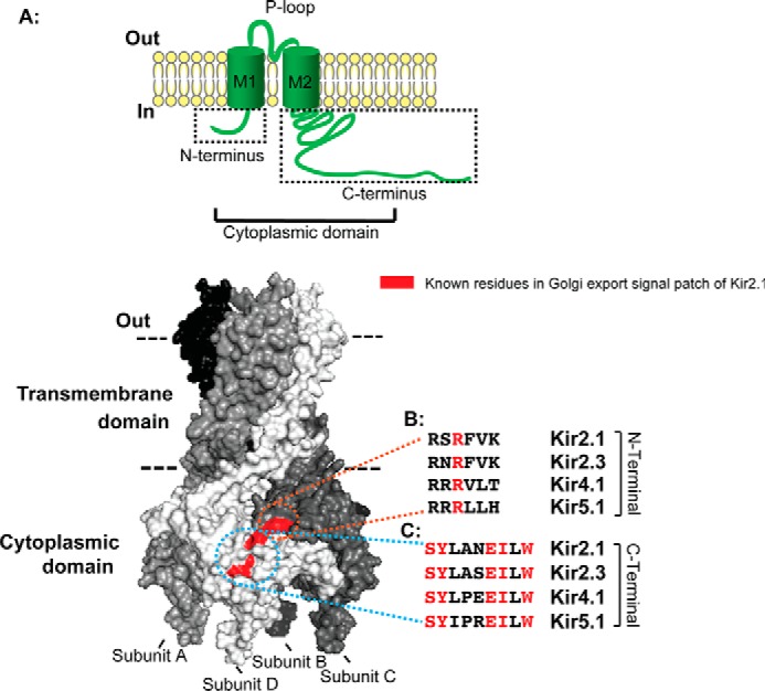
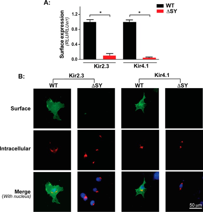
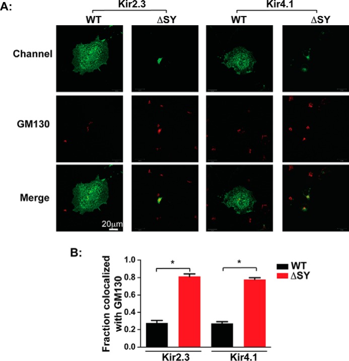
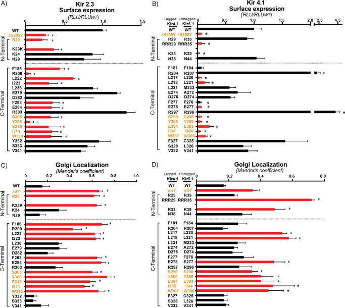
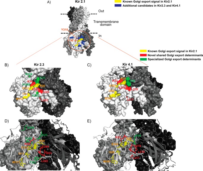
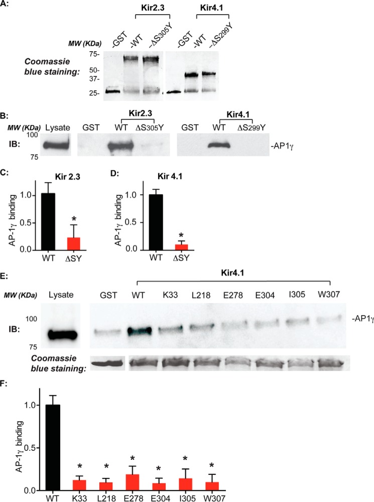
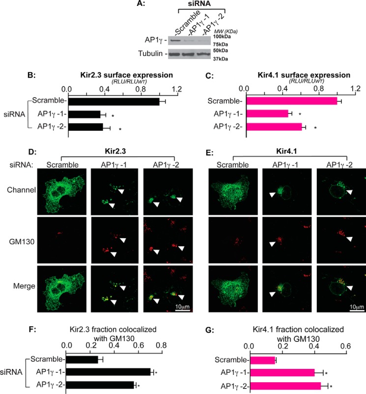
Nearly all members of the inwardly rectifying potassium (Kir) channel family share a cytoplasmic domain structure that serves as an unusual AP-1 clathrin adaptor-dependent Golgi export signal in one Kir channel, Kir2.1 (KCNJ2), raising the question whether Kir channels share a common Golgi export mechanism. Here we explore this idea, focusing on two structurally and functionally divergent Kir family members, Kir2.3 (KCNJ4) and Kir4.1/5.1 (KCNJ10/16), which have ∼50% amino identity. We found that Golgi export of both channels is blocked upon siRNA-mediated knockdown of the AP-1 γ subunit, as predicted for the common AP-1-dependent trafficking process. A comprehensive mutagenic analysis, guided by homology mapping in atomic resolution models of Kir2.1, Kir2.3, and Kir4.1/5.1, identified a common structure that serves as a recognition site for AP-1 binding and governs Golgi export. Larger than realized from previous studies with Kir2.1, the signal is created by a patch of residues distributed at the confluence of cytoplasmic N and C termini. The signal involves a stretch of hydrophobic residues from the C-terminal region that form a hydrophobic cleft, an adjacent cluster of basic residues within the N terminus, and a potential network of salt bridges that join the N- and C-terminal poles together. Because patch formation and AP-1 binding are dependent on proper folding of the cytoplasmic domains, the signal provides a common quality control mechanism at the Golgi for Kir channels. These findings identify a new proteostatic mechanism that couples protein folding of channels to forward trafficking in the secretory pathway.

Keywords: adaptor protein; clathrin; intracellular trafficking; potassium channel; protein trafficking (Golgi).


Figures

FIGURE 1.
FIGURE 1.
The Kir2.1 Golgi export signal patch is conserved in diverse Kir channels. A, schematic of a single Kir channel subunit. M1 and M2, transmembrane helix domains 1 and 2 with cytoplasmic N and C termini, which fold into a cytoplasmic domain. B and C, surface representation of a Kir2.1 tetramer model based on the crystal structure of chicken Kir2.2 (PDB code 3JYC) (47). The previously identified residues in the Golgi export signal patch of Kir2.1 with a surface-exposed side chain are marked in red. The Golgi export signal patch of Kir2.1 is embedded in the tertiary structure, minimally comprised of an arginine residue (R) from the N-terminal pole of the cytoplasmic domain of subunit C (B), juxtaposing a track of residues [SY]XXX[EI]X[W] from the C-terminal pole of the cytoplasmic domain of subunit D (C). Note that the known residues (red) of the Kir2.1 Golgi export signal patch are conserved in Kir2.1, 2.3, Kir4.1, and Kir5.1 (B and C).
FIGURE 2.
FIGURE 2.
The common ΔSY mutation blocks channel surface expression. A, surface expression quantified by HA antibody binding and luminometry in COS7 cells transiently expressing external HA-tagged WT and ΔSY mutants of Kir2.3 and Kir4.1. *, p < 0.05 by unpaired t test. RLU, relative light unit. B, immunocytochemical analysis of external HA-tagged Kir2.3 and Kir4.1 channels in intact (green) and permeabilized (red) COS7 cells, counterstained with DAPI (blue) to show nuclei.
FIGURE 3.
FIGURE 3.
Kir2.3 and Kir4.1 bearing the ΔSY mutation accumulate in the Golgi. A, co-localization of Kir2.3 and Kir4.1 bearing the ΔSY mutation (green) with the Golgi marker GM130 (red) in permeabilized COS7 cells. B, quantification of co-localization. The fraction of co-localized channel is presented as Mander's coefficient (n = 30 cells from three separate transfections; *, p < 0.05 by one-way randomized ANOVA followed by Tukey's post hoc test).
FIGURE 4.
FIGURE 4.
Golgi export sequence determinants of Kir2.3 and Kir4.1 elucidated by structure-guided mutagenesis. Residue candidates for the Golgi export sequence determinants were subjected to alanine replacement mutagenesis. A and B, cell surface expression of external HA-tagged Kir2.3 and Kir4.1 channels as quantified by surface HA antibody binding (n = 3). RLU, relative light unit. C and D, quantification of Kir2.3 and Kir4.1 co-localization with the Golgi marker GM130 (n = 30 cells from three individual transfections, the fraction of co-localized channel is presented as Mander's coefficient). Red bars, Golgi export sequence determinants; orange text, previously identified residues in the Kir 2.1 Golgi export signal. *, p < 0.05 by one-way randomized ANOVA followed by Dunnett's post hoc test.
FIGURE 5.
FIGURE 5.
The Golgi export signal patch in Kir2.3 and Kir4.1. A, surface representation of a Kir2.1 tetramer model. Tyr315 is marked (orange) to show the center of the Golgi export signal patch in Kir2.1. Yellow, known surface-exposed residues in the Golgi export signal patch of Kir2.1; blue, additional candidates with a surface-exposed side chain for Golgi export sequence determinants (∼30-Å radius). B and C, surface representation of the entire cytoplasmic domain of Kir2.3 and Kir4.1 tetramer models. D and E, ribbon display of two subunits against a surface rendering of the other two subunits for Kir2.3 and Kir4.1. Relevant residues are color-coded.
FIGURE 6.
FIGURE 6.
Common Golgi export sequence determinants in Kir channels are required for AP-1 interaction. A, GST and GST fusion proteins of the entire Kir2.3 and 4.1 cytoplasmic domain (WT and ΔSY mutants) following gel electrophoresis and Coomassie Brilliant Blue staining. MW, molecular weight. B, ΔSY mutations abolished AP-1 γ subunit binding. AP-1 clathrin adaptors bound to the Kir2.3 and Kir4.1 cytoplasmic domains were detected by immunoblot (IB) with an AP-1 γ subunit-specific antibody. ΔSY mutations abolished AP-1 γ subunit binding. C and D, quantification of relative AP-1 binding to Kir2.3 and Kir4.1 (n = 3; *, p < 0.05 by unpaired t test). E, mutations of the common Golgi export sequence determinants in Kir4.1 disrupt AP-1 γ subunit interaction. F, quantification of relative AP-1 binding to Kir4.1 (n = 3; *, p < 0.05 by one-way randomized ANOVA followed by Dunnett's post hoc test).
FIGURE 7.
FIGURE 7.
Golgi export of Kir channels requires the AP-1 clathrin adaptor. A, Western blots of AP-1, γ subunit and alpha-tubulin in COS-7 cells treated with two different AP-1 γ siRNA probes (AP1 γ-1 and AP1 γ-2) as well as scramble siRNA as the negative control. MW, molecular weight. B and C, AP-1 γ knockdown by each probe reduced the surface expression of Kir2.3 and Kir4.1 as measured by surface antibody binding (n = 6; *, p < 0.05 by one-way randomized ANOVA followed by Dunnett's post hoc test). RLU, relative light unit. D and E, cellular localization of HA-tagged Kir2.3 and Kir4.1 (green) and the Golgi marker GM130 (red) in permeabilized COS-7 cells after transfection with the indicated RNAi probes. Arrowheads, cells where channel co-localized with GM130. F and G, quantification of Kir2.3 and Kir4.1 co-localization with the Golgi marker GM130, presented as the fraction of co-localized channel with GM130, in COS-7 cells transfected with the indicated RNAi probes (n = 20 cells from three individual transfections; *, p < 0.05 by one-way randomized ANOVA followed by Dunnett's post hoc test).

References

    1. Deborde S., Perret E., Gravotta D., Deora A., Salvarezza S., Schreiner R., and Rodriguez-Boulan E. (2008) Clathrin is a key regulator of basolateral polarity. Nature 452, 719–723 - PMC - PubMed
    1. Farías G. G., Cuitino L., Guo X., Ren X., Jarnik M., Mattera R., and Bonifacino J. S. (2012) Signal-mediated, AP-1/clathrin-dependent sorting of transmembrane receptors to the somatodendritic domain of hippocampal neurons. Neuron 75, 810–823 - PMC - PubMed
    1. Gravotta D., Carvajal-Gonzalez J. M., Mattera R., Deborde S., Banfelder J. R., Bonifacino J. S., and Rodriguez-Boulan E. (2012) The clathrin adaptor AP-1A mediates basolateral polarity. Dev. Cell 22, 811–823 - PMC - PubMed
    1. Traub L. M., and Kornfeld S. (1997) The trans-Golgi network: a late secretory sorting station. Curr. Opin. Cell Biol. 9, 527–533 - PubMed
    1. Traub L. M., and Bonifacino J. S. (2013) Cargo recognition in clathrin-mediated endocytosis. Cold Spring Harb. Perspect. Biol. 5, a016790. - PMC - PubMed

Publication types

MeSH terms

Substances

Associated data

LinkOut - more resources