Review
. 2016 May 25:20:13.
doi: 10.1186/s40824-016-0060-8. eCollection 2016.

Antifibrotic therapies to control cardiac fibrosis

Affiliations
Review

Antifibrotic therapies to control cardiac fibrosis

Zhaobo Fan et al. Biomater Res. .

Abstract

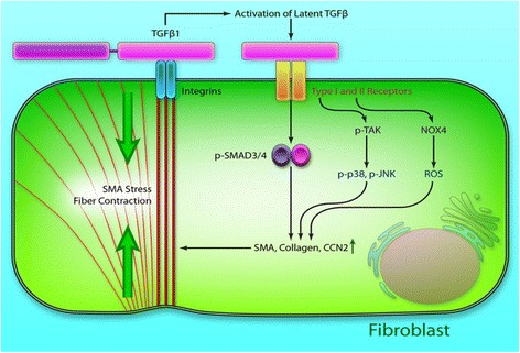
Cardiac fibrosis occurs naturally after myocardial infarction. While the initially formed fibrotic tissue prevents the infarcted heart tissue from rupture, the progression of cardiac fibrosis continuously expands the size of fibrotic tissue and causes cardiac function decrease. Cardiac fibrosis eventually evolves the infarcted hearts into heart failure. Inhibiting cardiac fibrosis from progressing is critical to prevent heart failure. However, there is no efficient therapeutic approach currently available. Myofibroblasts are primarily responsible for cardiac fibrosis. They are formed by cardiac fibroblast differentiation, fibrocyte differentiation, epithelial to mesenchymal transdifferentiation, and endothelial to mesenchymal transition, driven by cytokines such as transforming growth factor beta (TGF-β), angiotensin II and platelet-derived growth factor (PDGF). The approaches that inhibit myofibroblast formation have been demonstrated to prevent cardiac fibrosis, including systemic delivery of antifibrotic drugs, localized delivery of biomaterials, localized delivery of biomaterials and antifibrotic drugs, and localized delivery of cells using biomaterials. This review addresses current progresses in cardiac fibrosis therapies.

Keywords: Antifibrotic therapy; Cardiac fibroblasts; Cardiac fibrosis; Myocardial infarction; Myofibroblasts.


Figures

Fig. 1
Fig. 1
TGF-β signaling in fibroblasts. Latent TGF-β binds to its type I and II receptors, and activates the canonical Smad3/4 pathway and the noncanonical TGF-β–activated kinase-1 (TAK1)/p38/c-Jun N-terminal kinase (JNK) and NADPH oxidase 4 (NOX4)/reactive oxygen species (ROS) pathway resulting in induction of fibrogenic genes, such as α-smooth muscle actin (α-SMA) and collagen. (Reprinted from Leask A. [10])
Fig. 2
Fig. 2
Effect of time and hydrogel injection on the expression of TNF-α (a), IL-1β (b) and IL-6 (c) in infarcted left ventricle. * indicates significant differences between groups. Rats were divided into 3 injection treatment groups (immediately after MI (IM), 3 d after MI (3D) and 2 w after MI (2 W)) and 2 control groups (healthy and MI without treatments). (Reprinted from Yoshizumi et al. [47])
Fig. 3
Fig. 3
Ventricular wall histology for rat hearts 10 w after MI. Rats were divided into 3 injection treatment groups (immediately after MI (IM), 3 d after MI (3D) and 2 w after MI (2 W)) and 2 control groups (healthy and MI without treatments). Representative Masson’s trichrome stained cross-sections: a Healthy control, b MI control, c, f, i IM group, d, g, j 3D group, e, h, k 2 W group. A-H scale bars = 1 mm. Orange arrows point to the hydrogel residues, black arrows point to foreign body giant cells. Wall thickness (l) and infarction size (m) were measured from the complete set of these images. * indicates significant differences between groups. (Reprinted from Yoshizumi et al. [47])
Fig. 4
Fig. 4
The sequential IGF-1/HGF delivery using alginate hydrogel reduces fibrosis. a Representative photomicrographs of Masson’s trichrome staining (collagen-rich areas in blue and healthy myocardium in red), scar area. Bar = 500 μm. b Fibrotic content of the scar. * p < 0.05. (Reprinted from Ruvinov et al. [66])
Fig. 5
Fig. 5
Left ventricular (LV) myocardial histopathological changes on post-infarct day 42. a to d Microscopic identification of myocardial fibrosis over LV after Masson’s Trichrome staining. e Mean fibrotic area. fi Protein expressions of fibrotic (TGF-β and Smad3) and antifibrotic (Smad1/5 and BBMP-2) biomarkers in infarct area of LV myocardium. * vs. other groups with different symbols (*, †, ‡, §), p < 0.05 for all groups. AMI: acute MI. ADMSC: adipose derived MSC. PRF: platelet-rich fibrin. (Reprinted from Sun et al. [123])

References

    1. Dobaczewski M, de Haan JJ, Frangogiannis NG. The extracellular matrix modulates fibroblast phenotype and function in the infarcted myocardium. J Cardiovasc Transl Res. 2012;5:837–47. doi: 10.1007/s12265-012-9406-3. - DOI - PMC - PubMed
    1. Gonzalez A, Ravassa S, Beaumont J, Lopez B, Diez J. New targets to treat the structural remodeling of the myocardium. J Am Coll Cardiol. 2011;58:1833–43. doi: 10.1016/j.jacc.2011.06.058. - DOI - PubMed
    1. van den Borne SW, Diez J, Blankesteijn WM, Verjans J, Hofstra L, Narula J. Myocardial remodeling after infarction: the role of myofibroblasts. Nat Rev Cardiol. 2010;7:30–7. doi: 10.1038/nrcardio.2009.199. - DOI - PubMed
    1. Yarbrough WM, Mukherjee R, Stroud RE, Rivers WT, Oelsen JM, Dixon JA, et al. Progressive induction of left ventricular pressure overload in a large animal model elicits myocardial remodeling and a unique matrix signature. J Thorac Cardiovasc Surg. 2012;143:215–23. doi: 10.1016/j.jtcvs.2011.09.032. - DOI - PMC - PubMed
    1. Kong P, Christia P, Frangogiannis NG. The pathogenesis of cardiac fibrosis. Cell Mol Life Sci. 2014;71:549–74. doi: 10.1007/s00018-013-1349-6. - DOI - PMC - PubMed