Review
. 2016 May;16(2):e132-41.
doi: 10.18295/squmj.2016.16.02.002. Epub 2016 May 15.

Mechanisms of Diabetes-Induced Liver Damage: The role of oxidative stress and inflammation

Affiliations
Review

Mechanisms of Diabetes-Induced Liver Damage: The role of oxidative stress and inflammation

Jamaludin Mohamed et al. Sultan Qaboos Univ Med J. 2016 May.

Abstract

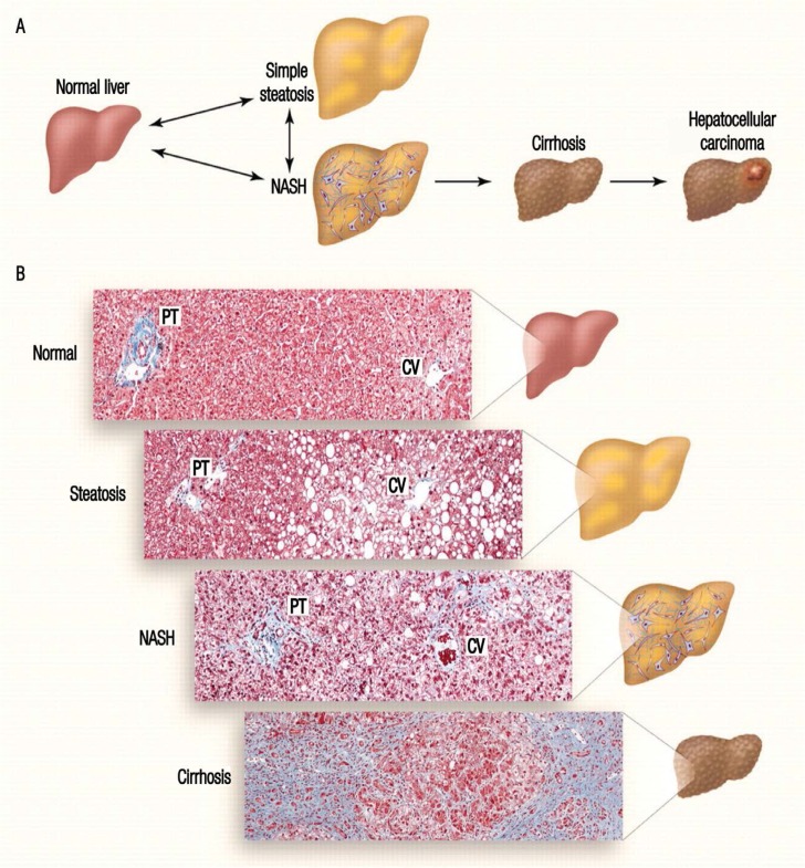
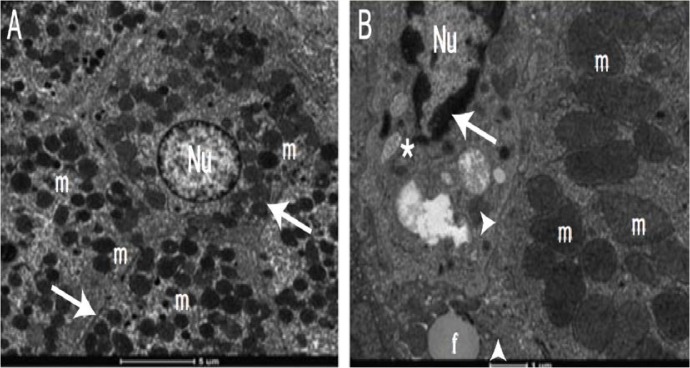
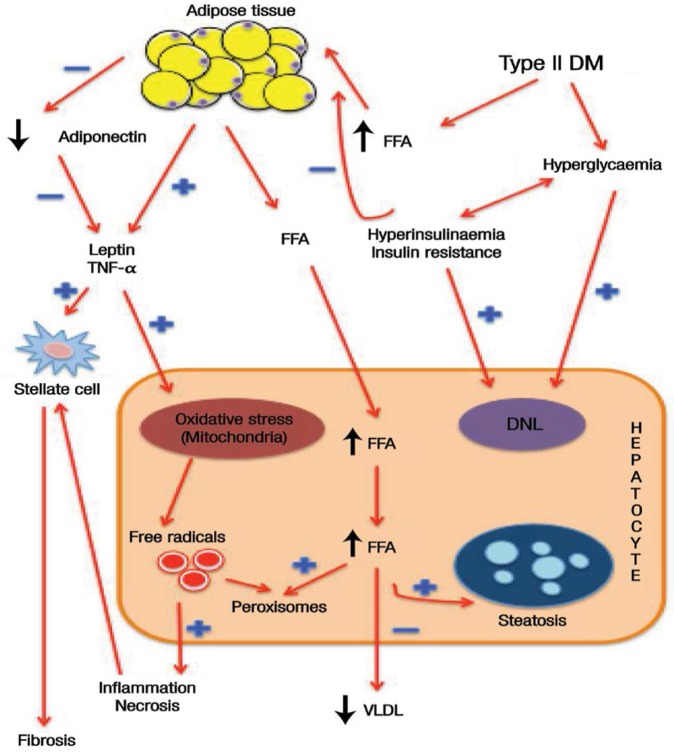
Diabetes mellitus is a non-communicable disease that occurs in both developed and developing countries. This metabolic disease affects all systems in the body, including the liver. Hyperglycaemia, mainly caused by insulin resistance, affects the metabolism of lipids, carbohydrates and proteins and can lead to non-alcoholic fatty liver disease, which can further progress to non-alcoholic steatohepatitis, cirrhosis and, finally, hepatocellular carcinomas. The underlying mechanism of diabetes that contributes to liver damage is the combination of increased oxidative stress and an aberrant inflammatory response; this activates the transcription of pro-apoptotic genes and damages hepatocytes. Significant involvement of pro-inflammatory cytokines-including interleukin (IL)-1β, IL-6 and tumour necrosis factor-α-exacerbates the accumulation of oxidative damage products in the liver, such as malondialdehyde, fluorescent pigments and conjugated dienes. This review summarises the biochemical, histological and macromolecular changes that contribute to oxidative liver damage among diabetic individuals.

Keywords: Diabetes Mellitus; Inflammation; Liver Diseases; Oxidative Stress.


Figures

Figure 1:
Figure 1:
Progression of liver damage. In diabetes mellitus, insulin resistance and hyperinsulinaemia cause non-alcoholic fatty liver disease and progress to non-alcoholic steatohepatitis (NASH) which manifests as inflammation and necrosis. Prolonged NASH will lead to liver fibrosis, known as cirrhosis, and finally hepatocellular carcinomas and end-stage liver disease. NASH = non-alcoholic steatohepatitis; PT = portal triad; CV = central vein. Reproduced with permission from Cohen JC, Horton JD, Hobbs HH. Human fatty liver disease: Old questions and new insights.
Figure 2A&B:
Figure 2A&B:
Electron micrographs of a (A) healthy and (B) diabetic liver. Note the difference in appearance between the hepatocyte nuclei (Nu), organelles, mitochondria (m) and Disse’s space (arrows). In the diabetic liver, there is accumulation of homogenous substances in the nuclear chromatin (*), reduction in the number of organelles, loss of mitochondrial cristae (▲) and granular degeneration (►) in the hepatocytes. Reproduced with permission from Lucchesi, AN, Cassettari LN, Spadella CT. Alloxan-induced diabetes causes morphological and ultrastructural changes in rat liver that resemble the natural history of chronic fatty liver disease in humans.
Figure 3:
Figure 3:
Diagram showing the mechanism of liver damage in diabetes mellitus. Insulin resistance causes peripheral adipocytes to undergo lipolysis. Free fatty acids are then released to the bloodstream and eventually accumulate in the liver. At the same time, adipocytokines release tumour necrosis factor-α and leptin, worsening the hepatocyte damage by increasing oxidative stress in the mitochondria. The combined action of mitochondrial oxidative stress, hyperinsulinaemia and hyperglycaemia produce free radicals which in turn induce inflammation and cellular necrosis. Tissue inflammation stimulates the hepatic stellate cells to produce collagen, leading to fibrosis, cirrhosis and, finally, hepatocellular carcinomas. DM = diabetes mellitus; FFA = free fatty acids; TNF = tumour necrosis factor; DNL = de novo lipogenesis; VLDL = very-low-density lipoprotein.

References

    1. Danaei G, Finucane MM, Lu Y, Singh GM, Cowan MJ, Paciorek CJ, et al. National, regional, and global trends in fasting plasma glucose and diabetes prevalence since 1980: Systematic analysis of health examination surveys and epidemiological studies with 370 country-years and 2.7 million participants. Lancet. 2011;378:31–40. doi: 10.1016/s0140-6736(11)60679-x. - DOI - PubMed
    1. Reid AE. Non-alcoholic fatty liver disease. In: Feldman M, Friedman LS, Brandt LJ, editors. Sleisenger and Fordtran’s Gastrointestinal and Liver Disease: Pathophysiology/diagnosis/management. 8th ed. St Louis, Missouri, USA: Saunders; 2006. pp. 1772–99.
    1. Levinthal GN, Tavill AS. Liver disease and diabetes mellitus. Clin Diabetes. 1999;17:73.
    1. Guven A, Yavuz O, Cam M, Ercan F, Bukan N, Comunoglu C, et al. Effects of melatonin on streptozotocin-induced diabetic liver injury in rats. Acta Histochem. 2006;108:85–93. doi: 10.1016/j.acthis.2006.03.005. - DOI - PubMed
    1. Larter CZ, Farrell GC. Insulin resistance, adiponectin, cytokines in NASH: Which is the best target to treat? J Hepatol. 2006;44:253–61. doi: 10.1016/j.jhep.2005.11.030. - DOI - PubMed

LinkOut - more resources